대한소화기학회지 2000;36:473 - 482
1)
접수: 2000년 6월 17일, 승인: 2000년 9월 8일 연락처: 김재광, 150-713, 서울시 영등포구 여의도동 62
가톨릭대학교 의과대학부속 성모병원 내과 Tel: (02) 3779-1079, Fax: (02) 780-3132
※ 본 논문은 1999년 대한소화기학회 추계학술대회에서 구연발표되었음.
※ 본 논문은 1999년 가톨릭중앙의료원 연구보조비의 일 부로 이루어졌음.
서 론
E-cadherin은 칼슘의존성 세포간 접착분자로 막 당단백질로 구성되어 있으며, 정상 조직 구조의 세 포막 형성과 유지에 매우 중요한 역할을 담당하고
위와 대장의 선암 및 선종에서 E - ca dh er in / - ca t en in 복합체의 발현
가톨릭대학교 의과대학 내과학교실, 임상병리학교실*
문성배・김재광・박수헌・한준열・최명규・이교영*・강창석*・정규원・선희식
E x p r e s s i o n o f E -c a d h e r i n a n d -c a t e n i n c o m p l e x i n H u m a n Ad e n o c a r c i n o m a a n d Ad e n o m a o f S t o m a c h a n d Co lo n
S u n g B a e M o o n , M .D ., J a e Kw a n g Ki m , M .D ., S o o H e o n P a r k , M .D ., J o o n Ye o l H a n , M .D., My u n g Gy u Ch o i , M .D., Ky o Yo u n g Le e , M.D.*, Ch a n g S u k Ka n g , M.D.*, Ky u Wo n Ch u n g , M.D. a n d He e S i k S u n , M.D.
Departments of Internal Medicine and Clinical Pathology*, College of Medicine, The Catholic University of Korea, Seoul, Korea
Background/Aims: Poor expressions of E-cadherin and β-catenin complex in the immunohisto-
chemical staining were frequently observed in the tumors. We investigated the correlation between the expressions of E-cadherin and β-catenin and the clinicopathologic factors of adenocarcinomas and adenomas in the stomach and colon. Methods: Immunohistochemical staining of E-cadherin and β-catenin was performed for the adenocarcinomas of stomach (65) and colon (98) and adenomas of stomach (39) and colon (47). Results: The abnormal stainings of E-cadherin and β-catenin were observed in the most adenocarcinomas of the stomach and colon, and in the certain numbers of adenomas of the stomach and colon. This abnormality was frequently observed in the undif- ferentiated and infiltrative adenocarcinoma. The abnormal staining of E-cadherin and β-catenin in gastric cancer was closely correlated with poor survival, but not in the colon cancer. Conclusions:These findings show that the decreased expression of E-cadherin and β-catenin are involved in progress of stomach and colon cancer. The cytoplasmic expression of E-cadherin and β-catenin in some adenocarcinoma seems to be also involved in the carcinogenesis but needs further study. (Kor
J Gastroenterol 2000;36:473 - 482)
Key Words: E-cadherin, β-catenin, Adenocarcinoma, Adenoma, Stomach, Colon
474 대한소화기학회지 : 제 36 권 제 4 호 2000
있다.1 암의 침윤과 전이는 여러 과정이 관련되지만 초기 단계에서는 세포간 결합의 붕괴가 관련된다.
E-cadherin (120 Kd)은 세포막의 접착대(zonula adherens)에 집중되어 있고, 세포질에 있는 α-catenin (102 Kd), β-catenin (95 Kd), γ-catenin (80 Kd) 등 과 결합하여 E-cadherin/catenin 복합체를 형성하고 있다.2-4 또한 catenin들은 E-cadherin과 세포골격의 actin filament 사이의 연결 역할을 하여 세포구조를 유지하고 있다. 이러한 복합체의 변이는 세포간 결 합력을 저하시키고 암의 전이 및 침윤을 일으킨 다.5-7 E-cadherin과 α-catenin의 발현 감소는 여러 암 조직(식도암, 위암, 대장암, 폐암, 유방암, 자궁경부 암 등)의 분화 정도와 침윤, 전이, 예후와 밀접하게 연관되며, β-catenin의 발현 감소는 위암 및 대장암 에서 높은 침윤도 및 불량한 예후와 연관된다.8-12
β-catenin은 세포막에 있는 E-cadherin/catenin 복 합체의 기능을 조절하고 있고, 세포질에서 APC 단 백질, GSK3β, axin(또는 conductin) 등과 상호작용 을 통하여 세포 내외 간의 신호전달 역할을 담당하 고 있다. 정상 조직에서 세포 내 유리 β-catenin은 APC 단백질 결합 여부에 의하여 조절되지만 APC 유전자 또는 β-catenin에 변이가 발생하면 유리 β- atenin이 증가되어 악성화를 일으킨다.13,14 β-atenin 은 위암, 대장암, 간암 및 췌장암의 발생에 일부 관 여하며, 특히 대장암에서 adenoma-carcinoma se- quence의 초기 단계에 밀접하게 관련됨이 알려져 있다.4,15
여러 암조직에서 E-cadherin과 β-catenin의 발현 정도에 대한 많은 보고가 있으나 위 및 대장의 선종 에서 E-cadherin과 β-catenin의 발현에 대한 보고는 드물다. 이에 저자들은 위와 대장의 선암 및 선종에 서 E-cadherin과 β-catenin의 발현 양상과 임상병리 학적인 인자들과의 연관성에 대하여 알아보았다.
대상 및 방법
1. 대 상
위 및 대장의 선암으로 절제술을 받은 환자의 암 조직에서 파라핀 포매 조직의 보관 상태가 양호한 위선암 65예와 같은 환자의 암 주변 정상 부위 23
예, 대장선암 98예와 같은 환자의 암 주변 정상 부 위 28예, 위 및 대장의 선종으로 점막절제술을 시행 받은 환자의 위선종 39예, 대장선종 47예를 대상으 로 하였다. 암 주변 정상 부위는 선암의 경계 부위 에서 최소한 5 cm 이상 떨어진 정상 조직에서 얻었 다. 위선암 및 대장선암의 임상적 병기는 American Joint Committee on Cancer (1993)의 분류에 따랐다.
위 및 대장의 선암 환자는 모두 수술 전에 항암제 또는 방사선치료를 받지 않았다.
2. 면역조직화학적 염색
E-cadherin과 β-catenin에 대한 일차항체(Zymed Laboratories Inc., U.S.A.)를 이용하여 labeled-[strept]- avidin-biotin (LAB-SA)방법으로 면역조직화학적 염 색을 시행하였다. 10% 중성 완충 포르말린에 고정 시킨 조직의 파라핀 포매 조직을 4 μm 두께로 박절 한 후 탈파라핀과 함수과정을 거쳐 수세하였고, 10 분간 peroxidase quenching 용액으로 처리하였다. 이 후 차단혈청용액에 10분간 작용시켜 비특이성 염색 을 차단하였다. E-cadherin과 β-catenin에 대한 일차 항체는 각각 1:50배, 1:40배로 희석한 후 약 1시간 실온에서 반응시켰고, 이차항체인 biotinylated rabbit anti mouse immunoglobin (HISTOSTAINTM SP kit, Zymed Laboratories Inc., U.S.A)을 10분간 반응시켰 다. Horse radish peroxidase와 색소원으로 반응시킨 후 헤마톡실린으로 염색하였다.
조직 표본은 200배율로 관찰하였으며, 정상 조직 을 기준으로 모든 세포의 세포막에서 최고강도의 70%이상의 발현을 보이는 경우는 정상, 70% 이하 의 발현 강도나 음성 발현, 70% 이상의 발현 강도 를 보이지만 세포질 또는 핵 내에 발현을 보이는 경 우는 비정상으로 분류하였다(Fig. 1). 판독의 정확도 를 기하기 위하여 이미 판독한 조직을 무작위 추출 하여 반복 관찰하였다.
3. 통계 분석
각 조직의 발현 정도는 chi-sqaure test와 Fisher' s exact test를 사용하여 비교, 분석하였다. 위 및 대장 의 선암에서 조직 분화 정도, 암 침윤, 림프절 전이 에 따른 발현은 chi-sqaure test를 사용하여 분석하였
문성배 외 8인. 위와 대장의 선암 및 선종에서 E-cadherin/ -catenin 복합체의 발현 475
고, 예후는 Kaplan-Meier' s product estimates를 사용 하여 분석하였다. 유의성은 5% 이하로 하였다.
결 과
1. 위선암 및 위선종에서 E - ca dh er in 과 β- ca t e n in 의 발현 정도
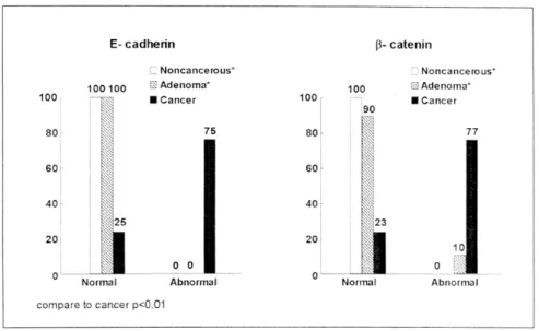
E-cadherin은 암 주변 정상 부위와 위선종에서 모 두(23/23; 39/39) 정상으로 발현되었으나, 위선암의 경우 24% (16/67)에서만 정상으로 발현되었다(Fischer exact test, p<0.01). β-catenin의 경우 암 주변 정상 부위에서는 100% (23/23), 위선종에서는 90% (35/
39)에서 정상으로 발현되었으나, 위선암에서는 16%
(11/67)만 정상으로 발현되었다(Fischer exact test,
p<0.01)(Fig. 2).
2. 위선암에서 조직학적 유형과 암 침윤, 림프절 전이, 예후와 E- cadherin 및 β- catenin의 발현 정도의 관계
E-cadherin은 분화도가 고분화에서 저분화로 갈 수록, 암 침윤이 진행될수록 정상 발현은 감소하였 다(chi-sqaure test, p<0.05; chi-sqaure test, p<0.01).
그러나 림프절 전이 여부와 E-cadherin 발현 사이에 통계학적인 유의성은 없었다(chi-sqaure test, p>0.05) (Table 1).
β-catenin은 분화도가 고분화에서 저분화로 갈수 록, 암 침윤이 진행될수록, 림프절 전이가 있을수록 정상 발현은 감소하였다(chi-sqaure test, p<0.05;
Fig. 1. Immunohistochemical staining of E-cadherin and β-cateninin on normal epithelium and cancer. (A) normal tissue was located 5 cm from the boundary of adenocarcinoma. (B) and (C) moderately differentiated gastric adenocarcinoma. D: well differentiated gastric adenocarcinoma. A was expressed as normal staining. (B), (C) and (D) were expressed as abnormal staining. In specimen (A), arrow indicate strong E-cadherin staining in the cytomembrane. Specimen (B) showed inhomogenous staining of E-cadherin, which was expressed less than 70%
of normal staining. Specimen C was negative for β-cateninin staining. Specimen D reveals intense cytoplasmic staining (arrow) of β-cateninin. {Hematoxylin stain, ×200 (A-C), ×400 (D)}.
476 The Korean Journal of Gastroenterology : Vol. 36, No. 4, 2000
chi-sqaure test, p<0.01; chi-sqaure test, p<0.05)(Table 2).
E-cadherin 및 β-catenin의 발현 정도(정상과 비정 상)에 따라 장기간 생존율을 비교하였을 때 정상 발 현군에서 비정상 발현군에 비하여 통계적으로 유의 하게 생존율이 높았다(Kaplan-Meier' s product estimates,
p<0.05)(Fig. 3).
3. 대장선암 및 대장선종에서 E - ca dh er in 과 β- cat en in 의 발현 정도
E-cadherin은 암 주변 정상 부위에서는 100% (28/
Table 1. Staining Intensity of E-cadherin and Clinicopathologic Features in Gastric Cancer
Pathologic features No.
E-cadherin (%) Normal Abnormal Differentiaton*
Well Moderately Poorly T categories†
T1/T2 T3/T4 N categories
N0 N1-3
6 23 38 15 52 25 42
4 (67) 6 (26) 6 (16) 8 (53) 8 (16) 8 (32) 8 (19)
2 (23) 17 (74) 32 (84) 7 (47) 44 (84) 17 (68) 34 (81)
* p<0.05.
†p<0.01.
p>0.05.
Fig. 2. Immunohistochemical staining intensity of E-cadherin and β-catenin in the gastric cancer and adenoma. The abnormal stainings of E-cadherin and β-catenin were more observed in gastric cancer than adenoma and normal tissue (Fischer exact test, p<0.01).
Table 2. Staining Intensity of β-catenin and Clini- copathologic Features in Gastric Cancer
Pathologic features No.
β-catenin (%) Normal Abnormal Differentiaton*
Well Moderately Poorly T categories†
T1/T2 T3/T4 N categories
N0 N1-3
6 23 38 15 52 25 42
4 (67) 6 (26) 5 (13) 7 (47) 8 (16) 9 (36) 6 (14)
2 (23) 17 (74) 33 (87) 8 (53) 44 (84) 16 (64) 36 (86)
* p<0.05.
†p<0.01.
p<0.05.
Moon, et al. E-cadherin/ -catenin in Human Adenocarcinoma and Adenoma of Stomach and Colon 477
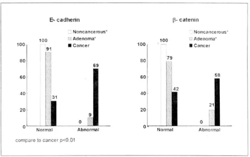
28), 대장선종에서는 91% (43/47)에서 정상으로 발 현되었으나, 대장선암의 경우 31% (30/98)에서만 정 상으로 발현되었다(Fischer exact test, p<0.01). β- catenin은 암 주변 정상 부위에서는 100% (23/23),
대장선종에서는 79% (37/47)에서 정상으로 발현되 었으나, 대장선암의 경우 42% (41/98)에서만 정상으 로 발현되었다(Fischer exact test, p<0.01)(Fig. 4).
Fig. 4. Immunohistochemical staining intensity of E-cadherin and β-catenin in the colon cancer and adenoma. The abnormal stainings of E-cadherin and β-catenin were more observed in colon cancer than adenoma and normal tissue (Fischer exact test, p<0.01).
Fig. 3. Survial curves in gastric cancer. Overall survival rates of normal staining (dotted line) of E-cadherin and β-catenin in gastric cancer presented significant differences from abnormal staining (solid line) (Kaplan-Meier' s product estimates, p<0.05).
478 대한소화기학회지 : 제 36 권 제 4 호 2000
4. 대장선암에서 조직학적 유형과 암 침윤, 림프절 전이, 예후와 E - cadh er in 및 β- c a t e n in 의 발현 정도의 관계
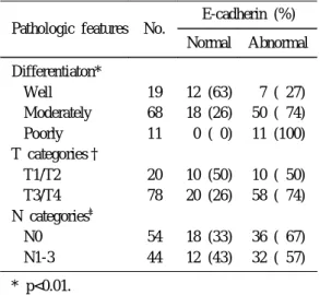
E-cadherin 및 β-catenin은 분화도가 고분화에서
저분화로 갈수록, 암 침윤이 진행될수록 정상 발현 이 감소하였다(chi-sqaure test, p<0.01; chi-sqaure test, p<0.05). 그러나 림프절 전이 여부와 발현 정도 사이에 통계학적인 유의성은 없었다(chi-sqaure test, p>0.05) (Table 3, 4).
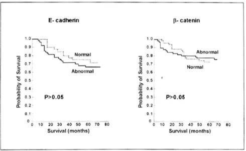
E-cadherin 및 β-catenin의 발현 정도(정상과 비정 상)에 따라 장기간 생존율을 비교하였을 때 통계적 인 유의성은 없었다(Kaplan-Meier' s product estima- tes, p>0.05)(Fig. 5).
5. 위선암과 대장선암에서 E- ca dherin 및 β- ca t e n in 의 발현 비교
위선암과 대장선암 사이에 E-cadherin의 발현 정 도는 유의한 차이가 없었다(chi-sqaure test, p>0.05).
그러나 β-catenin은 위선암에서 대장선암에 비하여 통계적으로 유의하게 발현이 감소하였다(chi-sqaure test, p<0.01)(Fig. 6).
6. 위선암과 대장선암에서 E- cadherin 및 β- c a t en in 의 세포질 발현
위선암에서 E-cadherin 및 β-catenin의 세포질 발 현은 각각 13% (9/67), 9% (6/67)에서 관찰되었으나 암주 변 정상 부위에 비하여 통계적 의미는 없었다 (chi-sqaure test, p>0.05). 대장선암에서 E-cadherin 및 β-catenin의 세포질 발현은 각각 14% (14/98), 18% (18/98)로 암 주변 정상 부위에 비하여 통계적 으로 유의하게 많았다(chi-sqaure test, p<0.05). β- catenin의 세포질 발현은 위선암보다 대장선암에서 많이 관찰되었으나 통계적인 유의성은 없었다(chi- sqaure test, p>0.05). E-cadherin 및 β-catenin의 세포 질 발현 유무에 따라 장기간 생존율을 비교하였을 때 통계적인 유의성은 없었다(Kaplan-Meier' s product estimates, p>0.05).
고 찰
E-cadherin과 β-catenin은 세포의 형태 유지와 세 포간 결합에 중요한 역할을 담당하며, 암조직에서 이들의 발현 감소는 암세포의 침윤과 전이에 관련되 는 것으로 알려져 있고, 여러 암조직에서 침윤이 심 Table 3. Staining Intensity of E-cadherin and
Clinicopathologic Features in Colon Cancer
Pathologic features No.
E-cadherin (%) Normal Abnormal Differentiaton*
Well Moderately Poorly T categories†
T1/T2 T3/T4 N categories
N0 N1-3
19 68 11 20 78 54 44
12 (63) 18 (26) 0 ( 0) 10 (50) 20 (26) 18 (33) 12 (43)
7 ( 27) 50 ( 74) 11 (100) 10 ( 50) 58 ( 74) 36 ( 67) 32 ( 57)
* p<0.01.
†p<0.05.
p>0.05.
Table 4. Staining Intensity of β-catenin and Clinicopathologic Features in Colon Cancer
Pathologic features No.
β-catenin(%) Normal Abnormal Differentiaton*
Well Moderately Poorly T categories†
T1/T2 T3/T4 N categories
N0 N1-3
19 68 11 20 78 54 44
8 (42) 33 (49) 0 ( 0) 13 (65) 28 (36) 23 (43) 18 (41)
11 ( 58) 35 ( 51) 11 (100) 7 ( 35) 50 ( 64) 31 ( 57) 26 ( 59)
* p<0.01.
†p<0.05.
p>0.05.
문성배 외 8인. 위와 대장의 선암 및 선종에서 E-cadherin/ -catenin 복합체의 발현 479
할수록, 원격 전이가 있을수록 E-cadherin과 β-catenin 의 발현이 감소되고 예후가 나쁘다고 한다.5,10-12,16,17 본 연구에서 위 및 대장의 선종이나 암 주변 정상 조직의 E-cadherin과 β-catenin은 강하게 발현되었으 나 선암에서는 현저히 발현이 감소되었다. 위선암의
임상병리학적인 병기와 E-cadherin과 β-catenin과의 관계를 살펴보면 정상 발현은 분화 정도가 고분화 에서 저분화로 진행될수록 그리고 침윤이 심해질수 록 감소하였다. 림프절 전이의 경우 β-catenin은 정 상 발현이 감소하였으나 E-cadherin은 상관이 없었 Fig. 6. Immunohistochemical staining intensity of E-cadherin and β-catenin in the colon and
gastric cancer. There was not significant difference in E-cadherin, but significant difference in β-catenin (chi-sqaure test, p<0.01).
Fig. 5. Survial curves in colon cancer. Overall survival rates of normal staining (dotted line) of E-cadherin and β-catenin in colon cancer presented no differences from abnormal staining (solid line) (Kaplan-Meier' s product estimates, p>0.05).
480 The Korean Journal of Gastroenterology : Vol. 36, No. 4, 2000
다. 대장선암의 임상병리학적인 병기와 E-cadherin 과 β- catenin과의 관계를 살펴보면 정상 발현은 분 화 정도가 고분화에서 저분화로 진행될수록 그리고 침윤이 심해질수록 감소되었으나, 림프절 전이 여부 와는 상관이 없었다. 위 및 대장선암의 원격 전이는 예가 부족하여 알아보지 못하였다. 위선암에서 E-cadherin과 β-catenin의 발현 감소가 있을 경우 예 후가 나쁘다고 알려져 있으며,12 본 연구에서도 위선 암에서 비정상 발현은 정상 발현에 비하여 예후가 불량하였으나, 대장선암에서는 정상과 비정상 발현 의 예후 차이는 없었다. 대장선암의 경우 예후에 관 한 다른 보고가 없어 직접 비교할 수 없었다. 종양 에서 세포간 결합력 상실은 E-cadherin, α-catenin 및 β-catenin의 변이로 E-cadherin/catenin 복합체의 기 능이 상실되어 발생한다.18,19 최근 E-cadherin 및 α -catenin의 변이보다는 β-catenin의 변이가 더 중요 한 역할을 한다고 알려져 있다. β-catenin의 변이는 세포막에서 E-cadherin의 기능 상실을 유발하고, 세 포질에서 신호전달의 변화로 암진행에 관여한다.6,20 본 연구에서 E-cadherin 및 β-catenin의 세포질 및 핵 내 발현이 일부 조직에서 관찰되었다. E-cadherin 은 위선암 및 대장선암에서만 관찰되었으며, β- catenin의 경우 위 및 대장선암뿐 아니라 위선종 1예, 대장선종 5예에서도 관찰되었다. 정상 세포의 β- catenin은 세포질 내에서 APC 단백질, GSK3β, axin(또는 conductin) 등과 결합하여 복합체를 형성 하고, APC 유전자 및 GSK3β에 의해 유리 β-catenin 이 조절된다.19 가족성 선종성 용종증이나 대장암에 서는 주로 APC 유전자의 변이 또는 β-catenin 및 axin의 변이로 인하여 세포 내에 유리 β-catenin이 크게 증가되고 핵 내로 들어가 TCF-4 (T cell factor 4)/LEF (lymphoid-enhance-factor)와 결합하여 C-MYC 유전자를 활성화하여 악성화로 진행한다.19,21 이러 한 APC 유전자와 β-catenin과의 관련성은 대장암 뿐 아니라 위암, 간암, 췌장암, 자궁암 등 여러 암조 직에서도 밝혀지고 있다.6,21-24 또한 β-catenin- TCF/LEF 복합체의 활성화는 세포 내 uPAR (uro- kinase-type plasminogen activator receptor)의 활성화 시키고, 세포막에서 ZO-1 (zonula occludens-1) 유전 자의 작용을 억제한다.21 uPAR은 세포 표면에서
plasmin을 활성화시켜 세포 외 기질의 단백질분해능 을 조장하고 세포이동에 관여한다. 또한 접착대에 있는 ZO-1 단백질은 세포막에 있는 E-cadherin 및 β-catenin과 상호작용하며, ZO-1의 감소는 극성 상 실을 유발한다. 즉, β-catenin-TCF/LEF 복합체의 활 성화는 세포막의 극성 상실과 단백분해능 증가를 초래하여 세포간 결합력 상실과 침윤을 일으킨다.
이는 β-catenin이 E-cadherin/catenin 복합체를 통한 기능 조절 외에 세포막에 직접 작용하여 세포운동, 세포이동, 침윤, 전이 등과 같은 세포간 결합을 조절 한다. 본 연구에서 위 및 대장선암의 β-catenin의 세 포질 발현이 일부에서 핵 내까지 관찰된 것으로 보 아 이러한 기전을 추측할 수 있다.
발암과정에서 세포질에 증가된 β-catenin이 세포 막에 존재하는 E-cadherin/catenin 복합체에 어떠한 영향을 미치는지 아직 밝혀져 있지 않다. β-catenin- TCF/LEF 복합체의 작용을 통한 ZO-1의 감소가 E-cadherin/catenin 복합체의 감소에 영향을 미친다 고 추정은 할 수 있으나, 확실히 밝혀진 바 없다. 향 후 세포막에서 E-cadherin/catenin 복합체의 발현 감 소의 기전에 대하여 연구가 필요하겠다.
선종에서 선암으로 이행하는 adenoma-carcinoma sequence에는 여러 기전(WNT/Wingless, K-ras, Trans- forming growth factor/β-catenin, P53)이 관여하는 것으로 알려져 있으며,25 선종에서 악성화의 초기 진 행에 주로 APC 유전자와 β-catenin의 변이로 인한 세포질 및 핵 내 β-catenin의 증가가 중요한 역할을 하고 있다.4,15,26,27 본 연구의 대장선암에서 β-catenin 의 세포질 발현은 18%로 위선암에 비하여 통계적으 로 의미 있게 양성률이 높았으며, 일부의 대장선종 에서도 세포질 발현이 관찰되어 adenoma-carcinoma sequence에 관련이 있을 것으로 생각되지만 예가 적 어 확신할 수 없었다. E-cadherin의 세포질 발현에 대한 보고도 있으나 임상병리학적인 연관성을 알 수는 없었다.28 본 연구에서도 대장선암에서 E- cadherin의 세포질 발현이 암 주변 정상 부위에 비 하여 통계적으로 의미 있게 관찰되었으나, E- cadherin의 세포질 발현에 대한 분자생물학적인 역 할에 대하여 아직 알려진 바가 없어 추후 연구가 필 요할 것으로 생각된다. E-cadherin 및 β-catenin의 세
Moon, et al. E-cadherin/ -catenin in Human Adenocarcinoma and Adenoma of Stomach and Colon 481
포질 발현 유무에 따른 5년 이상 생존율의 차이는 없었다(자료 생략). 참고적으로 위선종 및 선암에서 의 E-cadherin과 β-catenin의 발현 양상은 이전에 본 교실에서 시행하였던 결과와 유사하였으며, 세포질 발현에 대한 연구로 위암에서 APC 유전자 exon 11 의 이형접합성 상실과 β-catenin의 세포질 발현과의 관계를 알아보았으나 직접적인 관련이 없었다.29,30
결론적으로 위 및 대장의 선암에서 E-cadherin과 β-catenin의 정상 발현 감소는 선암의 분화도, 침윤 성과 관련이 있으며, 위암 및 대장암의 진행에 일부 관여할 것이다. β-catenin의 세포질 발현은 세포의 악성화에 관련됨이 밝혀지고 있으나 E-cadherin의 세 포질 발현과 역할은 더 많은 연구가 필요할 것이다.
요 약
목적: 위 및 대장의 선암과 선종에서 E-cadherin 과 β-catenin의 역할을 알아보기 위하여 면역조직화 학염색을 실시하여 발현 강도와 임상병리학적인 연 관성을 알아보았다. 대상 및 방법: 수술을 시행한 위선암 환자의 암조직 67예와 같은 환자의 암 주변 정상 부위 23예, 위선종 39예, 대장선암 환자의 암 조직 98예와 같은 환자의 암 주변 정상 부위 28예, 대장선종 47예를 대상으로 하였다. 파라핀 포매 조 직에서 E-cadherin과 β-catenin의 면역조직화학적 염 색을 시행하였다. 결과: 위 및 대장에서 선암의 E-cadherin 및 β-catenin의 정상 발현은 선종이나 암 주변 정상 조직에 비하여 현저히 감소하였다. 위암에 서 E-cadherin 및 β-catenin의 정상 발현은 조직 분 화가 나쁠수록, 침윤이 진행될수록 감소되었으며, 림프절 전이의 경우 β-catenin에서만 정상 발현이 감소하였다. 대장암에서 E-cadherin과 β-catenin의 정상 발현은 조직 분화가 나쁠수록, 침윤이 진행될 수록 감소되었다. 위암 및 대장암 사이에 E-cadherin 의 발현 감소율은 차이가 없었으며, β-catenin은 위 암과 대장암에서 모두 발현이 감소하였으나 위암에 서 더 감소하였다. 위암과 대장암의 일부에서 E-cadherin과 β-catenin의 세포질 발현이 관찰되었 다. 위암에서 E-cadherin과 β-catenin의 정상 발현은 비정상 발현에 비하여 예후가 좋았으나 대장암에서
는 차이가 없었다. 결론: E-cadherin과 β-catenin의 발현은 위암과 대장암의 진행에 관여할 것으로 생각 된다. 향후 E-cadherin과 β-catenin의 세포질 발현에 대한 추가적인 연구가 필요하겠다.
색인단어: E-cadherin, β-catenin, 위선암, 위선종, 대 장선암, 대장선종
참 고 문 헌
1. Hatta K, Okada TS, Yakeichi M. A monoclonal antibody disrupting calcium-dependent cell-cell adhesion of brain tissues: possible role of its target antigen in animal pattern formation. Proc Natl Acad Sci USA 1985;82:2789-2793.
2. Boller K, Vestweber D, Kemler R. Cell-adhesion molecule uvomorulin is localized in the interme- diate junctions of adult intestinal epithelial cells. J Cell Biol 1985;100:327-332.
3. Ozawa M, Kemler R. Molecular organization of the uvomorulin-catenin complex. J Cell Biol 1992;116:
989-996.
4. Rubinfeld B, Souza B, Alvert I, et al. Association of the APC gene product with beta-catenin. Science 1993;262:1731-1734.
5. Breen E, Steele G, Mercurio AM. Role of E-cad- herin/α-catenin complex in modulating cell-cell and cell-matrix adhesive properties of invasive colon carcinoma cells. Ann Surg Oncol 1995;2:378-385.
6. Caca K, Kolligs FT, Ji X, et al. β-catenin and γ -catenin mutations, but not E-cadherin inactivation, underlie T-cell factor/lymphoid enhancer factor transcriptional deregulation in gastric and pancreatic cancer. Cell Growth Differ 1999;10:369-376.
7. Mareel M, Boterberg T, Noe V, et al. E-cadherin/
catenin/cytoskeleton complex: a regulator of cancer invasion. J Cell Physiol 1997;173:271-274.
8. Takayama T, Shiozaki H, Shibamoto S, et al. β- Catenin expression in human cancers. Am J Pathol 1996;148:39-46.
9. Shimoyama Y, Nagafuchi A, Fujita S, et al. Cad- herin dysfunction in a human cancer cell line:
482 대한소화기학회지 : 제 36 권 제 4 호 2000
possible involvement of loss of α-catenin expres- sion in reduced cell-cell adhesiveness. Cancer Res 1992;52:5770-5774.
10. Oyama T, Kanai Y, Ochiai A, et al. A truncated β- Catenin disrupts the interaction between E-caherin and α-catenin: A cause of loss of intercellular adhesiveness in human cancer cell lines. Cancer Res 1994;54:6282-6287.
11. Oka H, Shiozaki H, Kobayashi K, et al. Expression of E-cadherin cell adhesion molecules in human breast cancer tissues and its relationship to me- tastasis. Cancer Res 1993;53:1696-1701.
12. Jawhari A, Jordan S, Poole S, et al. Abnormal immunoreactivity of the E-cadherin-catenin complex in gastric carcinoma: relationship with patient survival. Gastroenterology 1997;112:46-54.
13. Peifer M. β-Catenin as oncogene: the smoking gun.
Science 1997;275:1752-1753.
14. He TC, Sparks AB, Rago C, et al. Identification of c-MYC as a target of the APC pathway. Science 1998;281:1509-1512.
15. Sheng H, Shao J, Williams CS, et al. Nuclear translocation of β-catenin in hereditary and car- cinogen-induced intestinal adenomas. Carcinoge- nesis 1998;19:543-549.
16. Kadowaki T, Shiozaki H, Inoue M, et al. E- cadherin and α-catenin expression in human esophageal cancer. Cancer Res 1994;54:291-296.
17. Shino Y, Watanabe A, Yamada Y, et al. Clini- copathologic evaluation of immunohistochemical E-cadherin expression in human gastric carcinomas.
Cancer 1995;76:2193-2201.
18. Becker KF, Atkinson MJ, Reich U, et al. E- cadherin gene mutations provide clues to diffuse type gastric carcinomas. Cancer Res 1994;54:3845- 3852.
19. Ilyas M, Tomlinson PM, Rowan A, Pignatelli M, Bodmer WF. β-catenin mutations in cell lines established from human colorectal cancers. Proc Natl Acad Sci USA 1997;94:10330-10334.
20. Kawanishi J, Kato J, Sasaki K, Fujii S, Watanabe N, Nitsu Y. Loss of E-cadherin-dependent cell-cell
adhesion in a human cancer cell line, HSC-39. Mol Cell Biol 1995;15:1175-1181.
21. Mann B, Gelos M, Siedow. Target genes of β- Catenin-T cell-factor/lymphoid-enhance-factor signa- ling in human colorectal carcinomas. Proc Natl Acad Sci USA 1999;96:1603-1608.
22. Park WS, Oh RR, Park JY, et al. Frequent somaticmutations of the β-Catenin gene in intestinal type gastric cancer. Cancer 1999;59:4257- 4260.
23. Legoix P, Bluteau O, Bayer J, et al. Beta-catenin mutations in hepatocellular carcinoma correlate with a low rate of loss of heterozygosity. Oncogene 1999;18:4044-4046.
24. Mirabelli-Primdahl L, Gryfe R, Kim H, et al. β- Catenin mutations are specific for colorectal carcinomas with microsatellite instability but occur in endometrial carcinomas irrespective of mutator pathway. Cancer Res 1999;59:3346-3351.
25. Laurent-Puig P, Blons H, Cugnenc PH. Sequence of molecular genetic events in colorectal tumorige- nesis. Eur J Cancer Prev 1999;9(suppl 1):39-47.
26. Hao X, Palazzo JP, Ilyas M, Tomlinson I, Talbot IC. Reduced expression of molecules of the cad- herin/catenin complex in the transition from colo- rectal adenoma to carcinoma. Anticancer Res 1997;
17:2241-2247.
27. Herter P, Kuhnen C, Muller KM, Wittinghofer A, Muller O. Intracellular distribution of beta-catenin in colorectal adenomas, carcinomas and Peutz- Jeghers polyps. J Cancer Res Clin Oncol 1999;125:
297-304.
28. Shiozaki H, Tahara H, Oka H, et al. Expression of immunoreactive E-cadherin adhesion molecules in human cancers. Am J Pathol 1991;139:17-23.
29. 문성배, 김재광, 최 황 등. 위선암, 위선종 및 위궤 양에서 E-cadherin과 β-catenin의 발현. 대한소화기 학회지 1999;34:463-471.
30. 김재광, 문성배, 추교영 등. 위암에서 APC 유전자의 이형접합성 상실과 β-catenin 발현과의 관계. 대한소 화기학회지 2000;35:724-731.