www.krspine.org
Union Rates of Local Autobone and β-Tricalcium Phosphate Mixed Graft in Lumbar Posterolateral Fusion
Man-Jun Park, M.D., Young-Chul Ko, M.D., Chul-Young Jung, M.D., Il-Soo Eun, M.D., Chang-Kyu Kim, M.D., Min-Woo Kim, M.D., Keum-Min Hwang, M.D.
J Korean Soc Spine Surg 2013 Sep;20(3):71-76.
Originally published online September 30, 2013;
http://dx.doi.org/10.4184/jkss.2013.20.3.71
Korean Society of Spine Surgery
Department of Orthopedic Surgery, Inha University School of Medicine
#7-206, 3rd ST. Sinheung-Dong, Jung-Gu, Incheon, 400-711, Korea Tel: 82-32-890-3044 Fax: 82-32-890-3467
©Copyright 2013 Korean Society of Spine Surgery pISSN 2093-4378 eISSN 2093-4386
The online version of this article, along with updated information and services, is located on the World Wide Web at:
http://www.krspine.org/DOIx.php?id=10.4184/jkss.2013.20.3.71
This is an Open Access article distributed under the terms of the Creative Commons Attribution Non-Commercial License (http://
creativecommons.org/licenses/by-nc/3.0) which permits unrestricted non-commercial use, distribution, and reproduction in any medium, provided the original work is properly cited.
Journal of Korean Society of
Spine Surgery
Union Rates of Local Autobone and β-Tricalcium Phosphate Mixed Graft in Lumbar Posterolateral Fusion
Man-Jun Park, M.D., Young-Chul Ko, M.D., Chul-Young Jung, M.D., Il-Soo Eun, M.D., Chang-Kyu Kim, M.D., Min-Woo Kim, M.D., Keum-Min Hwang, M.D.
Department of Orthopedic Surgery, Busan Medical Center, Busan, Korea Study Design: A retroprospective study.
Objectives: We used a local autobone and β-tricalcium phosphate mixed graft with posterolateral fusion in spinal stenosis and spondylolisthesis and evaluated union rates to verify the efficacy.
Summary of Literature Review: Several reports have shown high union rates of posterolateral fusion using β-tricalcium phosphate.
However, in Korea, only one study reported a low union rate.
Materials and Methods: Forty-two patients who underwent lumbar posterolateral fusion with a local autobone and β-tricalcium phosphate mixed graft from September 2010 to July 2011 were followed up. There were 32 cases with spinal stenosis and 10 cases with spondylolisthesis. Bone fusion was determined along with the fusion rates based on Lenke’s criteria. Clinical outcomes were determined using Kim’s method.
Results: In spinal stenosis, bone union was presented in 19 cases(59.4%) out of 32 cases and in spondylolisthesis, bone union was presented in 7 (70.0%) out of 10. In spinal stenosis, 12 cases showed excellent outcome, 16 good, 3 fair and 1 poor, 27 cases(87.5%) were superior to the good. In spondylolisthesis, 2 cases showed excellent outcome, 5 good, 3 fair and 0 poor, 8 cases(70.0%) were superior to the good.
Conclusions: Posterolateral fusion using a local autobone and β-tricalcium phosphate mixed graft showed lower bone fusion rates. We need further studies to enhance the fusion rate when using local autobone and β-tricalcium phosphate mixed grafts.
Key Words: Spinal stenosis, Spondylolisthesis, β-tricalcium phosphate, Posterolateral fusion
Received: March 21, 2013 Revised: May 3, 2013 Accepted: September 10, 2013 Published Online: September 30, 2013 Corresponding author: Young Chul, Ko, M.D.
Department of Orthopedic Surgery, Busan Medical Center, 359 Worldcup- daero, Yeonje-gu, Busan, Korea
TEL: 82-51-607-2866, FAX: 82-51-607-2551 E-mail: [email protected]
“This is an Open Access article distributed under the terms of the Creative Commons Attribution Non-Commercial License (http://
creativecommons.org/licenses/by-nc/3.0/) which permits unrestricted non-commercial use, distribution, and reproduction in any medium, provided the original work is properly cited.”
서 론
척추 유합술에는 전방 추체간 유합술, 후방 추체간 유합술, 후측방 유합술 등이 있으며, 이 중 후측방 유합술은 Watkins1)가 1953년 처음으로 소개한 이후 널리 사용되고 있는 술식으로, 전 방 추체간 유합술후 척추경 나사못 고정술에 비해 술 후 방사선 학적인 추간판의 높이, 추간공의 높이 및 추간공의 면적의 증가 는 떨어지나,2) 비교적 쉬운 술기와 높은 골유합율, 인접 분절의 변화가 적다는 장점이 있다.3) 척추 유합술에 사용되는 골이식의 종류에는 자가골, 동종골, 이종골, 합성골이 있으며, 이중 자가 장골능(iliac crest)에서 채취한 자가골 이식이 가장 우수한 방법 으로 90% 이상의 골유합율이 보고되고 있다.4-6) 그러나 자가골 채취시 발생할 수 있는 실혈, 공여부의 감염, 신경손상, 만성 공 여부 통증 등의 합병증 및 고령 환자나 어린 환자에서는 충분한 자가골을 얻을 수 없다는 제한점이 있어,7) 이러한 문제를 해결 하기 위하여 케이지 충전물에 탈무기질화 골기질(demineralized
bone matrix, DBM)을 사용하는 등8) 다양한 골이식 대체물이 연 구 및 사용되고 있다. 그 중 β-Tricalcium phosphate(β-TCP) 는 골전도(osteoconduction) 기능을 가지고 있는 흡수성 물질로,
Man-Jun Park et al Volume 20 • Number 3 • September 2013
www.krspine.org
72
최근 골 대체물로 사용되어 임상적으로 유용함이 여러 연구를 통해 보고되고 있다.9-12)
이에 저자들은 척추관 협착증 및 척추전방전위증 환자에서, 후방 감압술시 채취한 국소 자가골과 β-TCP를 혼합한 이식술 을 이용한 척추 후측방 유합술을 시행하여, 그 유합율을 알아보 고자 한다.
대상 및 방법
2010년 9월부터 2011년 7월까지 본원에서 척추관 협착증 및 등급 II 이하의 척추 전방 전위증으로 진단 받은 환자 중 후방 감 압술 및 국소 자가골과 β-TCP 혼합 이식술을 이용하여 요추 부 후측방 유합술을 시행한 총 48례의 환자 중 최소 1년이상 추 시가 가능하였던 42례를 대상으로 하였다. 이 중 척추관 협착증 은 32례, 척추전방전위증은 10례였다. 환자의 평균 연령은 척 추관 협착증의 경우 63.3세(42~80세), 척추전방전위증의 경우 65.7세(38~79세)로 전체 환자의 평균은 63.9세이며, 남녀의 분 포는 척추관 협착증의 경우 남자 14명, 여자 18명이고 척추전방 전위증의 경우 남자 2명, 여자 8명이었다. 전체 환자의 체질량 지수(Body mass index, BMI) 평균은 24.4kg/m2 (17.4~34.24kg/
m2) 이었으며 42명중 흡연자가 9명, 당뇨병 환자는 9명이었다.
후측방 유합술은 전체 환자에서 양측에 시행하였으며 42명중 13명(31.0%)이 한 분절, 23명(54.8%)이 두 분절, 6명(14.3%) 이 세 분절을 유합하여 평균 1.83분절을 유합하였다. 수술은 한 명의 척추외과 전문의가 시행하였다. 수술은 정중선을 절개하 여 극돌기, 추궁판, 후방 관절 및 횡돌기를 노출 시킨 후 후방 감압술을 시행하였고 전례에서 척추경 나사못(ILIAD™ spinal thoracolumbar system, Medyssey, Korea)을 이용하여 해당 분
절을 내고정하였다. 유합 범위가 2분절 이상일 경우에는 축 성 회전(axial rotation) 안정성을 높이기 위하여 횡연결기기 (transverse connector)를 사용하였다.13-14) 그리고 후방 감압술에 서 얻어진 국소 자가골과 상품화된 β-TCP 물질인 PolyBoneⓇ (Kyungwon Medical, Korea)을 혼합 이식하여 후측방 유합술을 시행하였다. 후방관절 절제술 후 발생할 수 있는 분절간 불안정 성 및 전위의 증가를 방지하기 위하여 후방 관절의 아래 부위를 피질하 절제하여 신경근 감압술을 시행하여 후방 관절을 최대 한 보존하였다. 방사선 촬영은 수술 후 2주, 1개월, 3개월, 6개월, 12개월에 시행하였고 양측 상하 횡돌기간에 형성된 골괴의 크 기, 골주의 단절여부 및 이식골의 흡수 여부를 관찰하여 Lenke 등15)의 골유합 분류를 사용하여 2인의 영상의학과 전문의가 평 가였다. 양측에 견고하고 충분한 골괴가 형성되면 A, 한쪽에는 이식골괴가 충분하게 형성되었으나 반대편에는 이식골괴가 작 게 형성된 경우는 B, 양측에 이식골괴가 가늘고 작게 형성된 경 우는 C, 양측에 이식골의 흡수소견과 분명한 불유합의 소견이 보이면 D로 분류하였으며 평가자간 평가결과가 다를 경우, 컴퓨 터단층촬영을 시행하여 골유합을 평가하였다. 술 후 임상적 결 과는 의무기록과 추시면담을 통한 자료를 이용하여 김과 이16)의
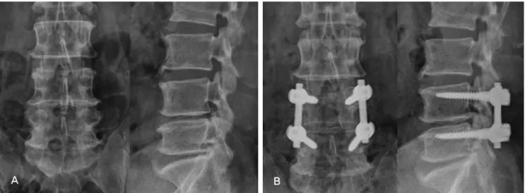
Fig. 1. (A) A 54-year-old male’s preoperative simple radiograph shows spinal stenosis L3-5. (B) At a postoperative 12 months, radiograph shows com- plete bony union of Lenke A degree.
Table 1. Bone fusion rates by Lenke’s criteria
Spinal stenosis Spondylolisthesis Total
A 16 (51.6%) 5 (45.4%) 21 (50.0%)
B 7 (22.6%) 3 (27.3%) 10 (23.8%)
C 8 (25.8%) 3 (27.3%) 11 (26.2%)
D 0 (%) 0 (0%) 0 (%)
Total 31 (100%) 11 (100%) 42 (100%)
방법으로 측정하여 우수, 양호, 보통, 불량으로 구분하였다. 통계 학적 분석은 SPSS (Version 18.0, SPSS Inc, Chicago, IL, USA)를 이용하였으며 유합에 영향을 줄 수 있는 나이, 체질량지수, 흡연 유무, 당뇨병 이환 여부에 대해 골유합군과 불유합군에서의 차 이를 알아보기 위하여 Fisher의 정확한 검정(Fisher’s exact test), student’s t-test를 시행하였다.
결 과
수술 후 추시 1년째 단순 방사선 사진을 Lenke 등의 골유합 기 준에 따라 분류한 결과 척추관 협착증에 시행한 후측방 유합술
의 경우 32례중 A 11례(34.4%)(Fig. 1), B 8례(25.0%)(Fig. 2), C 13례(40.6%)(Fig. 3), D 0례(0%)로 19례인 59.4%에서 B이상의 골유합을 보였으며, 척추전방전위증에서 시행한 후측방 유합술 의 경우 10례 중 A 5례(50.0%), B 2례(20.0%), C 3례(30.0%), D 0례(0%)로 7례인 70.0%에서 B이상의 골유합을 보여, 총 42례중 26례(61.9%)에서 B이상의 양호한 골유합을 보였다(Table 1). B 이상의 양호한 골유합이 관찰된 군과, 불유합군에서의 나이, 체 질량지수, 흡연 유무, 당뇨병 이환 여부는 두 군에서 유의한 차 이는 없었다(Table 2). 전례에서 심부 감염, 내고정물의 파괴, 경 막 파열, 신경학적 이상 등의 수술 후 합병증은 관찰되지 않았 다. 수술 후 임상 결과는 척추관 협착증의 경우 28례(87.5%)에
Fig. 2. (A) A 75-year-old female’s preoperative simple radiograph shows spinal stenosis L4-S1. (B) At a postoperative 12 months, radiograph shows complete bony union unilaterally of Lenke B degree.
Table 2. Patients’ demographics
Union group (n=26) Nonunion group (n=16) p-value
Mean Age (years) 65.7 60.6 0.143
Mean BMI (kg/m2) 24.07 25.06 0.449
Smoking 4 5 0.265
DM 5 4 0.711
Table 3. Clinical results by Kim’s criteria
Spinal stenosis Spondylolisthesis Total
Excellent 12 (37.5%) 2 (20.0%) 14 (33.3%)
Good 16 (50.0%) 5 (50.0%) 21 (50.0%)
Fair 3 (9.4%) 3 (30.0%) 6 (14.3%)
Poor 1 (3.1%) 0 (%) 1 (2.4%)
Total 32 (100%) 10 (100%) 42 (100%)
Man-Jun Park et al Volume 20 • Number 3 • September 2013
www.krspine.org
74
서 양호 이상의 임상 결과를 보였으며 척추전방전위증의 경우 7 례(70.0%)에서 양호 이상의 임상적 결과를 보여, 총 42례중 35 례(83.3%)에서 양호 이상의 임상적 결과를 보였다(Table 3).
고 찰
척추 후측방 유합술시 사용할 수 있는 유합 물질로는 자가골, 동종골, 이종골, 합성골이 있다. 이중 주로 장골능에서 얻을 수 있는 자가 해면골은 칼슘등의 골전도(osteoconduction) 물질과 골성장세포(osteoblast), 골성장단백질을 모두 제공하여 골 형성 능력이 가장 우수하므로 척추 후측방 유합술에서 선호되고 있으 며,17) 병의 전염 가능성이나 조직 적합성 면에서도 가장 이상적 인 이식골이다. 그러나 자가골 이식은 공여부의 정상구조를 약 화 또는 희생시킬 수 있고 실혈, 공여부의 감염, 신경손상, 만성 공여부 통증 등의 합병증 및 그 양이 한정적이며 미성숙 연령에 서는 장골능의 성장판 손상에 의한 성장 장애가 올 수 있다는 제 한점이 있다.7,18) 사체나 다른 환자로부터 얻은 골을 이용하는 동 종골 이식 및 소나 돼지의 골을 탈단백등의 방법으로 항원성을 제거하여 만든 이종골 이식의 경우에도 질환의 전파, 골유도 능 력 부족으로 인한 골 생성 불량, 면역학적 거부반응19,20) 등의 단 점이 있다.
저자들이 사용한 PolyBoneⓇ은 이러한 단점을 보완할 수 있 는 β-TCP 합성골로, 골전도능을 가진 인산칼슘(calcium phosphate) 기조의 세라믹 물질이다. 이는 인체의 해면골과 유 사하며 200~500μm의 공극 크기 (pore size)의 생체흡수성 (bioabsorbable) 및 생체적합성(biocompatible)을 가진 복합체 이다. β-TCP의 이러한 구조적 특징과 성질이 골수로부터 골 형성 성장인자의 전달을 쉽게하며21), 골성장(bony ingrowth)
및 골유합이 되는 동안 수개월에 걸쳐 흡수가 되면서 골지지체 (scaffold) 역할을 하므로 요추 유합술에 적합한 물질로 알려져 있다22). β-TCP를 이용한 척추 유합술에 대한 연구는 우수한 골 유합율이 보고되고 있고,11,23,24) Muschik 등25)이 척추측만증 환 자의 후측방 추체간 유합술에서 자가골과 β-TCP를 병용한 경 우 자가골 이식에 동종골 이식을 병용한 경우와 유사한 유합율 을 보고하였으나, 양 등26)은 27%의 낮은 유합율을 보고하기도 하였다.
본 연구에서는 척추 후측방 유합술시 유합율을 높이기 위하 여 천공기를 이용하여 후방 구조물의 확실한 피질골 박리, 골이 식 부위와 연부조직의 접촉 제거, 견고한 기기 고정술을 병행하 였고,27) β-TCP의 경우 골형성능이 없으므로 이를 보완하기 위 하여 후방 감압 중 발생한 국소 채취 자가해면골을 혼합하여 이 식하였으나 술 후 1년째 평가한 골유합율은 양호한 유합으로 판 정된 것이 61.9%로 낮게 관찰되었다. 이는 기존의 높은 유합율 이 보고된 자가골 이식4-6)에 비해 낮았으며, 같은 술자에 의해 시 행된 국소 자가골과 동종골 혼합 이식술의 골유합율인 85.5%에 비해서도 낮게 관찰되었다28). 수술 후 임상 결과는 83.3%에서 양호 이상으로, 이는 후방감압술 시 신경근이 압박받는 부위만 선택적으로 피질하 절제를 시행하고 후방 관절을 최대한 보존하 여 후방 감압술 시 발생 가능한 불안정성을 최소화한 것에 기인 한 것으로 사료된다.
본 연구에서 사용한 골이식 대체제인 β-TCP를 이용한 유합 술시 유합율을 높이기 위해서 최근 여러 연구가 보고되고 있는 데, Zhang 등29)은 쥐를 이용한 연구에서 β-TCP 와 골수기질세 포(bone marrow stromal cells)를 혼합하여 사용하였을 시 골형 성 능력이 높으므로 혼합 사용의 이점을 보고하였으며, Orii 등30) 도 원숭이를 이용한 연구에서 β-TCP 와 골수기질세포의 혼합
Fig. 3. (A) A 56-year-old male’s preoperative simple radiograph shows spinal stenosis L4-5. (B) At a postoperative 12 months, radiograph shows bony union of Lenke C degree.
사용이 척추 후측방 유합술시 자가골 이식을 대체할 수 있다고 하였다. 이러한 동물 연구 및 본 연구에서 관찰된 국소자가골과 β-TCP 혼합이식술의 낮은 유합율을 비추어 볼 때, β-TCP 이 식을 이용한 요추 후측방 유합술시 유합율을 높이기 위해 자가 장골능 등에서 골수기질세포 채취를 동반하는 것도 고려할 수 있을 것으로 사료된다.
본 연구의 제한점으로는 후향적 연구로서, 자가골 이식을 이 용한 유합술을 대조군으로 설정하여 직접 유합율을 비교하지 못 하고 β-TCP를 사용한 군의 유합율만을 연구하였다는 점이 있 다. 또한 유합율을 평가함에 있어 환자의 유합 범위와 2분절 이 상에서 사용한 횡연결기기가 유합율에 영향을 줄 수 있었으나 이를 고려하지 않았다. 마지막으로, 추시 기간이 1년으로 비교 적 짧아 본 연구에서 평가한 유합율이 더 낮아질 수 있으므로 향 후 지속적인 추시 관찰이 필요할 것으로 사료된다.
결 론
β-TCP는 자가골 이식이나 동종골 이식시 발생할 수 있는 단 점을 보완하여 척추 후측방 유합술 시 사용할 수 있는 유합 물질 이나, 상품화된 β-TCP와 국소 자가골을 혼합하여 이식한 본 연구에서는 낮은 유합율을 보였다. 척추 유합술에서 β-TCP 사 용의 적응을 높이고 유합율을 증가시키기 위해서는 더 많은 연 구가 필요할 것으로 사료된다.
REFERENCES
1. Watkins MB. Posterolateral fusion of the lumbar and lum- bosacral spine. J Bone Joint Surg. 1953;35:1014:8.
2. Jeon CH, Kim YC, Chang HG, Kim YW, Jung NS, Shin DS. The comparison of changes in the dimensions of the intervertebral disc and neural foramen between anterior lumbar interbody fusion and posterolateral fusion in the lumbar spine. J Kor Soc Spine surg. 2007;14:263-9.
3. Lee CK, Langrana NA. Lumbosacral spinal fusion. A bio- mechanical study. Spine (Phila Pa 1976). 1984;9:574-81.
4. Rombold C. Treatment of spondylolisthesis by postero- lateral fusion, resection of the pars interarticularis and prompt immobilization of the patients. J Bone Joint Surg.
1966;48:1282-300.
5. Turner JA, Ersek M, Herron L, et al. Patient outcomes after lumbar spinal fusion. JAMA. 1992;268:907-11.
6. Lee SU, Kim KT, Ahn OK, Ok JC. Posterolateral fusion in
spondylolisthesis. J of Korean Orthop. 1996;31:695-701.
7. Younger EX, Chapman MW. Morbidity at bone graft do- nor sites. Orthop Trans. 1986;10:494.
8. Park HJ, Kim WK, Shim YJ, Kang DH. Efficiency of ante- rior interbody fusion using cage packed with DBM in dis- tractive flexion injury of cervical spine –demineralized bone matrix vs autoiliac cancellous bone-. J Kor Soc Spine surg.
2010;17:111-9.
9. Galois L, Mainard D, Cohen P, Pfeffer F, Traversari R, Del- agoutte JP. Filling of bone defects with tricalcium phosphate beta in traumatology. Ann Chir. 2000;125:972-81.
10. Galois L, Mainard D, Delagoutte JP. Beta-tri-calcium phosphate ceramic as a bone substitute in orthopaedic sur- gery. Int Orthop. 2002;26:109-15.
11. Le Huec JC, Lesprit E, Delavigne C, Clement D, Chauveaux D, Le Rebeller A. Tri-calcium phosphate ce- ramics and allografts as bone substitutes for spinal fusion in idiopathic scoliosis comparative clinical results at four years.
Acta Orthop Belg. 1997;63:202-11.
12. Cosar M, Ozer AF, Iplikcioglu AC, et al. The results of beta-tricalcium phosphate coated hydroxyapatite (beta- TCP/HA) grafts for interbody fusion after anterior cervical discectomy. J Spinal Disord Tech. 2008;21:436-41.
13. Suk SI. Spinal surgery 2nded. Newest medical publishing company. 2004.126-7.
14. Wahba GM, Bhatia N, Bui CN, Lee KH, Lee TQ. Biome- chanical evaluation of short-segmental posterior instru- mentation with and without crosslinks in a human cadav- eric unstable thoracolumbar burst fracture model. Spine (Phila Pa 1976). 2010;35:278-85.
15. Lenke LG, Bridgwell KH, Baldus C and Blanke K. Contrel- Dubousset instrument tation for adolescent idiopathic sco- liosis. J Bone Joint surg Am. 1992;74:1056-69.
16. Kim NH, Lee HM. Usefulness of posterolateral fusion of lumbar spine with allograft bone(tuboplast). J Kor Soc Spine surg. 1998;5:198-204.
17. Lexer E. The History of Bone Graft. Ciln Orthop.
1998;226:292-8.
18. Kurz LT, Garfin SR, Booth RE Jr. Harvesting autogenous iliac bone graft. A review of complication and techniques.
Spine (Phila Pa 1976). 1989;14:1324-31.
19. Burwell RG. Studies in the transplantation of bone, V the
Man-Jun Park et al Volume 20 • Number 3 • September 2013
www.krspine.org
76
capacity of fresh and treated homograft of bone to evoke transplantation immunity. J Bone Joint Surg. 1963;45:386- 401.
20. Bonfiglio M, Jetter WS. Immunological response to bone.
Clin Orthop. 1972;87:19-27.
21. Nandi SK, Roy S, Mukherjee P, Kundu B, De DK, Basu D. Orthopaedic applications of bone graft & graft substitutues:a review. Indian J Med Res. 2010;132:15-30.
22. Rihn JA, Kirkpatrick K, Albert TJ. Graft options in pos- terolateral and posterior interbody lumbar fusion. Spine (Phila Pa 1976). 2010;35:1629-39.
23. Delecrin J, Takahashi S, Gouin F, Passuti N. A synthetic porous ceramic as a bone graft substitute in the surgical management of scoliosis: a prospective, randomized study.
Spine (Phila Pa 1976). 2001;25:563-9.
24. Meadows GR. Adjunctive use of ultra-porous beta- tricalcium phosphate bone void filler in spinal arthrodesis.
Orthopedics. 2002;25 Suppl:579-84.
25. Muschik M, Ludwig R, Halbhubner S, Bursche K, Stoll T.
Beta tricalcium phosphate as a bone substitute for dorsal
spinal fusion in adolescent idiopathic scoliosis : prelimi- nary results of a prospective clinical study. Eur spine J.
2001;10:178-84.
26. Yang JY, Lee JK, Kim DH, et al. The effect of β-trical- cium phosphate in lumbar posterolateral fusion surgery – A prospective study -. J Kor Musculoskelet Transplant Soc.
2006;6:13-8.
27. Knapp DR, Jones ET. Use of cortical cancellous allograft for posterior spinal fusion. Clin Orthop. 1988;229:99-106.
28. Kim JW, Ko YC, Jung CY, Eun IS, Kim OG, Kim CK.
Union rates of local autobone and femoral head allobone mixed graft in lumbar posterolateral fusion. J Kor Muscu- loskelet Transplant Soc. 2010;10:50-8.
29. Zhang M, Wang K, Shi Z, Yang H, Dang X, Wang W. Os- teogenesis of the construct combined BMCs with β-TCP in rat. J plast Reconstr Aesthet Surg. 2010;63:227-32.
30. Orii H. Sotome S, Chen J, Wang J, Shinomiya K. Beta- tricalcium phosphate(beta-TCP) graft combined with bone marrow stromal cells(MSCs) for posterolateral spine fusion.
J Med Dent Sci. 2005;52:51-7.
요추부 후측방 유합술시 국소 자가골 및 β-Tricalcium phosphate 혼합 이식술의 골유합율
박만준 • 고영철 • 정철용 • 은일수 • 김창규 • 김민우 • 황금민 부산의료원 정형외과
연구 계획: 후향적 연구
목적: 척추관 협착증 및 척추전방전위증에서 국소 자가골 및 β-Tricalcium phosphate 혼합 이식술을 시행한 요추부 후측방 유합술에서 골유합율을 조 사하여 유용성을 알아보고자 한다.
선행문헌의 요약: β-Tricalcium phosphate를 이용한 요추부 후측방 유합술은 국외에서는 높은 유합율 보고하였으나 국내에서는 낮은 유합율을 보고한 1개의 연구만이 존재하였다.
대상 및 방법: 2010년 9월부터 2011년 7월까지 국소 자가골 및 β-Tricalcium phosphate 혼합 이식술을 시행하여 요추부 후측방 유합술을 시행한 후 최 소 1년이상 추시가 가능하였던 42례를 대상으로 하였다. 이 중 척추관 협착증은 32례, 척추전방전위증은 10례이다. Lenke 등의 분류를 토대로 골유합 상태를 파악하였고 임상적 결과는 김 등의 방법으로 평가하였다.
결과: 척추관 협착증의 경우 32례 중 19례(59.4%)에서 골유합 소견을 보였고, 척추전방전위증의 경우 10례 중 7례(70.0%)에서 골유합 소견을 보였다.
수술 후 임상 결과는 척추관 협착증의 경우 우수 12례, 양호 16례 및 보통 3례, 불량 1례로 28례(87.5%)에서 양호 이상의 경과를 나타내었고, 척추전방 전위증의 경우 우수 2례, 양호 5례 및 보통 3례, 불량 0례로 8례(70.0%)에서 양호 이상의 임상적 경과를 보였다.
결론: 국소 자가골 및 β-Tricalcium phosphate 혼합 이식술은 낮은 유합율을 보이므로 국소 자가골 및 β-Tricalcium phosphate 혼합 이식술 시행시 유 합율을 높이기 위한 노력 및 연구가 필요할 것으로 사료된다.
색인 단어 : 척추관 협착증, 척추전방전위증, β-Tricalcium phosphate, 후측방 유합술 약칭 제목: β-TCP를 이용한 요추부 후측방 유합술