www.e-arms.org 97 Chronic renal failure (CRF) is a disease in which glomerular
filtration rate decreases irreversibly due to consistent renal dysfunction caused by continuous decrease of glomerular function. The normal range of the glomerular filtration rate is 100 to 120 mL/min; renal function is maintained without any symptoms even during 30% to 50% of normal function.
However, renal replacement such as dialysis and renal transplantation is required to excrete body waste if glomerular filtration rate decreases to 5% to 10% of the normal range;
this state is defined as CRF.1 The number of patients who require renal replacement therapy is increasing due to the increase of patients with CRF; dialysis is still the mainly used renal replacement therapy due to financial burden and donor
deficiency of renal transplantation. Dialysis is classified into hemodialysis and peritoneal dialysis. Hemodialysis is more efficient and requires less time than peritoneal dialysis; hence, it is the most commonly used renal replacement therapy.
The number of patients on dialysis for CRF in Korea is increasing steadily; half of them are on hemodialysis.2,3 The number of patients on long-term hemodialysis is increasing due to longer life expectancy and the development of medical technology. The number of CRF patients with blood-flow decreasing complications such as diabetes and hypertension is increasing; therefore, securing appropriate vascular access and maintaining its patency is rising as an important issue.1 Various arteriovenous fistulas are used in order to improve long-term
Arteriovenous Fistula Formation Using Microscope Rather than Surgical Telescope
Byeong Ho Lee, In Suck Suh, A Jin Cho1, Jung Woo Noh1, Hii Sun Jeong*
Departments of Plastic and Reconstructive Surgery, 1Medicine of Nephrology, Kangnam Sacred Heart Hospital, Hallym University College of Medicine, Seoul, Korea
CC This is an open-access article distributed under the terms of the Creative Commons Attribution Non-Commercial License (http://creativecommons.org/licenses/by-nc/3.0) which permits unrestricted noncommercial use, distribution, and reproduction in any medium, provided the original work is properly cited.
Copyright © 2014 by the Korean Society for Microsurgery. All Rights Reserved.
Received November 3, 2014 Revised November 4, 2014 Accepted November 4, 2014
*Correspondence to: Hii Sun Jeong Department of Plastic and Reconstructive Surgery, Kangnam Sacred Heart Hospital, Hallym University College of Medicine, 1 Singil-ro, Yeongdeungpo-gu, Seoul 150- 950, Korea
Tel: +82-2-829-5182 Fax: +82-2-847-5183 E-mail: [email protected] Financial support: None.
Conflict of interest: None.
The number of patients with chronic renal failure who require renal replacement therapy is increasing and dialysis is still the mainly used renal replacement therapy. The first choice of surgical technique currently used is side-to-end anastomosis of the radial artery and the cephalic vein. The authors report on a case of an effective arteriovenous shunt operation performed using microscopy. A 53-year-old male with chronic renal failure was referred to plastic and reconstructive surgery department to undergo an arteriovenous shunt operation. Venography was performed before surgery in order to find the appropriate vessel for the arteriovenous shunt operation. The cephalic vein on the wrist showed a diameter of over 4 mm, which was appropriate for an arteriovenous shunt operation.
Anastomosis of the vessels was performed under microscopy using Nylon #9-0. Blood flow and vessel diameter were evaluated by venography after surgery and showed well maintained function of the shunt. Complications such as bleeding, edema of the upper arm, and wound dehiscence did not occur. Many factors and certain complications may affect the long-term patency of an arteriovenous shunt; however, exquisite surgical technique is the most important factor in a successful operation. Thus, arteriovenous shunt operation using microscopy is thought to be a good treatment option.
Key Words: Chronic renal failure, Arteriovenous fistula, Shunt, Microscopy
ARMS
Archives of Reconstructive MicrosurgeryCase Report
pISSN 2383-5257 eISSN 2288-6184 Arch Reconstr Microsurg 2014;23(2):97-100 http://dx.doi.org/10.15596/ARMS.2014.23.2.97
Arch Reconstr Microsurg Vol. 23. No. 2. November 2014
98
patency; efforts are made to maintain blood flow without any complications such as infection or thrombosis. Many surgical techniques are used to improve long-term patency; processes differ depending on the condition of the patient’s vessels and the preference of the surgeon.
In principle, when selecting a vessel, anastomosis of the radial artery and the cephalic vein on a distal level is attempted.4 However, if those vessels are not appropriate because of stenosis or any other reason, vessels on a proximal level such as brachiocephlic and brachiobasilic fistulas are used.5 When microsurgery such as that of the radial forearm free flaps is done and two parts of a blood vessel are anastomosed, the authors think that using fine microscopy is more suitable for anastomosis than is the use of a surgical telescope. As mentioned earlier, comparison of data is needed to confirm the effectiveness of fine microscopy. For this reason, the authors will report a case of an effective arteriovenous shunt operation using fine microscopy.
CASE REPORT
A 53-year-old male with CRF and hypertension was referred to the department of nephrology of our hospital for long- term hemodialysis in July 2014. The patient was hospitalized in the department of nephrology for four days and had an ultrasonography guided perm catheter inserted in the right
internal jugular vein for hemodialysis. On the third day of the patient’s stay in the hospital, the patient was referred to plastic and reconstructive surgery department to undergo an arteriovenous shunt operation.
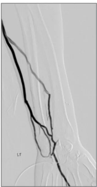
To find the appropriate vessel for the arteriovenous shunt operation, physical examination, laser Doppler mapping and venography were done one day before surgery. In the venography results, the cephalic vein on the left wrist showed a diameter of over 4 mm, which was appropriate for arteriovenous fistula formation (Fig. 1). Since the patient was right-handed, the left side was appropriate for the arteriovenous shunt operation. Before surgery, an Allen test was performed to check arterious patency; venous patency was checked by observing subcutaneous vein filling after applying a tourniquet to the humeral region. The operation was initiated by injecting 2 % lidocaine into the operation site and making a Z shape incision along the distribution of the vessel. A length of 2 to 3 cm of the vein was separated from the surrounding tissue and ligated at the distal end; a side-to-end anastomosis was performed with the radial artery. Before ligation of the vein, 5,000 units of heparin, diluted 1 to 25,000, were administered to the venous access to prevent thrombosis. Anastomosis of the vessels was done under fine microscopy, of which the magnifying power was ×3.0 with a simple interrupted suture using Nylon #9-0 (Fig. 2). After suturing, palpation was done to check thrill, and Doppler ultrasonography was used to confirm anastomosis. Electrocauterization of the branches of the vein used for anastomosis was done by bipolar cautery.
After surgery, venography and Doppler ultrasonography
Fig. 1. Preoperative venography. During venography, cephalic vein on left wrist showed a diameter of over 4 mm, which was appropriate for arteriovenous fistula formation.
Fig. 2. Aanastomosis of the vessels. Anastomosis of the vessels was done under fine microscopy, of which the magnifying power was ×3.0 with a simple interrupted suture using Nylon #9-0.
Byeong Ho Lee, et al. Arteriovenous Fistula Formation Using Microscope
www.e-arms.org 99
were performed to evaluate the blood-flow and to check for any obstruction in the arteriovenous fistula. Vessel diameter was evaluated by venography and blood flow was evaluated by Doppler ultrasonography one month after surgery, result showed optimal blood flow rate and well maintained function of arteriovenous fistula (Fig. 3). Complications that may occur after arteriovenous fistula formation such as bleeding, edema of the upper arm, bacterial infection, and wound dehiscence did not appear.
DISCUSSION
When CRF patients undergo hemodialysis, they need to secure a blood vessel. If they do not have an arteriovenous fistula, physicians will insert a central venous catheter and perform hemodialysis through it. The process of central venous catheterization is a comparatively safe, easy way to secure a blood vessel, but it has various side effects that are arrhythmia, pneumothorax, hemothorax, arteriopuncture, infection, arteriovenous fistula formation, and angiostenosis. Among these, angiostenosis is relatively common and difficult to treat.5
Therefore in 1966, Brescia et al.4 announced the process of arteriovenous fistula formation using the radial artery and the cephalic vein; this process made long-term repetitive hemodialysis with an arterialized vein possible, and this method is considered the primary surgical method for arteriovenous
fistula formation until now.
An ideal arteriovenous fistula requires three conditions. First, the arteriovenous fistula must be easy to use for hemodialysis;
second, it must maintain the target blood flow, rated at 200 to 300 mL/min, at all times; and third, it must be feasible for long- term use. However, many patients require a repetitive process of arteriovenous fistula formation for continuous hemodialysis in a certain period after surgery because complications such as obstruction or stenosis of the fistula occur due to injury of the vascular endothelium caused by repetitive vascular puncturing.
With the development of medical technology and longer human life spans, the number of patients who require long-term hemodialysis is increasing. Therefore, understanding factors that affect long-term patency prior to surgery is required to improve long-term patency after surgery.6
Artery expansibility decreases and resistance increases with age, both of which combine to cause less blood flow from the artery to the vein, which has a negative effect on the maturing of the arteriovenous fistula. The major cause of vessel immaturity is decrease of blood flow after surgery which is caused by problems in surgical techniques used in vessel anastomosis, calcification of the artery wall, small diameter of the vein, stiffening of the vein, and hypotension. Therefore, using Doppler ultra-sonography fot the selection of an artery that is capable of maintaining appropriate blood flow is desirable when performing an arteriovenous fistula.7 When an arteriovenous fistula is made, the site of anastomosis should be designed to achieve a sufficiently broad cross section to prevent stenosis and maintain the appropriate blood flow. According to this principle, other surgeons in other department join the two parts of vessels using continuous sutures that are tied by hand, using Nylon #7-0 via a surgical telescope that has a magnifying power of ×2.0. However, plastic surgeons are accustomed to doing simple interrupted suturing and tying the sutures using an instrument with Nylon #9-0 via a fine microscope of which magnifying power is ×3.0. The operation time differs according to suture method, but there is no difference in operation time from 1 to 2 hours. Through fine microscope, identifying the condition of intima (intimal tear, atherosclerotic plaques, friable calcified walls, etc) we could anastomos the vessels, efficiently.
In addition, continuous hand exercise to increase blood flow at the fistula site after surgery is important. In some cases in which it is hard to find autologous vessels appropriate for
Fig. 3. Postoperative 1 month venography. Blood flow and vessel diameter maintained the function of the arteriovenous fistula well.
Arch Reconstr Microsurg Vol. 23. No. 2. November 2014
100
arteriovenous fistula formation, an artificial vessel may be considered.8
Many factors such as age, sex, and complications (diabetes, hypertension, etc.) may affect the long-term patency of the arteriovenous fistula; however, exquisite surgical technique is the most important element of successful surgery. Thus, the arteriovenous shunt operation using fine microscopy is thought to be a good treatment option.
REFERENCES
1. Song CM, Ahn JB, Kim IS, Kim WS, Shin YC, Yoo HK, et al.
Clinical analysis of arteriovenous fistula in chronic renal failure patients. Korean J Thorac Cardiovasc Surg 2006;39:692-8.
2. Jo WM, Sohn YS, Rhu SM, Hwang JJ, Cho SJ, Choi YH, et al.
Clinical analysis of arteriovenous fistulas for hemodialysis.
Korean J Thorac Cardiovasc Surg 2002;35:369-74.
3. Ahn SJ, Choi EJ. Renal replacement therapy in Korea: Insan Memorial Registry 1997. Korean J Nephrol 1999;18:1-15.
4. Brescia MJ, Cimino JE, Appell K, Hurwich BJ. Chronic hemodialysis using venipuncture and a surgically created arteriovenous fistula. N Engl J Med 1966; 275:1089-92.
5. Yang JO, Lee SJ, Park KH, Jang YK, Kang MK, Sung IW, et al.
Percutaneous angioplasty and stenting in chronic hemodialysis patients with central venous stenosis. Korean J Nephrol 2001;20:
981-7.
6. Palder SB, Kirkman RL, Whittemore AD, Hakim RM, Lazarus JM, Tilney NL. Vascular access for hemodialysis. Patency rates and results of revision. Ann Surg 1985;202:235-9.
7. Finlay DE, Longley DG, Foshager MC, Letourneau JG. Duplex and color Doppler sonography of hemodialysis arteriovenous fistulas and grafts. Radiographics 1993;13:983-9.
8. Hirth RA, Turenne MN, Woods JD, Young EW, Port FK, Pauly MV, et al. Predictors of type of vascular access in hemodialysis patients. JAMA 1996;276:1303-8.