Ossification of the posterior longitudinal ligament (OPLL) is a condition of abnormal calcification in the posterior longitudinal ligament, and mostly involves the cervical
spine. Compression of the spinal cord by the OPLL mass can lead to neurological symptoms, and often operative treatments are required in the cases of severe neurological deficit.1) Despite a high degree of spinal cord compres- sion, symptoms do not manifest in many cases, because of which physicians often face the dilemma of choosing the most appropriate treatment strategy such as prophylactic surgery,2) early surgical intervention,3) or conservative treatment in mild symptomatic patients.4) In particular, unlike cervical spondylotic myelopathy, OPLL often fails to manifest clinical symptoms despite severely compressed
Significance of Intramedullary High Signal Intensity on Magnetic Resonance Imaging
in Patients with Cervical Ossification of the Posterior Longitudinal Ligament
Byung-Wan Choi, MD, Tae Woong Hum, MD
Department of Orthopedic Surgery, Inje University Haeundae Paik Hospital, Inje University College of Medicine, Busan, Korea
Background: The purpose of this study was to analyze the relation between intramedullary high signal intensity (IMHS) on mag- netic resonance imaging (MRI), radiographic parameters, and clinical symptoms in cervical ossification of the posterior longitudinal ligament (OPLL) patients.
Methods: Two hundred forty-one patients, who underwent simple radiography, computed tomography (CT), and MRI were includ- ed in the present study. As radiographic parameters, the OPLL occupying ratio and occupying area were measured on CT images.
Dynamic factors were assessed by measuring cervical range of motion (ROM) on simple radiographs. Visual analog scale (VAS) for neck and arm pain, and Japanese Orthopaedic Association (JOA) scores were evaluated for clinical analysis. The differences in radiographic and clinical findings were assessed between patients with IMHS on T2-weighted MRI findings (group A) and patients without IMHS (group B).
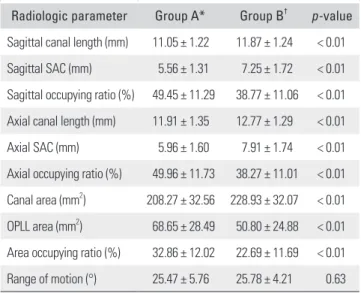
Results: Eighty-one patients were assigned to group A and 160 patients to group B. The occupying ratios were found to be higher in group A than in group B on both sagittal and axial views (p < 0.01). Group A also showed a higher area occupying ratio (p < 0.01).
The length and area of underlying spinal canal on the sagittal and cross-sectional planes were lower in group A than in group B (p
< 0.01). No significant difference in ROM was observed (p = 0.63). On the clinical findings, group A had a lower JOA score (p < 0.001), and no intergroup differences in VAS scores were observed.
Conclusions: In cervical OPLL cases, IMHS on MRI was associated with manifestation of myelopathic symptom. Occupying ratio was associated with high signal intensity on MRI, whereas no association was found with ROM. Occurrence of high signal inten- sity increased inversely with the length and area of underlying spinal canal.
Keywords: Ossification of posterior longitudinal ligament, Cervical spine, Intramedullary high signal change, Magnetic resonance imaging
Copyright © 2015 by The Korean Orthopaedic Association
This is an Open Access article distributed under the terms of the Creative Commons Attribution Non-Commercial License (http://creativecommons.org/licenses/by-nc/4.0) which permits unrestricted non-commercial use, distribution, and reproduction in any medium, provided the original work is properly cited.
Clinics in Orthopedic Surgery • pISSN 2005-291X eISSN 2005-4408 Received July 7, 2015; Accepted August 3, 2015
Correspondence to: Byung-Wan Choi, MD
Department of Orthopedic Surgery, Inje University Haeundae Paik Hospital, Inje University College of Medicine, 875 Haeun-daero, Haeundae-gu, Busan 48108, Korea
Tel: +82-51-797-0240, Fax: +82-51-797-0249 E-mail: alla1013@naver.com
radiographic findings, and sometimes goes undetected life-long.
Magnetic resonance imaging (MRI) is a useful ra- diographic method for the diagnosis of cervical myelopa- thy that evaluates the status of spinal stenosis and intra- medullary condition.5) In particular, intramedullary high signal intensity (IMHS) on T2-weighted MRI has been reported to be indicative of changes in spinal gray matter,6) associated with higher severity of myelopathy symptoms,7) and negative postoperative prognostic factor.8) Neverthe- less, not all OPLL cases with myelopathy show high signal intensity on MRI, and there are even cases where patients with high signal intensity findings exhibit no symptoms of myelopathy.
A large number of previous studies have investigated the relevance of high signal intensity on MRI to symptoms of cervical spondylotic myelopathy and its prognosis, and there is a lack of research on the significance of IMHS on MRI in OPLL patients in relation to clinical symptoms. To address this issue, we have analyzed the interrelations be- tween IMHS on MRI, radiographic parameters, and clini- cal symptoms in patients with cervical OPLL.
METHODS
Among 365 cervical OPLL patients from Inje University Haeundae Paik Hospital between 2010 and 2012, we se- lected 241 patients (146 men and 95 women; mean age, 52.4 years) as subjects for analysis in this study. The in- clusion criteria were as follows: (1) diagnosis of cervical OPLL; and (2) patients who underwent simple radiog- raphy, computed tomography (CT) scan and MRI. The exclusion criteria were history of trauma and prior history of cervical operation. If the radiological evaluations of CT or MRI were performed at different qualifications in another hospital, such cases were excluded from analysis.
Institutional Review Board approval was obtained for this study. The patient distribution according to OPLL type was as follows: local (n = 32), segmental (n = 79), mixed (n
= 102), and continuous (n = 28).
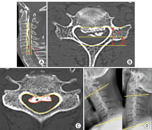
High signal intensity was defined as a case showing high signal intensity in both sagittal and cross-sectional planes on T2-weighted MRI scan as assessed by a radiolo- gist. The occupying ratio (the ratio of the length occupied by the OPLL to the normal spinal canal length) was deter- mined (Fig. 1A and B) by measuring the length available for the spinal cord and the length of the unaffected spinal canal on sagittal and axial view on the CT scan in maximal
A B
C
a b
c d
D
e f
Fig. 1. Radiological evaluations. (A) Sagittal occupying ratio: (b-a)/b. a:
sagittal space available for cord, b:
sagittal canal length. (B) Axial occupying ratio: (d–c)/d. c: axial space available for cord, d: axial canal length. (C) Area occupying ratio: f/e. e: area of spinal canal, f: area of ossification of posterior longitudinal ligament. (D) Range of motion (yellow line: endplate of C2 and C7).
compression area. The area occupying ratio (area occupied by the OPLL/area of the normal spinal cord) of the cross- section with the highest degree of cord compression was also determined (Fig. 1C).9) The entire measurement of CT scan was done using the axial cut which was made parallel to disc space. For the assessment of dynamic factors, we measured the cervical range of motion (ROM) by deter- mining the differences of the extension lines from the low- er edge of the second cervical vertebra and the lower edge of the seventh cervical vertebra, respectively, on simple flexion and extension radiographs (Fig. 1D). All the mea- surements were performed by the PACS system (m-view;
Maro Tech Inc., Seoul, Korea). Two blinded observers independently interpreted the radiological findings twice and the mean values were used for further measurements.
In order to verify the reliability of the measured values, the intra- and interobserver correlations were checked using intra- and interclass correlation coefficient (ICC).10) ICC value (Cronbach α) was analyzed by standardized confi- dence analysis and categorized as following: poor (α < 0.4), fair to good (0.4 to 0.7), and excellent (α > 0.7).
Clinical analysis was performed by using visual ana- log scale (VAS) for neck and arm pain, and Japanese Or- thopaedic Association (JOA) scores. Differences in clinical features were determined by comparing radiographic and clinical findings between patients showing high signal in- tensity on T2-weighted MRI findings (group A) and those who did not show high signal intensity (group B).
Statistical analysis included t-test and chi-square tests using the SPSS ver. 10.1 (SPSS Inc., Chicago, IL, USA).
RESULTS
The ICC for intra- and interobserver reliability showed that the data used for this study were reliable (0.63 and 0.62,
respectively).
Of the 241 enrolled patients, 81 (33.6%) were assigned to group A and 160 were assigned to group B (Table 1).
Group A had an older mean age (58.25 vs. 53.38 years; p <
0.001), and the men outnumbered the women with respect to occurrence of high signal intensity (group A, 75% and group B, 53%; p < 0.001). The occupying ratios determined by radiographic analysis were found to be higher in group A than in group B on both sagittal and axial planes (49.45%
and 49.96% vs. 38.77% and 38.27%; p < 0.01) (Table 2).
Group A also showed a higher area occupying ratio with statistical significance (32.86% vs. 22.69%; p < 0.01).
The length and area of the unaffected spinal canal on the sagittal and axial planes were measured to be statis- tically lower in group A (11.05 mm, 11.91 mm, and 208.27 mm2; p < 0.01) than in group B (11.87 mm, 12.77 mm, and 228.93 mm2; p < 0.01). Among the OPLL types, the mixed type was found to be most frequent in group A (p < 0.001)
Table 1. Demographic and Clinical Characteristics of Two Groups Characteristic Group A* (n = 81) Group B (n = 160)† p-value Age (yr) 58.25 ± 8.98 53.38 ± 8.92 < 0.001
Sex (male:female) 61:20 85:75 0.001
JOA score 13.53 ± 3.01 15.93 ± 1.77 < 0.001
Neck VAS 4.16 ± 2.51 4.30 ± 2.51 0.68
Arm VAS 4.28 ± 2.77 3.72 ± 2.97 0.15
Values are presented as mean ± standard deviation.
JOA: Japanese Orthopaedic Association, VAS: visual analog scale.
*Group A: intramedullary high signal intensity group. †Group B: intramedullary normal signal intensity group.
Table 2. Difference of Radiologic Parameters between Two Groups Radiologic parameter Group A* Group B† p-value Sagittal canal length (mm) 11.05 ± 1.22 11.87 ± 1.24 < 0.01 Sagittal SAC (mm) 5.56 ± 1.31 7.25 ± 1.72 < 0.01 Sagittal occupying ratio (%) 49.45 ± 11.29 38.77 ± 11.06 < 0.01 Axial canal length (mm) 11.91 ± 1.35 12.77 ± 1.29 < 0.01 Axial SAC (mm) 5.96 ± 1.60 7.91 ± 1.74 < 0.01 Axial occupying ratio (%) 49.96 ± 11.73 38.27 ± 11.01 < 0.01 Canal area (mm2) 208.27 ± 32.56 228.93 ± 32.07 < 0.01 OPLL area (mm2) 68.65 ± 28.49 50.80 ± 24.88 < 0.01 Area occupying ratio (%) 32.86 ± 12.02 22.69 ± 11.69 < 0.01 Range of motion (°) 25.47 ± 5.76 25.78 ± 4.21 0.63 Values are presented as mean ± standard deviation.
SAC: space available for cord, OPLL: ossification of the posterior longitudinal ligament.
*Group A: intramedullary high signal intensity group. †Group B: intramedullary normal signal intensity group.
Table 3. Radiological Patterns of Ossification of the Posterior Longi- tudinal Ligament in Each Group
Group Local Segmental Mixed Continuous Total
A* 9 15 52 5 81
B† 23 64 50 23 160
*Group A: intramedullary high signal intensity group. †Group B: intramedullary normal signal intensity group.
(Table 3).
No significant difference was seen in ROM (group A, 25.47° and group B, 25.78°; p = 0.63). As clinical condi- tions, the mean JOA score was 15.13 ± 2.52, and the VAS scores for neck pain and arm pain were 3.4 ± 2.50 and 3.91
± 2.90, respectively. Group A showed a lower JOA score (13.53, p < 0.001), and no intergroup difference in neck and arm VAS was observed (Table 1).
DISCUSSION
MRI is a useful tool for diagnosing cervical myelopathy, which reveals not only the degree of spinal cord compres- sion but also intramedullary conditions in detail.5) Since Takahashi et al.11)’s report on the IMHS on MRI in cervical spondylotic myelopathy, findings of low and high signal intensities on T1- and T2-weighted MRI, respectively, have been reported as frequent findings in severe myelopathy and indicate poor postoperative prognosis.7,8) In the case of myelopathy patients, who need surgical treatment, good clinical outcomes can be expected with timely surgical intervention before development of intramedullary signal change on MRI. So, it is essential to analyze the risk fac- tors for development of high signal intensity in MRI. Early surgical intervention can be recommended to the patients demonstrating risk factors; thus, poor clinical outcomes from delay in surgical treatment can be prevented. Wang et al.12) reported that high signal intensity on T2-weighted MRI and pyramidal sign in patients with cervical OPLL are associated with reduced ability to recover from spinal cord damage and poor postoperative prognosis. Qizhi et al.9) identified long-term symptoms, high occupying ratios, low preoperative JOA scores, kyphosis, and cervical in- stability as factors related to IMHS in patients with OPLL and recommended early surgical treatment to patients with these factors. In the present study, intramedullary sig- nal change was more frequently present in older patients and men. In radiographic findings, high signal intensity increased in inverse proportion to the length and area of underlying spinal canal diameters on sagittal and cross- sectional planes. The OPLL occupying ratio, a static fac- tor, was found to be associated with high signal intensity on MRI, but no association was confirmed with regards to ROM, a dynamic factor. Matsunaga et al.13) reported that in 156 cervical OPLL patients with a mean follow-up duration of 10.3 years, myelopathy occurred in all cases exhibiting 60% spinal stenosis or higher and in 49% of cases exhibiting less than 60% spinal stenosis, with aggra- vating tendency in large ROM and laterally tilted shape.
Mochizuki et al.7) reported that myelopathy symptoms
were more frequent in cases of large ROM and segmental OPLL. In the present study, however, ROM was found to have no significant relation with myelopathic symptoms, and the area-related degree of stenosis showed a statisti- cally significant association. In our opinion, these results are ascribed to the findings that the degree of compression in OPLL was influenced more by the occurrence of high signal intensity than by cervical ROM. Most cases of con- tinuous or mixed OPLL showed severe compression and narrow cervical ROM. Among previous studies, Chang et al.14) also reported that myelopathy symptoms in patients with OPLL were not related to ROM and tended to occur in accordance with the degree of maximum spinal cord compression. For more accurate determination as to which factor is more related to the occurrence of myelopathy symptoms, further evaluation on the degree of compres- sion, ROM, and findings of IMHS is needed.
Apart from the unsettled discussion about prophy- lactic and early-phase surgery, IMHS can serve as an indi- cation for timely surgery to prevent further damage to the spinal cord. This aspect will have to be explored in further studies.
The limitation of this study includes its retrospec- tive design. Furthermore, evaluation according to other diverse radiological factors on MRI such as signal change on T1-weighted image or length of signal change was not evaluated. However, we evaluated a relatively large number of OPLL patients and this result can serve as useful clini- cal information in proper management of cervical OPLL patients.
Finally in cervical OPLL, IMHS on T2-weighted MRI was associated with the manifestation of symptoms of cervical myelopathy. The high signal intensity was found to be associated with the occupying ratio, a static factor, but was not associated with ROM, a dynamic fac- tor of OPLL. Moreover, occurrence of high signal intensity increased in inverse proportion to the length and area of underlying spinal canal stenosis on sagittal and cross- sectional planes.
CONFLICT OF INTEREST
No potential conflict of interest relevant to this article was reported.
REFERENCES
1. Jeon TS, Chang H, Choi BW. Analysis of demographics, clinical, and radiographical findings of ossification of pos- terior longitudinal ligament of the cervical spine in 146 Korean patients. Spine (Phila Pa 1976). 2012;37(24):E1498- 503.
2. Epstein N. Diagnosis and surgical management of cervical ossification of the posterior longitudinal ligament. Spine J.
2002;2(6):436-49.
3. Ogawa Y, Chiba K, Matsumoto M, et al. Long-term results after expansive open-door laminoplasty for the segmental- type of ossification of the posterior longitudinal ligament of the cervical spine: a comparison with nonsegmental-type lesions. J Neurosurg Spine. 2005;3(3):198-204.
4. Matsumoto M, Toyama Y, Ishikawa M, Chiba K, Suzuki N, Fujimura Y. Increased signal intensity of the spinal cord on magnetic resonance images in cervical compressive myelop- athy: does it predict the outcome of conservative treatment?
Spine (Phila Pa 1976). 2000;25(6):677-82.
5. Ohshio I, Hatayama A, Kaneda K, Takahara M, Nagashima K. Correlation between histopathologic features and mag- netic resonance images of spinal cord lesions. Spine (Phila Pa 1976). 1993;18(9):1140-9.
6. Matsuda Y, Miyazaki K, Tada K, et al. Increased MR signal intensity due to cervical myelopathy: analysis of 29 surgical cases. J Neurosurg. 1991;74(6):887-92.
7. Mochizuki M, Aiba A, Hashimoto M, Fujiyoshi T, Yamazaki M. Cervical myelopathy in patients with ossification of the posterior longitudinal ligament. J Neurosurg Spine.
2009;10(2):122-8.
8. Sun Q, Hu H, Zhang Y, et al. Do intramedullary spinal cord changes in signal intensity on MRI affect surgical opportu- nity and approach for cervical myelopathy due to ossifica- tion of the posterior longitudinal ligament? Eur Spine J.
2011;20(9):1466-73.
9. Qizhi S, Lili Y, Ce W, Yu C, Wen Y. Factors associated with intramedullary MRI abnormalities in patients with ossifica- tion of the posterior longitudinal ligament. J Spinal Disord Tech. 2015;28(5):E304-9.
10. Henriksen M, Lund H, Moe-Nilssen R, Bliddal H, Dan- neskiod-Samsoe B. Test-retest reliability of trunk accelero- metric gait analysis. Gait Posture. 2004;19(3):288-97.
11. Takahashi M, Sakamoto Y, Miyawaki M, Bussaka H. In- creased MR signal intensity secondary to chronic cervical cord compression. Neuroradiology. 1987;29(6):550-6.
12. Wang LF, Zhang YZ, Shen Y, et al. Using the T2-weighted magnetic resonance imaging signal intensity ratio and clini- cal manifestations to assess the prognosis of patients with cervical ossification of the posterior longitudinal ligament. J Neurosurg Spine. 2010;13(3):319-23.
13. Matsunaga S, Nakamura K, Seichi A, et al. Radiographic predictors for the development of myelopathy in pa- tients with ossification of the posterior longitudinal liga- ment: a multicenter cohort study. Spine (Phila Pa 1976).
2008;33(24):2648-50.
14. Chang H, Song KJ, Kim HY, Choi BW. Factors related to the development of myelopathy in patients with cervical ossi- fication of the posterior longitudinal ligament. J Bone Joint Surg Br. 2012;94(7):946-9.