bloodresearch.or.kr Blood Res 2017;52:135-50.
Letters to the Editor 139
in AML is usually 190 Kd, compared to 210 Kd in most cases of CML. The presence of additional genetic alterations associated with the progression to the blast phase may also aid in establishing the correct diagnosis.
The exact mechanism of progression to BC is still unknown. Various studies speculate that stem cells undergo additional genetic alterations leading to transformation to BC. In a study done by Calabretta and Perrotti [4], they noted that the ectopic expression of the p210 bcr-abl tran- script resulted in growth factor independence and reduced susceptibility to apoptosis, leading to leukemic trans- formation of immortal hematopoietic stem cells. This abnor- mal transcript activated the Ras-MAP kinase and PI3Akt pathway leading to uncontrolled proliferation of leukemic cells [4]. The chromosomal abnormalities that have been found to be associated with progression to the blast phase include duplication of Ph+, trisomy 8 or 19, isochromosome 17q (leading to loss of the p53 gene on 17p), or translocations and inversions associated with MDS/AML [5]. The appear- ance of these mutations during treatment plays a significant role in the advancement of the disease. In addition, these genetic alterations lead to imatinib resistance in these pa- tients [5]. Not all patients will benefit from TKIs and many patients become resistant to it while on treatment, especially those who show a suboptimal response. A single specific point mutation, known as T315I kinase, in the bcr-abl gene has been isolated in association with resistance to imatinib.
Patients who are resistant to imatinib appear to benefit from SCT. Fruehauf et al. [6] observed a more promising hematological response for patients with CML in BC receiv- ing a combination of mitoxantrone with etoposide.
In conclusion, CML in megakaryocytic BC is rarely en- countered in clinical practice. It is important to correctly diagnose these cases as they benefit from a combination of conventional chemotherapy with imatinib, resulting in decreased mortality.
Jenna B Bhattacharya1, Richa Gupta1, Amit Samadhiya2
Departments of 1Pathology and 2Biochemistry, Maulana Azad Medical College, New Delhi, India
Correspondence to: Richa Gupta Department of Pathology, Maulana Azad Medical College, C 502, Prince Apartments, 54 I. P. Extension, Delhi - 92, India
E-mail: richagupta0209@gmail.com
Received on Sep. 6, 2016; Revised on Oct. 6, 2016; Accepted on Jan. 3, 2017 https://doi.org/10.5045/br.2017.52.2.137
AuthorsÊ Disclosures of Potential Conflicts of Interest No potential conflicts of interest relevant to this article were reported.
REFERENCES
1. Choi W, Kim M, Lim J, et al. Four cases of chronic myelogenous leukemia in mixed phenotype blast phase at initial presentation
mimicking mixed phenotype acute leukemia with t(9;22). Ann Lab Med 2014;34:60-3.
2. Pagano L, Pulsoni A, Vignetti M, et al. Acute megakaryoblastic leukemia: experience of GIMEMA trials. Leukemia 2002;16:
1622-6.
3. Al-Shehri A, Al-Seraihy A, Owaidah TM, Belgaumi AF. Mega- karyocytic blast crisis at presentation in a pediatric patient with chronic myeloid leukemia. Hematol Oncol Stem Cell Ther 2010;3:42-6.
4. Calabretta B, Perrotti D. The biology of CML blast crisis. Blood 2004;103:4010-22.
5. Luatti S, Castagnetti F, Marzocchi G, et al. Additional chromoso- mal abnormalities in Philadelphia-positive clone: adverse prog- nostic influence on frontline imatinib therapy: a GIMEMA Working Party on CML analysis. Blood 2012;120:761-7.
6. Fruehauf S, Topaly J, Buss EC, et al. Imatinib combined with mi- toxantrone/etoposide and cytarabine is an effective induction therapy for patients with chronic myeloid leukemia in myeloid blast crisis. Cancer 2007;109:1543-9.
Hairy cell leukemia: a case report of atypical presentation without
splenomegaly
TO THE EDITOR: Hairy cell leukemia (HCL) is a rare B-cell neoplasm of middle age. Pancytopenia, particularly mono- cytopenia, splenomegaly, and hairy cells in the bone marrow represent the classical triad of HCL [1, 2]. Splenomegaly accounts for 80–90% of associated features in HCL [3]. The bone marrow aspirations of HCL patients frequently show dry tap due to marrow fibrosis induced by hairy cell [4].
However, careful examination of diluted cytology prepara- tions is useful because of the characteristic morphology of the hairy cells [3, 4]. The exact prevalence of atypical pre- sentation of HCL is difficult to be determined because most cases were published as incidental reports. Herein, we pres- ent an atypical case of HCL with only arthralgia but no typical symptoms such as splenomegaly and bone marrow fibrosis. This report of an atypical presentation of such a rare disease could lead to earlier recognition and diagnosis of HCL, even without a classical clinical presentation.
A 43-year-old Saudi woman was incidentally found to have leukopenia for 3 months. She had a history of arthralgia, but neither other significant complaints nor remarkable findings on physical examination. The initial complete blood count revealed a hemoglobin concentration of 10.6 g/dL, white blood cell count of 1.95×10⁹/L, platelet count of 110×109/L, absolute neutrophil count of 0.82×10⁹/L, mono- cyte count of 0.02×10⁹/L, and lymphocyte count of 1.07×10⁹/L. On peripheral blood smear, a few atypical cells with oval nuclei and hairy circumferential cytoplasmic pro- jections were observed. On bone marrow aspiration, there
Blood Res2017;52:135-50. bloodresearch.or.kr
140 Letters to the Editor
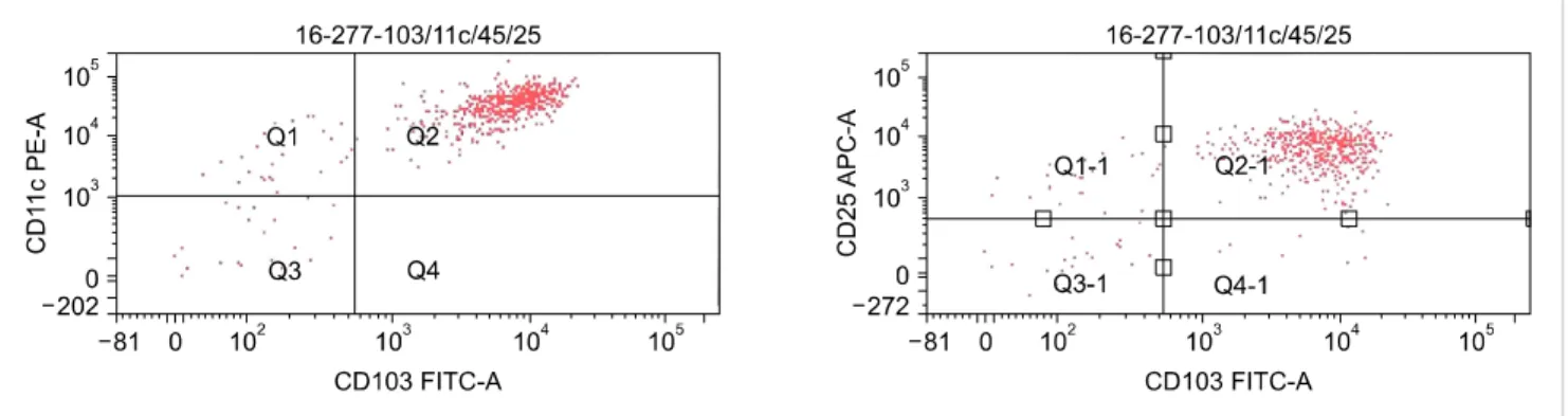
Fig. 2. Flow cytometry analysis of hairy cells (CD103+/CD11c+/CD25+).
Fig. 1. Hairy cells in bone marrow aspiration (Grunwald-Giemsa stain
×1,000).
were many abnormal medium-sized lymphoid cells with oval nuclei, homogenous, ground glass chromatin, incon- spicuous nucleoli, and abundant cytoplasm with hairy pro- jections similar to those observed in the peripheral blood smear (Fig. 1). Trephine biopsy revealed the typical fried egg appearance of hairy cells with no increased fibrosis.
Immunophenotyping revealed the population expressing CD19, CD20, CD22, CD25, CD103, CD11c, FMC7 and HLA-DR (Fig. 2) with kappa chain restriction. The patient was diagnosed with HCL and referred to a tertiary care hospital for further evaluation prior to administration of purine analogue and cladribine chemotherapy.
Arthralgia can occur at any time during the course of the disease, unrelated to the tumor burden [2]. Most HCL cases, without splenomegaly, are associated with a hypo- cellular marrow [3]. The absence of both splenomegaly and marrow fibrosis suggested that this patient may be in the early phase of the disease. Since arthralgia can present during any phase of HCL, this finding did not oppose this suggestion.
It is important to distinguish HCL, requiring different ther- apeutic protocols, from other chronic B-cell neoplasms with circulating villous cells such as splenic marginal zone lym-
phoma (SMZL) or HCL variant (HCL-V) [5]. In im- munophenotypic assay, HCL-V cells are negative for CD25 and ANNEXIN 1, and SMZL cells are negative for CD25, CD103, and CD11c [6]. Like this, confirming im- munophenotypic characteristics is helpful in differential di- agnosis and selecting therapeutic options [7]. Especially, the BRAF V600E mutation, recently identified as a charac- teristic biomarker of HCL, could be incorporated on a rou- tine diagnostic work-up of HCL in the near future [8].
Although these sophisticated diagnostic methods are avail- able, this case report highlights the importance of basic morphological examination for diagnosis of atypical HCL.
Mona Alfaraj1, Hussain Alsaeed2
1Consultant Hematopathologist, Hematology Section of Medical Laboratory Department, 2Consultant Hematologist, Department of Internal Medicine, Qatif Central Hospital, Saudi Arabia Correspondence to: Mona Alfaraj Consultant Hematopathologist, Hematology Section of Medical Laboratory Department, Qatif Central Hospital, PO Box 10463, 3287, Al Qatif 32654-7302, Al Qatif 32654, Saudi Arabia E-mail: mona23mf@hotmail.com
Received on Jun. 5, 2016; Revised on Sep. 27, 2016; Accepted on Nov. 18, 2016 https://doi.org/10.5045/br.2017.52.2.139
AuthorsÊ Disclosures of Potential Conflicts of Interest No potential conflicts of interest relevant to this article were reported.
REFERENCES
1. Jones G, Parry-Jones N, Wilkins B, Else M, Catovsky D; British Committee for Standards in Haematology. Revised guidelines for the diagnosis and management of hairy cell leukaemia and hairy cell leukaemia variant. Br J Haematol 2012;156:186-95.
2. Quest GR, Johnston JB. Clinical features and diagnosis of hairy cell leukemia. Best Pract Res Clin Haematol 2015;28:180-92.
3. Venkatesan S, Purohit A, Aggarwal M, et al. Unusual pre-
bloodresearch.or.kr Blood Res 2017;52:135-50.
Letters to the Editor 141
Table 1. Concordance between morphology and PCR-determined clonality of bone marrow, based on histology in cases of non- Hodgkin lymphoma.
Histology by WHO classification
BM involvement (+) BM involvement (-) PCR (+) PCR (-) PCR (+) PCR (-)
DLBL 19 6 0 1 12
MZBL 1 1 0 0 0
MCL 1 1 0 0 0
FL 2 0 0 0 2
MALT 4 0 0 0 4
SLL 1 1 0 0 0
Total 28 9 0 1 18
Abbreviations: DLBL, diffuse large B-cell lymphoma; FL, follicular lymphoma; MALT, MALT type lymphoma; MCL, mantle cell lymphoma; NMZL, nodal marginal zone B-cell lymphoma; PCR, polymerase chain reaction; SLL, small lymphocytic lymphoma;
WHO, World Health Organization.
sentation of hairy cell leukemia: a case series of four clinically un- suspected cases. Indian J Hematol Blood Transfus 2014;30(Suppl 1):413-7.
4. Bethel KJ, Sharpe RW. Pathology of hairy-cell leukaemia. Best Pract Res Clin Haematol 2003;16:15-31.
5. Del Giudice I, Matutes E, Morilla R, et al. The diagnostic value of CD123 in B-cell disorders with hairy or villous lymphocytes.
Haematologica 2004;89:303-8.
6. Bamanikar S, Kumar H, Verma A, Buch A. Hairy cell leukemia:
a case report. JMS 2012;2:200-3.
7. Grever MR. How I treat hairy cell leukemia. Blood 2010;115:
21-8.
8. Robak T, Matutes E, Catovsky D, Zinzani PL, Buske C; ESMO Guidelines Committee. Hairy cell leukaemia: ESMO Clinical Practice Guidelines for diagnosis, treatment and follow-up. Ann Oncol 2015;26(Suppl 5):v100-7.
Utility of an immunoglobulin gene rearrangement assay based on multiplex PCR in detecting bone marrow involvement in B-cell non-Hodgkin lymphoma
TO THE EDITOR: B-cell non-Hodgkin lymphoma (B-NHL) comprises a heterogeneous group of neoplasms [1].
Traditionally, marrow involvement of NHL has been de- tected by examining bone marrow (BM) samples by micro- scopy [2]. However, even if BM samples do not show malig- nant lymphoid aggregates, this may not reflect the status of the entire bone marrow. Recently, diagnostic hematology has expanded with the introduction of new molecular technologies. BIOMED-2 multiplex polymerase chain re- action (PCR) (InVivoScribe Technologies, San Diego, CA, USA) has been applied to identify immunoglobulin (Ig) genes and T cell receptor (TCR) gene clonality in clinical specimens [3, 4].
The purpose of this study was to compare the utility of an Ig rearrangement assay based on BIOMED-2 multiplex PCR in detecting BM involvement in B-NHL with that of microscopic examination and positron emission tomog- raphy with computed tomography (PET-CT). A total of 28 patients (median age, 59 yrs; range, 37–95 yrs) who were diagnosed with B-NHL between January 2007 and March 2010 at Kangdong Sacred Heart Hospital, Seoul, Korea were enrolled in the study, which included 17 men and 11 women, whose histopathological characteristics are listed in Table 1. DNA was extracted from the BM aspirations from each patient according to a previously reported method [5].
We performed a BIOMED-2 multiplex PCR assay using an IdentiClone Ig (IgH: immunoglobulin heavy chain, IGK:
immunoglobulin kappa, IGL: immunoglobulin lambda) Gene Clonality Assay Kit (InVivoScribe Technologies, San Diego, CA, USA). The full study comprised the use of five kits targeting IGH (IGHA: VHFR1-JH; IGHB: VHFR2-JH; IGHC: VHFR3-JH; IGHD:DH1–6-JH; and IGHE: DH7-JH), two kits target- ing IGK (IGKA: Vκ-Jκ; IGKB: Vκ-Kde; and JκCκ intron-Kde), and one kit targeting IGL (Vλ-Jλ). When the first IGH test performed was negative, the IGK and IGL tests followed.
Generally, PCR sensitivity is influenced by the primers used, and in the past, the most common PCR method for B-cell Ig genes amplified CDR3 regions, using FR3 and JH con- sensus primers [6]. Unlike other PCR methods that target only the FR3 region for IGH gene rearrangement, the BIOMED-2 multiplex PCR test uses variable target gene rearrangement of 5' primers to target FR1, 2, and 3 of the V-J and D-J regions of IGH. After PCR amplification, a heteroduplex analysis was performed. In this method, PCR products are heat-denatured and cold-renatured to form duplexes (hetero- or homo-duplexes). Then, polyacrylamide gels were used to visualize monoclonal bands upon electro- phoresis at room temperature.
In our study, the overall concordance rate between the results of microscopic BM examination and IG rearrange- ment assay was 96.4% (27/28). Ten cases were found positive for Ig gene rearrangement (Table 1). Nine cases revealed microscopic BM infiltration; case 10 did not (Table 2). Eight of the 10 Ig rearrangements were IGH-positive, 5 were positive for IGHA, and 6 were positive for IGHC (Table 2). Similarly, a study by Abbas et al. [7], using a BIOMED-2 kit to assess B-cell gene rearrangements in leukemias and lymphomas, showed that 92% of cases had IGH clonality, with 74% detected by IGHA, 75.5% by IGHB, 65.1% IGHC, 26% by IGHD, and 2.1% by IGHE.
In our study, two DLBCL cases (33.3%, 2/6) with negative IGH tests showed positive results in IGK tests. This result is in line with a report by van Krieken et al. [4], which