ORIGINAL ARTICLE
Received: September 27, 2018, Revised: December 26, 2018, Accepted: December 26, 2018 Corresponding author: Yoonseok Heo, 27 Inhang-ro, Jung-Gu, Incheon 22332, Korea
Department of General Surgery, Inha University Hospital
Tel: +82-32-890-3431, Fax: +82-32-890-3087, E-mail: [email protected]
CC This is an open access article distributed under the terms of the Creative Commons Attribution Non-Commercial License (http://creativecommons.org/licenses/by-nc/4.0) which permits unrestricted non-commercial use, distribution, and reproduction in any medium, provided the original work is properly cited.
Continuous Positive Airway Pressure Therapy Can Prevent Pulmonary Atelectasis after Laparoscopic Roux-en-Y Gastric Bypass Surgery in Obese Patients
Departments of 1General Surgery and 2Neurology, Inha University Hospital, 3Department of Biomedical Sciences, College of Medicine, Inha University, Departments of 4Radiology and 5Family Medicine, Inha University Hospital, Incheon, Korea
Jong-hyuk Ahn1, Eun-Kee Bae2, Young-Ju Suh3, Yong Sun Jeon4, Yeon Ji Lee5, Yoonseok Heo1
Purpose: To compare the prophylactic effects of postoperative continuous positive airway pressure (CPAP) therapy plus conventional postoperatively pulmonary physiotherapy (CPP) and postoperative CPP alone on the development of pulmonary atelectasis after laparoscopic Roux-en-Y gastric bypass (LGBP) in obese patients. Materials and Methods: Patients with BMIs>27.5 kg/m2 aged between 20 and 65 years were enrolled in the present study. All subjects received LGBP and were divided into 2 groups. Patients in the CPAP group received both CPAP and CPP therapy postoperatively, and patients in the conventional group received CPP alone. The primary outcome was the incidence of postoperative pulmonary atelectasis as determined by chest X-ray after LGBP, and the secondary outcome was duration of postoperative hospital stay (HS). Results: Seventy-three patients were enrolled in this study. Fifty-seven patients received CPAP plus CPP, and 16 patients received CPP. The CPAP group had an atelectasis incidence of 40.4% (23/57) and the conventional group an incidence of 62.5% (10/16). Multivariate analysis showed the incidence of atelectasis after LGBP was significantly lower in the CPAP group (OR 0.198, 95% CI 0.045-0.874; P=0.033) and that HS was significantly correlated with the developments of atelectasis, pneumonia, and complications (partial correlation coefficients 0.271, 0.444 and 0.382; P-values 0.025, <0.05 and <0.05, respectively). Conclusion: Patients that received continuous positive airway pressure therapy plus conventional pulmonary physiotherapy postoperatively were at significantly less risk of developing pulmonary atelectasis after LGBP than patients that received conventional pulmonary physiotherapy postoperatively.
Key Words: Pulmonary atelectasis, Continuous positive airway pressure, Gastric bypass, Postoperative complications, Postoperative care
INTRODUCTION
Anesthetic procedures sometimes cause postoperative pulmonary complications (PPCs) that may lead to other severe complications, and thus, surgical patients are at a risk of developing PPCs postoperatively. PPCs are a
spectrum of pulmonary diseases and include pulmonary atelectasis, pneumonia, exacerbation of previous chronic lung disease, acute respiratory distress syndrome, and respiratory failure [1], and also increase the incidence of morbidity and hospital stays (HSs) [1]. In fact, patients that develop respiratory failure postoperatively have a 30-day
mortality rate of 20% [1]. PPCs are a common and serious problem, and thus, surgeons continue to try to prevent them after general anesthesia, and this is especially true of atelectasis because it can be the initial manifestation of PPCs [1].
Obesity is a risk factor of PPCs due to changes in physiologic status, such as the increased effort required to breathe, reduced pulmonary function (total lung capacity, functional residual capacity (FRC), and vital capacity (VC), and hypoxemia and hypercapnia [2]. Furthermore, reduced FRC leads to atelectasis resulting in severe PPCs, such as pneumonia and respiratory failure [3].
It has been established that obstructive sleep apnea syndrome (OSAS) is a potential cause of pulmonary dysfunction and respiratory failure in obese patients [1].
However, few studies have investigated the prevalence of atelectasis after bariatric surgery. According to a study on patients suffering from obstructive sleep apnea syndrome (OSAS), the prevalence of atelectasis diagnosed by chest radiography after bariatric surgery was 17% [4].
Many prophylactic lung expansion methods have been developed to prevent PPCs, such as incentive spirometry, chest physical therapies involving deep breathing, coughing, and limb movements, and non-invasive intermittent or continuous positive airway pressure (CPAP) therapy [5]. The beneficial effects of pulmonary physical therapy on pulmonary function and on the prevention of pulmonary complications has been demonstrated in several clinical studies [6].
CPAP is a noninvasive pressure therapy that delivers constant pressure during inspiration and expiration and has been demonstrated to improve oxygenation and reduce atelectasis after abdominal surgery [7]. Furthermore, CPAP treatment after abdominal surgery has been shown to achieve higher FRCs than pulmonary physiotherapy based on coughing and deep breathing and to be associated with a lower incidence of atelectasis during the first 72 postoperative hours than pulmonary physiotherapy or incentive spirometry (23% vs. 42% and 41%, respectively) [7].
The purpose of this study was to compare the abilities of CPAP plus conventional pulmonary physiotherapy (CPP) and of CPP to prevent pulmonary atelectasis after
laparoscopic Roux-en-Y gastric bypass (LGBP) in obese patients. In addition, we sought to identify factors that increase postoperative hospital stays (HSs).
MATERIALS AND METHODS
1. Patients
This retrospective study was conducted on 73 obese patients (body mass index [BMI]>27.5 kg/m2) aged 20 to 65 years that underwent LGBP from June 2007 to August 2017. The study subjects were divided into 2 groups, that is, a CPAP group in which patients received perioperative CPAP plus postoperative CPP, and a conventional group in which patients received postoperative CPP alone (Table 1).
The study was approved beforehand by our institutional review board (INHAUH 2017-10-013).
2. Procedures
All patients were assessed preoperatively by chest X-ray and by measuring pulmonary functions. Pulmonary function testing (PFT) was conducted using Elite DX and CPFS/D (Medgraphics, St. Paul, Minnesota, USA). One surgeon conducted LGBP in all patients under general anesthesia in a routine manner.
After LGBP, all subjects received CPP based on deep breathing, coughing, and limb moving for pulmonary atelectasis prophylaxis. Members of the CPAP group received CPAP immediately after surgery until discharge in addition to CPP.
1) Continuous positive airway pressure (CPAP) treatment
CPAP therapy provides a constant pressure throughout inspiration and expiration to the respiratory tracts of spontaneously breathing people and requires that various types of masks be applied over the nose or mouth. The goal of this technique is to prevent postoperative complications and to improve the oxygenation, especially in smokers and obese people [8].
Indications for CPAP treatment were as follows: an apnea-hypopnea index (AHI) of >15 or an AHI of >5 with symptoms of sleep apnea [9]. Patients were provided CPAP temporarily for 2 weeks before LGBP to determine the appropriate pressure setting for each patient. The initial
Table 1. Patients’ demographics and characteristics of the continuous positive airway pressure and conventional pulmonary physiotherapy groups
Variables Total (n=73) CPAP treatment (n=57) Conventional pulmonary
physiotherapy (n=16) P-value Patient demographics
Sex 1.000
Male 25 (34.2%) 20 (35.1%) 5 (31.2%)
Female 48 (65.8%) 37 (64.9%) 11 (68.8%)
Age (year, mean±SD) 39.1±11.25 38.7±11.04 40.2±12.30 0.649
BMI (kg/m2, mean±SD) 37.0±5.84 37.5±5.70 34.9±6.05 0.111
Social and Past history
Smoking history 12 (16.4%) 89 (15.8%) 3 (18.8%) 0.718
CAOD 3 (4.1%) 3 (5.3%) 0 1.000
Cardiac valvular disease 1 (1.4%) 1 (1.8%) 0 1.000
Asthma 2 (2.7%) 1 (1.8%) 1 (6.2%) 0.393
OSAS* (n=70) (n=57) (n=13) <0.05
Mild 17 (23.3%) 13 (22.8%) 4 (30.8%)
Moderate 15 (20.5%) 14 (24.6%) 1 (7.7%)
Severe 33 (45.2%) 30 (52.6%) 3 (23.1%)
Preoperative evaluation
Albumin<3.0 g/dL 0 0 0
PFT** (n=69) (n=54) (n=15)
FVC (L, mean±SD) 3.54±0.80 3.55±0.78 3.50±0.88 0.846
FEV1 (L, mean±SD) 2.96±0.67 2.98±0.67 2.88±0.68 0.584
FEV1/FVC (%, mean±SD) 83.9±5.16 84.2±4.67 82.8±6.73 0.464
(n=65) (n=50) (n=15)
VC (L, mean±SD) 3.60±0.81 3.62±0.79 3.53±0.88 0.701
IC (L, mean±SD) 2.80±0.61 2.85±0.63 2.65±0.52 0.276
ERV (L, mean±SD) 0.82±0.41 0.80±0.36 0.88±0.56 0.594
ASA class 1.000
2 37 (50.7%) 29 (50.9%) 8 (50.0%)
3 36 (49.3%) 28 (49.3%) 8 (50.0%)
Perioperative management
Anesthetic time (min, mean±SD) 254.0±91.28 236.0±47.48 318.4±160.98 0.060
PCA 66 (90.4%) 50 (87.7%) 16 (100.0%) 0.335
CPAP 57 (78.1%) 57 (100%) 0 (0%)
HS (day, median [IQR]) 7 [7-8] 7 [7-7] 8 [7-9] 0.055
Adverse events
Atelectasis 33 (45.2%) 23 (40.4%) 10 (62.5%) 0.157
Pneumonia 2 (2.7%) 1 (1.8%) 1 (6.2%) 0.393
Postoperative complications*** 11 (15.1%) 8 (14.0%) 3 (18.8%) 0.697
CPAP = continuous positive airway pressure; SD = standard deviation; BMI = body mass index; CAOD = coronary artery obstructive disease;
OSAS = obstructive sleep apnea syndrome; PFT = pulmonary function test; FVC = forced vital capacity; FEV1 = forced expiratory volume in 1 second; VC = vital capacity; IC = inspiratory capacity; ERV = expiratory reserve volume; ASA = American Society of Anesthesiologists;
PCA = patient-controlled analgesics; HS = length of postoperative hospital stay; IQR = interquartile range.
*Seventy patients were evaluated for the severity of OSAS by polysomnography.
**A total of 69 patients were evaluated for PFT. Four of 54 patients were evaluated for FVC, FEV1 and FEV1/FVC.
***Postoperative complications: left gastric artery pseudoaneurysm (n=2, 2.7%), small bowel obstruction (n=2, 2.7%), marginal ulcer bleeding (n=2, 2.7%), fever of unknown origin (n=1, 1.4%), postoperative acute tubular necrosis of kidney (n=1, 1.4%), anastomosis site stricture (n=1, 1.4%) and anastomosis site perforation (n=1, 1.4%).
amount of positive airway pressure was determined by reviewing full night polysomnography results. Compliance with CPAP treatment was evaluated 2 weeks after CPAP commencement. Non-compliant patients were treated by
expiratory pressure relief (EPR; n=7, 12.3% of CPAP users), which involved application of the prescribed pressure during inspiration but a pressure lower than prescribed pressure during expiration [10].
Voluntary respiratory motion is generally limited by postoperative pain and resulting lung volume reductions increase the risk of pulmonary atelectasis [2]. To minimize the effect of pain, all participants received intravenous medication when pain was rated at over 4 using a Numeric Rating Scale (NRS), and patient-controlled analgesia (PCA) was also provided if a patient required more powerful pain control [11]; 66 patients (90.4%) were allowed PCA in addition to intravenous medication.
All patients were re-evaluated by chest X-ray on the third postoperative day, and if necessary, every other day thereafter. Chest radiographs were reviewed by an experimental radiologic physician unaware of the treatment being received.
3. Outcome measurements
The primary outcome measure was the incidence of atelectasis as determined by chest X-ray after LGBP.
Atelectasis can be evaluated using pulmonary symptoms, signs, postoperative pulmonary functions, and by chest X-ray and/or computed tomography (CT) of the lungs [12].
We considered chest X-ray an objective diagnostic measure because CT was not routinely performed and because of the subjective natures of symptoms and signs.
No patient underwent a chest CT scan or PFT after the operation, because these modalities were performed only when there was some evidence of a serious complication.
Chest radiographs were evaluated by an experimental radiologic physician unaware of patient group identities.
Atelectasis was evaluated by chest X-ray [13], and defined as the presence of; 1) crowded pulmonary vessels and bronchi in an atelectatic lesion; 2) displacement of interlobar fissures toward an atelectatic lesion; 3) opacification of an atelectatic lesion; 4) upward displacement of the diaphragm; 5) compensatory overexpansion of the unaffected lung; and 6) when severe, as displacement of thoracic structures, such as trachea, heart, and mediastinum.
4. Statistical analysis
Data was analyzed using SPSS Ver. 20 (SPSS Inc., Chicago, IL, USA). Data normality was verified using the Kolmogorov-Smirnov and Shapiro-Wilk tests. The
Chi-square test and Fisher's exact test were used to determine the significances of intergroup differences for categorical variables, and the Student's t-test and the Mann-Whitney test were used to analyze continuous variables. Non-normally distributed variables were analyzed using the Mann-Whitney test. Multivariate logistic regression analysis was performed to identify independent predictors of pulmonary atelectasis development. Because HS data was not normally distributed, Spearman’s correlation analysis and partial correlation analysis were used to analyze relations between HSs and other variables.
Statistical significance was accepted for P values <0.05.
RESULTS
Seventy-three patients participated in the study. There were 25 men (34.2%) and 48 women (65.8%) of mean overall age 39.1±11.25 years and mean BMI 37.0±5.84 kg/m2 (Table 1).
Seventy patients (95.9%) were evaluated for sleep patterns by attended full night polysomnography during the preoperative period. A total of 57 of the 73 study subjects (78.1%) received CPAP plus CPP, 16 study subjects (21.9%) received CPP alone because 7 failed to meet indications for CPAP (5 patients had an AHI of <5 and 2 had an AHI of <15 without symptoms of OSAS), 6 patients refused CPAP, and 3 patients refused attended full night polysomnography.
The only significant difference observed between the characteristics of patients in the CPAP and conventional groups was the severity of obstructive sleep apnea syndrome (OSAS; P<0.05), as determined by polysomno- graphy. Mean BMI was higher in the CPAP group, but not significantly so (P=0.111), and mean anesthetic time and HS were non-significantly shorter in the CPAP group (P=0.060 and 0.055, respectively).
1. Atelectasis
Thirty-three of the 73 study subjects (45.2%) experienced postoperative pulmonary atelectasis after LGBP. The incidences of pulmonary atelectasis in the CPAP and conventional groups were 40.4% (23/57) and 62.5%
(10/16), respectively (Table 1).
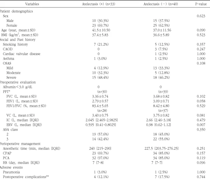
Table 2. Univariate analysis of risk factors for the development of pulmonary atelectasis
Variables Atelectasis (+) (n=33) Atelectasis (−) (n=40) P-value
Patient demographics
Sex 0.623
Male 10 (30.3%) 15 (37.5%)
Female 23 (69.7%) 25 (62.5%)
Age (year, mean±SD) 41.5±10.50 37.0±11.56 0.090
BMI (kg/m2, mean±SD) 37.4±5.83 36.6±5.89 0.523
Social and Past history
Smoking history 7 (21.2%) 5 (12.5%) 0.357
CAOD 0 3 (7.5%) 0.247
Cardiac valvular disease 0 1 (2.5%) 1.000
Asthma 1 (3.0%) 1 (2.5%) 1.000
OSAS 0.108
Mild 4 (12.9%) 13 (33.3%)
Moderate 10 (32.3%) 5 (12.8%)
Severe 15 (48.4%) 18 (46.2%)
Preoperative evaluation
Albumin<3.0 g/dL 0 0
PFT* (n=30) (n=39)
FVC (L, mean±SD) 3.36±0.74 3.68±0.82 0.102
FEV1 (L, mean±SD) 2.79±0.57 3.09±0.71 0.058
FEV1/FVC (%, mean±SD) 83.4±5.65 8.42±4.80 0.529
(n=28) (n=37)
VC (L, mean±SD) 3.40±0.75 3.75±0.82 0.081
IC (L, median [IQR]) 2.645 [2.465-2.9825] 2.66 [2.46-3.18] 0.479
ERV (L, median [IQR]) 0.595 [0.41-0.8025] 0.98 [0.62-1.12] 0.007
ASA class 0.350
2 19 (57.6%) 18 (45.0%)
3 14 (42.4%) 22 (55.0%)
Perioperative management
Anesthetic time (min, median [IQR]) 240 [215-290] 227.5 [203.75-276.25] 0.251
CPAP 23 (69.7%) 34 (85.0%) 0.157
PCA 32 (97.0%) 34 (85.0%) 0.119
HS (day, median [IQR]) 7 [7-8] 7 [7-7] 0.096
Adverse events
Pneumonia 1 (3.0%) 1 (2.5%) 1.000
Postoperative complications** 4 (12.1%) 7 (17.5%) 0.744
SD = standard deviation; BMI = body mass index; CAOD = coronary artery obstructive disease; OSAS = obstructive sleep apnea syndrome;
PFT = pulmonary function test; FVC = forced vital capacity; FEV1 = forced expiratory volume in 1 second; VC = vital capacity; IC = inspiratory capacity; IQR = interquartile range; ERV = expiratory reserve volume; ASA = American Society of Anesthesiologists; CPAP = continuous positive airway pressure; PCA = patient-controlled analgesics; HS = length of postoperative hospital stay.
*A total of 69 patients were evaluated for PFT. Four of 54 patients were evaluated for FVC, FEV1 and FEV1/FVC.
**Postoperative complications: left gastric artery pseudoaneurysm (n=2, 2.7%), small bowel obstruction (n=2, 2.7%), marginal ulcer bleeding (n=2, 2.7%), fever of unknown origin (n=1, 1.4%), postoperative acute tubular necrosis of kidney (n=1, 1.4%), anastomosis site stricture (n=1, 1.4%) and anastomosis site perforation (n=1, 1.4%).
Univariate analysis showed the incidence of atelectasis increased significantly as expiratory reserve volume (ERV) reduced (P=0.007). CPAP treatment was not found to influence the incidence of atelectasis significantly (P=0.157; Table 2).
OSAS severity was considered an inappropriate variable for the multivariate model. AHI results obtained by
polysomnography were used as an indication for CPAP.
OSAS grades were found to be significantly associated with CPAP therapy. Of the PFT variables, only ERV was entered into the multi-variate analysis, because forced vital capacity (FVC), forced expiratory volume in 1 second (FEV1), and VC showed only moderate correlations with ERV (correlation coefficients (rho) 0.664, 0.617 and 0.629;
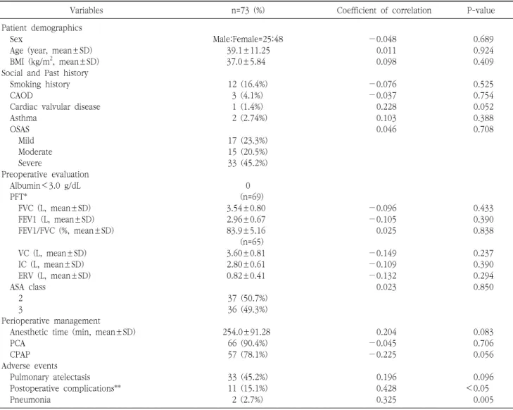
Table 4. Univariate analysis of the correlation between the length of hospital stay after laparoscopic Roux-en-Y gastric bypass and study variables
Variables n=73 (%) Coefficient of correlation P-value
Patient demographics
Sex Male:Female=25:48 −0.048 0.689
Age (year, mean±SD) 39.1±11.25 0.011 0.924
BMI (kg/m2, mean±SD) 37.0±5.84 0.098 0.409
Social and Past history
Smoking history 12 (16.4%) −0.076 0.525
CAOD 3 (4.1%) −0.037 0.754
Cardiac valvular disease 1 (1.4%) 0.228 0.052
Asthma 2 (2.74%) 0.103 0.388
OSAS 0.046 0.708
Mild 17 (23.3%)
Moderate 15 (20.5%)
Severe 33 (45.2%)
Preoperative evaluation
Albumin<3.0 g/dL 0
PFT* (n=69)
FVC (L, mean±SD) 3.54±0.80 −0.096 0.433
FEV1 (L, mean±SD) 2.96±0.67 −0.105 0.390
FEV1/FVC (%, mean±SD) 83.9±5.16 0.025 0.838
(n=65)
VC (L, mean±SD) 3.60±0.81 −0.149 0.237
IC (L, mean±SD) 2.80±0.61 −0.109 0.390
ERV (L, mean±SD) 0.82±0.41 −0.132 0.294
ASA class 0.023 0.850
2 37 (50.7%)
3 36 (49.3%)
Perioperative management
Anesthetic time (min, mean±SD) 254.0±91.28 0.204 0.083
PCA 66 (90.4%) −0.045 0.706
CPAP 57 (78.1%) −0.225 0.056
Adverse events
Pulmonary atelectasis 33 (45.2%) 0.196 0.096
Postoperative complications** 11 (15.1%) 0.428 <0.05
Pneumonia 2 (2.7%) 0.325 0.005
SD = standard deviation; BMI = body mass index; CAOD = coronary artery obstructive disease; OSAS = obstructive sleep apnea syndrome;
PFT = pulmonary function test; FVC = forced vital capacity; FEV1 = forced expiratory volume in 1 second; VC = vital capacity; IC = inspiratory capacity; ERV = expiratory reserve volume; ASA = American Society of Anesthesiologists; PCA = patient-controlled analgesics;
CPAP = continuous positive airway pressure.
*A total of 69 patients were evaluated for PFT. Four of 54 patients were evaluated for FVC, FEV1 and FEV1/FVC.
**Postoperative complications: left gastric artery pseudoaneurysm (n=2, 2.7%), small bowel obstruction (n=2, 2.7%), marginal ulcer bleeding (n=2, 2.7%), fever of unknown origin (n=1, 1.4%), postoperative acute tubular necrosis of kidney (n=1, 1.4%), anastomosis site stricture (n=1, 1.4%) and anastomosis site perforation (n=1, 1.4%).
Table 3. Multi-variate analysis of independent predictors of pulmonary atelectasis
Variables OR 95% CI P-value
Age 1.028 0.974-1.085 0.314
ERV 0.149 0.030-0.740 0.020
CPAP 0.198 0.045-0.874 0.033
PCA 2.177 0.214-22.117 0.511
OR = odds ratio; CI = confidential interval; ERV = expiratory reserve volume; CPAP = continuous positive airway pressure; PCA = patient- controlled analgesics.
P<0.05), and because univariate analysis showed ERV significantly predicted atelectasis development (P=0.007;
Table 2). We suggest this is because reduced FRC, that is, lung volume at the end of passive expiration (the sum of ERV and residual volume), during surgery leads to alveolar dead space development, impaired oxygenation, and atelectasis.
In multivariate analysis, statistical significance should be
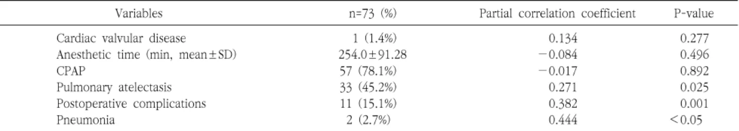
Table 5. Multi-variate analysis of partial correlations between duration of hospital stay after laparoscopic Roux-en-Y gastric bypass and study variables
Variables n=73 (%) Partial correlation coefficient P-value
Cardiac valvular disease 1 (1.4%) 0.134 0.277
Anesthetic time (min, mean±SD) 254.0±91.28 −0.084 0.496
CPAP 57 (78.1%) −0.017 0.892
Pulmonary atelectasis 33 (45.2%) 0.271 0.025
Postoperative complications 11 (15.1%) 0.382 0.001
Pneumonia 2 (2.7%) 0.444 <0.05
SD = standard deviation; CPAP = continuous positive airway pressure.
applied to factors with P-values of <0.1. However, the purpose of this study was to investigate the usefulness of CPAP therapy to prevent postoperative complications, especially pulmonary complications like atelectasis in obese patients, so we included CPAP therapy variables in the multivariate analysis. Therefore, we analyzed the presence or absence of CPAP treatment and PCA treatment, including P-value greater than 0.1, in multivariate analysis.
Multivariate analysis was conducted by adjusting for ERV, age, CPAP therapy, and PCA treatment. The results obtained showed CPAP (odds ratio [OR], 0.198, 95%
confidential interval [CI] 0.045-0.874; P=0.033) and ERV (OR, 0.149, 95% CI 0.030-0.740; P=0.020; Table 3) independently predicted atelectasis development.
2. Durations of postoperative hospital stays
Univariate analysis showed HS was significantly associated with the incidence of pneumonia (rho 0.325, P=0.005) and overall complications (rho 0.428, P<0.05; Table 4). HS was not found to be significantly associated with receipt of CPAP therapy, and HSs in the CPAP and conventional groups were not significantly different (rho −0.225, P=0.056).
Partial correlation analysis was conducted by using multivariate analysis to examine relations between HSs and variables shown by univariate analysis to be associated with atelectasis development and a P-value of <0.1. It was found the development of HS was significantly and partially correlated with the developments of pulmonary atelectasis (partial correlation coefficient (r) 0.271, P=
0.025), pneumonia (r 0.444, P<0.05), and overall complications (r 0.382, P=0.001) (Table 5).
DISCUSSION
In 1991, the National Institute of Health Consensus Development Panel concluded bariatric surgery provides an effective and acceptable treatment for morbidly obese patients [14]. Bariatric surgery involves modification of gastrointestinal tract anatomy to cause gastric restriction and/or nutrient mal-absorption, and it has been shown LGBP provides durable weight loss for up to 20 years and reduces overall mortality [15].
PPCs constitute a pulmonary disease spectrum that includes atelectasis, pneumonia, respiratory failure, and prolonged mechanical ventilation and are a common, serious problem [1]. For this reason, many studies have been undertaken to identify PPC risk factors, and to reduce their incidences, especially that of atelectasis, after general anesthesia [1].
After upper abdominal surgery, multiple factors, such as FRC, VC, anesthetic agents, neuromuscular blocking agents, postoperative analgesics, pain, increased intra-abdominal pressure, and greater inflammatory response, restrict pulmonary function [16].
During anesthetic procedures, unusual patient positioning increases ventilation to perfusion ratio mismatch and alters pulmonary blood distribution, which results in increased alveolar dead space (DS) [16], and it has been shown pulmonary function deterioration and increased alveolar DS lower ERV and increase the incidence of pulmonary atelectasis [16].
Before commencing this study, we believed anesthetic time would probably be associated with a higher incidence of atelectasis. However, the mean anesthetic times of patients that developed or did not develop postoperative
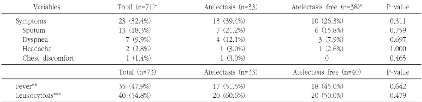
Table 6. Analysis of the relationship between the development of pulmonary atelectasis after laparoscopic Roux-en-Y gastric bypass and patients’ symptoms and signs
Variables Total (n=71)* Atelectasis (n=33) Atelectasis free (n=38)* P-value
Symptoms 23 (32.4%) 13 (39.4%) 10 (26.3%) 0.311
Sputum 13 (18.3%) 7 (21.2%) 6 (15.8%) 0.759
Dyspnea 7 (9.9%) 4 (12.1%) 3 (7.9%) 0.697
Headache 2 (2.8%) 1 (3.0%) 1 (2.6%) 1.000
Chest discomfort 1 (1.4%) 1 (3.0%) 0 0.465
Total (n=73) Atelectasis (n=33) Atelectasis free (n=40) P-value
Fever** 35 (47.9%) 17 (51.5%) 18 (45.0%) 0.642
Leukocytosis*** 40 (54.8%) 20 (60.6%) 20 (50.0%) 0.479
*Seventy-one patients were evaluated for symptoms; the medical records of 2 patients in the atelectasis free group were incomplete and excluded from the analysis.
**Fever was diagnosed when body temperature was ≥38.0oC during the three days after surgery.
***Leukocytosis was diagnosed when the white blood cell count was ≥10,000/μL during the 3 days after surgery.
atelectasis were 240.4 min±51.02 and 273.8 min±121.01, respectively, which were not significantly different (P=0.150).
Obese patients are at greater risk of PPCs, and in a previous study, a BMI ≥27 kg/m2 was found to predict PPC development independently [17]. In another study, it was suggested diminished lung volumes, caused by excessive abdominal fat, anesthetic procedures, and postoperative pain, may have caused prolonged reductions in lung volumes and capacities and led to postoperative complications [18]. Actually, it has been reported that after operations, FRC values are less than 50% of normal in obese patients [18].
To prevent postoperative pulmonary changes, it was recommended; (1) pain-induced reflexes should be blocked and that (2) intensive physiotherapy and (3) postoperative CPAP be implemented [5,19]. CPAP therapy applies constant positive pressure to airways during inspiration and expiration, re-expands collapsed alveoli, and may normalize postoperative FRC and VC values, which if left unchecked may diminish pulmonary function [20]. Furthermore, CPAP therapy may improve pulmonary ventilation and gas exchange and prevent the developments of pulmonary atelectasis and PPCs [21].
Pulmonary atelectasis-related symptoms, that is, fever, cough, shortness of breath, tachypnea, and chest pain, have weak diagnostic values because they are nonspecific, subjective, and depend on patient condition [22]. In the
present study, 23 (32.4%) of the study subjects had atelectasis-related symptoms up to the third postoperative day; 13 experienced excessive sputum production, 7 dyspnea, 2 headache, and one chest discomfort. However, no significant relationship was observed between these symptoms and the development of pulmonary atelectasis.
We consider fever and leukocytosis incomplete factors in terms of diagnosing pulmonary atelectasis. Thirty-five of our study subjects (47.9%) had a fever (body temperature of
≥38.0oC) up to the third postoperative day and 40 patients (54.8%) had leukocytosis (white blood cell count of
> 10,000/μl; Table 6), but neither fever nor leukocytosis was significantly associated with postoperative pulmonary atelectasis (P-values 0.642 and 0.479, respectively).
This study demonstrates perioperative CPAP treatment reduces the incidence of postoperative pulmonary atelectasis in obese patients after LGBP; the incidences of pulmonary atelectasis in the CPAP and conventional groups were 40.4% (23/57) and 62.5% (10/16), respectively (Table 1). Furthermore, we found the OR of CPAP plus conventional therapy was 0.198 (95% CI 0.045-0.874;
P=0.33) and that of ERV was 0.149 (95% CI 0.030-0.740;
P=0.020; Table 3). These results show CPAP treatment should be considered a powerful means of preventing pulmonary atelectasis, because it can reduce the risk of atelectasis by one fifth (OR 0.198) and because ERV is a non-adjustable risk factor.
Complications and their severities can be used as
secondary endpoints. However, we did not consider using complications as secondary endpoints for the following reasons: 1) It was difficult to identify risk factors associated with complications because the number of patients (n=73) included in the study was small and because only 16 patients received conventional pulmonary physiotherapy, and 2) Only 11 complications occurred and complication rates were similar in the CPAP and CPP groups. Therefore, we defined the secondary endpoint as duration of HS.
The study also showed HSs were correlated with the developments of pulmonary atelectasis, pneumonia, and postoperative complications (r 0.271, 0.444, and 0.382, respectively; P<0.05; Table 5), which concurs with a significant relation observed between postoperative complications and HSs after abdominal surgery in a previous study [23].
Based on the results of the present study, we suggest CPAP therapy after LGBP offers a preventive strategy for postoperative atelectasis and recommend that clinicians evaluate risk factors of complications preoperatively, reduce procedure-related factors, detect postoperative complications at the earliest opportunity, and treat complications properly [24].
Several limitations of this study warrant consideration.
First, it was inevitably limited by its retrospective nature. In particular, patient allocations to the two study groups were subject to selection bias. However, the study shows atelectasis, pneumonia, and complications were less frequent in the CPAP group, members of which were considered to be at high risk of pulmonary complications.
It is impossible to deny the results although the composition of patients in the conventional pulmonary physiotherapy group is diverse and the number of patients is limited.
Second, the number of patients enrolled was relatively small, and thus, our results cannot properly represent the general population. Furthermore, because only 33 patients developed pulmonary atelectasis, the multivariate logistic analysis was underpowered. In addition, few postoperative complications (n=11, 15.1%) occurred, and thus, it was not possible to access risk factors of postoperative complications or to statistically investigate relations between CPAP, atelectasis, and complications.
Third, the absence of a standard diagnostic definition for pulmonary atelectasis constitutes another limitation.
Atelectasis is diagnosed based on considerations of pulmonary symptoms, signs, postoperative pulmonary functions, and chest X-ray and/or pulmonary CT findings [2,12]. As explained above, we decided to evaluate the development of atelectasis using chest radiographs.
Unfortunately, this could not be performed using PFT and CT results because tests were not conducted on a routine basis. Accordingly, diagnostic differences between our study and previous studies that used CT and PFT may have resulted in a higher reported incidence of atelectasis (45.2% vs. 37.84% and 13.49%) [4,25].
A fourth limitation involved the difference between predicted efficacy of CPAP treatment and actual efficacy.
Compliance with CPAP therapy has been reported to range from 46.6 to 86% [26-30], and thus, compliance with CPAP, which was not evaluated in the present study, may have affected our results. Actually, 4 of the 6 patients (66.7%) that refused CPAP developed atelectasis.
Summarizing, the present study shows CPAP is a useful modality for preventing pulmonary atelectasis after LGBP in obese patients. The study also shows low preoperative ERV predicts the development of pulmonary atelectasis and that efforts to reduce postoperative complications, including atelectasis, may reduce hospital stays.
ACKNOWLEDGMENTS
This work was supported by grants from the Korean Health Technology R & D Project (HC15C1322), Ministry of Health & Welfare, Republic of Korea (for YH).
REFERENCES
1. Smetana GW, Lawrence VA, Cornell JE. Preoperative pulmonary risk stratification for noncardiothoracic surgery: systematic re- view for the American College of Physicians. Ann Intern Med 2006;144:581-95.
2. Eichenberger A, Proietti S, Wicky S, et al. Morbid obesity and post- operative pulmonary atelectasis: an underestimated problem.
Anesth Analg 2002;95:1788-92, table of contents.
3. Tusman G, Böhm SH, Warner DO, Sprung J. Atelectasis and peri- operative pulmonary complications in high-risk patients. Curr Opin Anaesthesiol 2012;25:1-10.
4. Baltieri L, Peixoto-Souza FS, Rasera-Junior I, Montebelo MI, Costa D, Pazzianotto-Forti EM. Analysis of the prevalence of atelectasis in patients undergoing bariatric surgery. Braz J Anesthesiol 2016;66:577-82.
5. Lawrence VA, Cornell JE, Smetana GW. Strategies to reduce post- operative pulmonary complications after noncardiothoracic sur- gery: systematic review for the American College of Physicians.
Ann Intern Med 2006;144:596-608.
6. Hanekom SD, Brooks D, Denehy L, et al. Reaching consensus on the physiotherapeutic management of patients following upper abdominal surgery: a pragmatic approach to interpret equivocal evidence. BMC Med Inform Decis Mak 2012;12:5.
7. Stock MC, Downs JB, Gauer PK, Alster JM, Imrey PB. Prevention of postoperative pulmonary complications with CPAP, incentive spirometry, and conservative therapy. Chest 1985;87:151-7.
8. Ireland CJ, Chapman TM, Mathew SF, Herbison GP, Zacharias M.
Continuous positive airway pressure (CPAP) during the post- operative period for prevention of postoperative morbidity and mortality following major abdominal surgery. Cochrane Database Syst Rev 2014;(8):CD008930.
9. Swedish Council on Health Technology Assessment. Obstructive sleep apnoea syndrome: a systematic literature review.
Stockholm: Swedish Council on Health Technology Assessment (SBU), 2007.
10. Zhu K, Aouf S, Roisman G, Escourrou P. Pressure-relief features of fixed and autotitrating continuous positive airway pressure may impair their efficacy: evaluation with a respiratory bench model.
J Clin Sleep Med 2016;12:385-92.
11. Hawker GA, Mian S, Kendzerska T, French M. Measures of adult pain: Visual Analog Scale for Pain (VAS Pain), Numeric Rating Scale for Pain (NRS Pain), McGill Pain Questionnaire (MPQ), Short-Form McGill Pain Questionnaire (SF-MPQ), Chronic Pain Grade Scale (CPGS), Short Form-36 Bodily Pain Scale (SF-36 BPS), and Measure of Intermittent and Constant Osteoarthritis Pain (ICOAP). Arthritis Care Res (Hoboken) 2011;63 Suppl 11:S240-52.
12. Dripps RD, Deming MV. Postoperative atelectasis and pneumonia. Ann Surg 1946;124:94-110.
13. Woodring JH, Reed JC. Types and mechanisms of pulmonary atelectasis. J Thorac Imaging 1996;11:92-108.
14. NIH conference. Gastrointestinal surgery for severe obesity.
Consensus Development Conference Panel. Ann Intern Med 1991;115:956-61.
15. Choi YB. Current status of bariatric and metabolic surgery in Korea. Endocrinol Metab (Seoul) 2016;31:525-32.
16. Miskovic A, Lumb AB. Postoperative pulmonary complications. Br J Anaesth 2017;118:317-34.
17. Brooks-Brunn JA. Predictors of postoperative pulmonary compli-
cations following abdominal surgery. Chest 1997;111:564-71.
18. Blouw EL, Rudolph AD, Narr BJ, Sarr MG. The frequency of respi- ratory failure in patients with morbid obesity undergoing gastric bypass. AANA J 2003;71:45-50.
19. Canet J, Mazo V. Postoperative pulmonary complications.
Minerva Anestesiol 2010;76:138-43.
20. Denehy L, Carroll S, Ntoumenopoulos G, Jenkins S. A randomized controlled trial comparing periodic mask CPAP with physi- otherapy after abdominal surgery. Physiother Res Int 2001;6:
236-50.
21. Lindner KH, Lotz P, Ahnefeld FW. Continuous positive airway pressure effect on functional residual capacity, vital capacity and its subdivisions. Chest 1987;92:66-70.
22. Mavros MN, Velmahos GC, Falagas ME. Atelectasis as a cause of postoperative fever: where is the clinical evidence? Chest 2011;
140:418-24.
23. Xie YB, Wang CF, Zhao DB, et al. Risk factors associated with postoperative hospital stay after pancreaticoduodenectomy: a retrospective study. Chin Med J 2013;126:3685-9.
24. Pessaux P, Msika S, Atalla D, Hay JM, Flamant Y. Risk factors for postoperative infectious complications in noncolorectal abdomi- nal surgery: a multivariate analysis based on a prospective multi- center study of 4718 patients. Arch Surg 2003;138:314-24.
25. Al Jaaly E, Fiorentino F, Reeves BC, et al. Effect of adding post- operative noninvasive ventilation to usual care to prevent pulmo- nary complications in patients undergoing coronary artery bypass grafting: a randomized controlled trial. J Thorac Cardiovasc Surg 2013;146:912-8.
26. Hussain SF, Irfan M, Waheed Z, Alam N, Mansoor S, Islam M.
Compliance with continuous positive airway pressure (CPAP) therapy for obstructive sleep apnea among privately paying pa- tients- a cross sectional study. BMC Pulm Med 2014;14:188.
27. Johnson MK, Carter R, Nicol A, Paton R, Banham SW. Long-term continuous positive airway pressure (CPAP) outcomes from a sleep service using limited sleep studies and daycase CPAP titra- tion in the management of obstructive sleep apnoea/hypopnoea syndrome. Chron Respir Dis 2004;1:83-8.
28. McArdle N, Devereux G, Heidarnejad H, Engleman HM, Mackay TW, Douglas NJ. Long-term use of CPAP therapy for sleep ap- nea/hypopnea syndrome. Am J Respir Crit Care Med 1999;159:
1108-14.
29. Hollandt JH, Mahlerwein M. Nasal breathing and continuous pos- itive airway pressure (CPAP) in patients with obstructive sleep ap- nea (OSA). Sleep Breath 2003;7:87-94.
30. Sin DD, Mayers I, Man GC, Pawluk L. Long-term compliance rates to continuous positive airway pressure in obstructive sleep ap- nea: a population-based study. Chest 2002;121:430-5.