Received: December 13, 2017 Revised: December 19, 2017 Accepted: December 22, 2017
Copyright © 2017. The Korean Academy of Oral &
Maxillofacial Implantology
This is an Open Access article distributed under the terms of the Creative Commons Attrib- ution Non-Commercial License (http://creative- commons.org/licenses/by-nc/4.0/) which permits unrestricted non-commercial use, distribution, and pISSN : 1229-5418
Implantology 2017; 21(4): 266-276 https://doi.org/10.12972/implantology.20170020
eISSN : 0000-0000 OPEN ACCESS
골유도 재생술에 티타늄 메쉬 사용 시 노출을 줄이기 위한 콜라테입의 사용: 증례보고
김남윤*
김남윤 치과의원
Use of Colla-tape to Reduce Exposure When Using Titanium Mesh in Guided Bone
Regeneration: A Case Report
Nam Yoon Kim*
Kim Nam Yoon Dental Office, Seongnam, Korea
*Corresponding author: Nam Yoon Kim, knyperio@gmail.com
Abstract
The purpose of this study is to evaluate the use of colla tape over the titanium mesh have same effectiveness compare to other materials to reduce exposure of the titanium mesh in guided bone regeneration (GBR). One of the most frequent complications in GBR procedure is early exposure of the barrier membrane. The exposure of membrane could make poor result in GBR procedure. Moreover, In the case of non-resorbable membranes like as a titanium mesh, the frequency of the exposure rates increase more than the resorbable membranes. The stiffness of barrier membrane could help to maintain the space of the bony defects, but it has more exposure frequency in the early stage of healing time. The advantage of using colla-tape over the titanium mesh as a reservoir is 1) to stabilize the wound 2) to release tension at the superficial layer of the flap. 3) to prevent soft tissue ingrowth beneath the titanium mesh at the beginning of the healing period. It is also useful that the non-resorbable or resorbable barrier membrane, PRP, PRF are capable to cover the titanium mesh but colla-tape has cost-effectiveness and simple procedure compare to other materials.
Keywords: Colla-tape, Guided bone regeneration, Implant, Non-resorbable membrane, Titanium mesh
I. 서론
임플란트 수술 시 골결손부를 만나는 것이나, 부족한 골량에 임플란트를 식립하는 것
은 보편적인 술식이다. 부족한 골량을 만들거나 골결손부를 채우는 술식이 골유도재생술
이다. 하지만 많은 임상가들이 경험이 많아질수록 어렵게 느껴지는 것 또한 골유도재생
은 아직도 불분명하다. 무치악 부위로 장기간 흡수된 치조골의 경우 골결손 부위의 형태와 골 흡수량까 지 정확하게 예측할 수 없다. 2달 이상 충분한 발치와의 치유기간이 지나도 골형성이 제대로 되지 않은 경우도 5% 이상에서 만날 수 있다
1. 발치와 골이식술이 치조골 흡수를 줄일 수 있는 대체적인 술식으 로 소개되고 있다.
골유도 재생술에 사용되는 골 이식재로 자가골이 gold standard이지만 구강 내에서 채취하는 데 양 이 제한되어 있고, 흡수되는 속도를 예측하기 어려우며, 감염의 확률이 높고 부가적인 수술부위가 더 필요하게 되어, 요즘에는 드릴링 시 얻어지는 소량의 자가골과 이종골을 섞어 사용하는 경우가 자가골 의 장점과 이종골의 늦은 흡수속도와 골격유지능력 때문에 선호하는 경향이 있다
2.
차폐막의 경우에는 비흡수성 차폐막과 흠수성 차폐막 모두가 골유도재생술에 이용되고 결과도 우 수하지만 2벽성 골결손부나 수직적인 골증대가 필요한 경우 혹은 수직 수평적으로 혼합된 골결손부의 경우 생체친화성이 있고, 3차원적으로 적절한 공간을 유지해주며 하방의 골이식재가 수축을 하더라도 그대로 공간을 유지하는 titanium mesh의 사용을 추천한다
3. 최근에는 임플란트 식립후 골결손부의 형 태에 맞게 디자인된 titanium mesh가 개발되어 예전보다 쉬운 임상적용이 가능하다.
그러나 이러한 공간유지능력 때문에 표재성 잇몸의 두께가 얇아진다 든지 10-30% 정도의 막의 조 기노출이 titanium mesh의 사용을 꺼리게 된 이유라고 할 수 있다
4. 많은 연구자들이 titanium mesh 사 용 시 차폐막의 조기 노출을 줄이기 위해 titanium mesh위에 비흡수성 차단막인 Gore-tex나 PRP, 흡수 성 콜라겐 차폐막 등 다양한 재료와 방법들을 적용하였다
5. 본 증례에서는 진료실에서 쉽게 구할 수 있 는 colla-tape을 사용하였다. Colla-tape은 창상을 보호하고 지혈효과가 있으며 혈병을 잘 유지하여 각 종 수술 시 치유를 돕는 역할을 한다. Titanium mesh위에 덮어 창상을 보호하고 판막에 가해지는 장력 을 완충하는 역할에 있어 가장 가성비가 좋은 재료라고 할 수 있다. 본 증례에서는 중등도 결손부가 있 는 임플란트 수술 증례에 골유도재생술의 차폐막으로 titanium mesh를 사용 시 resevoir 역할로 Colla-tape 을 사용한 증례에 대해 보고하고자 한다.
II. 증례보고
1. 증례 1
만 53세 여성환자가 #24, 25의 동요도와 불편함을 주소로 내원하였다. 파노라마 촬영과 치주낭측정
후 국소적으로 심한 골결손을 동반한 만성치주염으로 진단하였고, 치석제거와 치근활택술을 시행하
고 교합성 외상을 보이는 #24, 25치아의 교합 조정술을 시행하였다. 4주 정도 재평가 기간 후에 동요도
가 줄어들지 않고 환자의 불편감이 심해져 #24, 25의 발치를 결정하였다(Fig. 1).
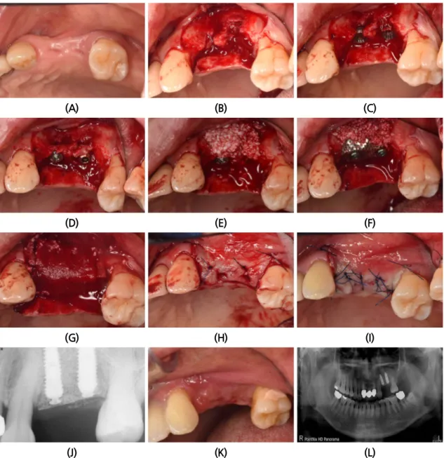
(A) (B)
Fig. 1. (A) Case 1. Initial Panoramic view, See the clinical attachment loss of #24,25 area due to trauma from occlusion. (B) 2 months after extraction. See the tremendous alveolar bony defects of #24,25 area.
Nam Yoon Kim : Use of Colla-tape to Reduce Exposure When Using Titanium Mesh in Guided Bone Regeneration: A Case Report. Implantology 2017
발치 후 약 9주 정도의 치유기간을 기다려 임플란트 식립을 위해 국소마취하에 판막형성을 하였다.
발치와의 치유가 완전하지 않았고, 임플란트 식립 후에는 수직적으로 2-3개의 나사선이 노출 되었다.
구개측 치조골쪽으로 밀듯이 드릴링을 하고 임플란트의 플랫폼(Platform) 부위를 치조골정에서 1.5 mm 가량 깊게 식립하였다. 제조자의 추천은 치조골정에서 약 0.5 mm 깊게 식립하는 것을 추천하나 수 평적 수직적 골유도재생을 위해서 제조자의 추천보다 약간 더 깊이 식립하는 것이 유리하기 때문이다.
노출된 나사선에는 드릴링 시 얻어진 자가골을 덮고 바깥쪽에는 임플란트 직경의 협설측으로 2 mm 이 상의 폭을 만들 수 있도록 충분한 양의 이종골을 덮었다. 상악골의 치밀도가 하악골에 비해 떨어지기 때문에 별도의 피질골제거(decortication)는 실시하지 않았다
(Fig. 2A-2E).본 증례에서는 ㈜네오바이오텍에서 제조한 GBR system을 사용하였고 높이 1.5 mm의 스페이서
(Spacer)를 임플란트에 연결한 후에 80도 정도로 접은 무치악용 titanium mesh (E2)를 적용하였다. 상
부의 고정은 GBR 전용 덮개나사(Cover screw)를 사용하였다. Colla-tape은 titanium mesh를 덮을 만
큼 외과용 가위를 사용하여 절단하고 다듬었다. 그 후 Colla-tape은 마른(dry) 채로 환부에 덮고 창상부
위에서 스며 나온 혈액에 의해 자연스레 젖을 수 있도록 적용한 후에 잠시 기다렸다가 협측의 판막에
감장절개를 하여 판막의 긴장도를 낮춰준 후에 수평누상봉합을 2군데 실시하여 협설측의 판막이 인접
한 근처에 올 수 있도록 한 후에 단속봉합으로 마무리하였다. 이러한 이중-봉합법은 판막의 긴장도를
줄이며 일차유합의 확률을 높일 수 있는 방법으로 추천된다
(Fig. 2F-2I).수술 후 촬영한 구내 방사선
사진에서 #24 부위 임플란트의 GBR 전용 덮개나사가 제대로 고정이 되지 않은 것을 알 수 있다. 그러
나 높이 올라온 것도 아니고, 일차 유합에 방해되지 않아 그냥 두기로 했다(Fig. 2J). 봉합사는 4-0 나일
론사를 사용하였으며 2주 후에 봉합사 제거 시 완벽한 일차유합을 이루고 있었고 염증이나 2차적인 감
염의 징후가 없었다
(Fig. 2K, 2L).(A) (B) (C)
(D) (E) (F)
(G) (H) (I)
(J) (K) (L)
Fig. 2. (A) Pre-operative clinical photo. (B) In bucco-lingually, there are aberrant healing of extraction site. (C) Two of regular implants are placed. (D) Autogenous grafts are placed on the exposed threads of implant that they are from drilling procedure. (E) Deproteined bovine bone are placed enough in the outer site of implant. (F) As a barrier membrane, Titanium mesh (E2) are used. then 2 points are fixed on the coronal part and add more graft in the lower part. (G) Cover the titanium mesh with Colla tape.
(H) Approximation of the each flap with horizontal mattress sutures. (I) Get the primary closure by the perform of interrupted suture. (J) Intra-oral standard x-ray after the placement of implant and application of Titanium mesh. (K) 2 weeks after. Stiches are out. (L) Panoramic view after first surgery.
Nam Yoon Kim : Use of Colla-tape to Reduce Exposure When Using Titanium Mesh in Guided Bone Regeneration: A Case Report. Implantology 2017
4 개월 후까지 titanium mesh가 노출되지 않았음을 확인할 수 있었고 uncovering을 위해 국소마취하
에 titanium mesh를 제거하였다. 협설측으로 골양조직이 충분한 양이 생긴 것을 관찰할 수 있었으며
치유지대주를 연결한 후에 유경피판을 형성하여 협설측 판막을 회전하여 봉합하였다(Palacci technique).
상악 구치부에서 이 방식은 치유지대주 주변의 건강한 잇몸 형성에 도움이 된다
(Fig. 3).(A) (B) (C)
(D) (E) (F)
(G) (H)
Fig. 3. (A) After 4 months healing period. (B) after 4 months, the state of titanium mesh. (C) In bucco-lingually, there makes enough bone volume. (D) After removal of titanium mesh, GBR cover screw, spacer. (E) After the connection of healing abutment. there makes enough bone volume in bucco-lingually. (F) perfome the pedicle flap in buccally and rotate the flap to the proximal area (Palacci technique). (G) After the uncovering procedure and the suture it up. (H) Panoramic view, after the uncovering procedure.
Nam Yoon Kim : Use of Colla-tape to Reduce Exposure When Using Titanium Mesh in Guided Bone Regeneration: A Case Report. Implantology 2017
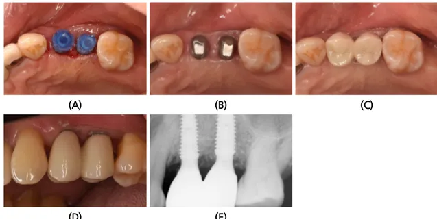
Uncovering 후에 약 3주간의 치유기간을 기다린 후 Pick Cap Impression coping을 사용하여 인상 채득과 교합인기를 채득한 후에 보철물을 제작하였다. 개별지대주를 position jig를 사용하여 장착한 후에 15분 정도의 시간 차를 두고 지대주 나사를 2번 이상 30 Ncm 이상으로 고정하고, 그 기간에는 지 대주의 침하를 유도하기 위해 환자에게 Cotton Roll을 물고 있게 하였다. 그 후 전부-지르코니아 보철 물을 이용하여 접촉점과 교합을 조정한 후에 영구 접착하였다(Intercem
®) (Fig. 4A-4D).
보철물 접착 후 잔존 접착제의 유무를 판단하기 위해 구내 방사선 촬영을 실시하였다
(Fig. 4E).(A) (B) (C)
(D) (E)
Fig. 4. (A) After the removal of healing abutment and connection of Pick Cap Impression coping for impression taking. (B) After the connection of customized abutment. (C) After the delivery of definitive prosthesis (Monolithic zirconia prosthesis). (D) See the supragingival margin of the prosthesis in bucal aspect. (E) Intra-oral x-ray after the delivery of definitive prosthesis.
Nam Yoon Kim : Use of Colla-tape to Reduce Exposure When Using Titanium Mesh in Guided Bone Regeneration: A Case Report. Implantology 2017
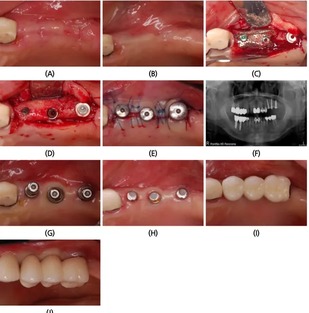
2. 증례 2
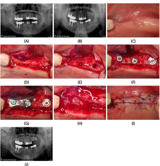
57 세의 여성 환자가 상악 좌측부위 시린 증상을 주소로 내원 하였다. 파노라마 촬영과 구강 내 검진 결과 #26의 보철물 하방의 우식이 관찰 되었고 #22, 23-26의 브릿지를 제거한 결과 #26의 우식이 심하 여 발치가 추천되었다
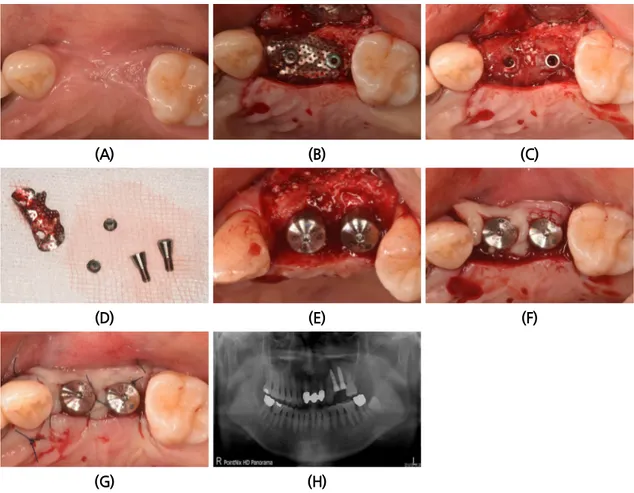
(Fig. 5A).발치 후 7주후 임플란트 식립을 위하여 환자가 재내원하였다. 재촬영한 파노라마 상에서 #26부위의 상악동의 함기화가 빠르게 진행되어 상악동 거상술과 골이식술이 필요하게 되었다. 예상과 달리 임플 란트 식립을 위한 판막 형성 후 #24, 25 부위에 임플란트 식립이 불가능할 정도의 좁은 치조제가 관찰 되었으며 #26부위도 빠르게 치조제의 흡수가 관찰되었다(Fig. 5B-5D).
임플란트 식립을 위한 드릴링 전에 협설 측으로 어느 정도 치조제 폭을 만들기 위하여 수평적인 골삭 제를 실시하였다
(Fig. 5E).그 후에 #24부위는 치조제 위축이 심하여 드릴링 시 협측으로 드릴이 빠져나 오게 되는데, 이때 핸드피스를 잡은 손을 구개측으로 밀면서 드릴링과 식립을 하여 25 Ncm의 초기고정 을 얻었다. #25부위는 치조제의 중간 부분을 약간 벌리면서 식립을 하였고 #26부위는 ㈜네오바이오텍 SCA kit 를 사용하여 상악동의 치조제 접근을 이용하여 상악동을 거상하고 별도의 골이식 없이 약 2.5 mm 의 상악동 점막을 거상하고 상악동 하연의 피질골을 이용하여 2중 피질골 고정을 얻었다
(Fig. 5F).그 후에 #24, 25부위는 높이 1 mm의 스페이서(Spacer)를 연결하고, 노출된 나사선 부위는 자가골을
한층 덮고, 외부에는 탈단백우골을 이식하였다. 협설 측으로 임플란트 직경에 2 mm 이상의 충분한 협
설측으로 골이식 후에 80도 이하로 구부린 무치악용 titanium mesh (E2)를 적용하였다. Titanium mesh 는 GBR전용 덮개나사로 고정 상부에 2부분 고정하였고 GBR system에 있는 고정나사(fixing screw)로 #24,25 사이의 근단부에 고정을 추가하였다. 후에 이전의 증례에서 했던 것과 같은 방법으로 Colla-tape 을 적용하였다. 봉합은 수평누상 봉합과 단속봉합을 2중으로 사용하였다. 봉합사는 4-0 나
(A) (B) (C)
(D) (E) (F)
(G) (H) (I)
(J)
Fig. 5. (A) Case 2. Initial Panoramic view. (B) 2 months after the extraction of #26. Panoramic view. (C) Pre-operative clinical photo. (D) See the narrow alveolar ridge in premolar area. (E) The sharp alveolar ridge trimmed horizontally to get some width for implant placement, and then start to drill in the osteotomy site. (F) Placement of the implant #24,25,26. #24,25 are connected 1mm height of the spacer, the #26 connected cover screw. (G) Titanium mesh(E2)was fixed with GBR cover screw, add buccal side fixation with small fixing screw. (H) After the application of colla tape. (I) Perform the horizontal mattress suture and interrupted suture for primary closure. (J) Panoramic view after the
일론사를 사용하였으며 2주 후에 제거하였다. 2주 후에 일차유합을 잘 이루고 있었으며 4개월까지 일 차유합을 유지하였다
(Fig. 5G-5J).4개월 후에 Uncovering을 위한 2차 수술 시 titanium mesh와 고정나사는 쉽게 제거가 되었으며
#24, 25 부위에 많은 골양 조직이 만들어진 것을 확인할 수 있다
(Fig. 6A-6D).적절한 크기와 높이의 치 유 지대주를 연결한 후에 단속봉합을 실시하였다(Fig. 6E, 6F). 충분한 치유가 일어날 수 있도록 약 4주
(A) (B) (C)
(D) (E) (F)
(G) (H) (I)
(J)
Fig. 6. (A) Clinical photo after the removal of stitches. (B) Clinical photo after 4 months. (C) After 4 months, perform the uncovering surgery. (D) There make enough volume of the bone-like tissue. (E) After the uncovering surgery, connecting the healing abutment and suture it up. (F) Panoramic view, after the uncovering surgery. (G) After the connecting of transfer impression coping. (H) Using the preformed abutment. (I) After the delivery of Porcelain Fused Metal (PFM) prosthesis. (J) After the delivery of the definitive prosthesis in buccal aspect.
간 기다린 후에 transfer type impression coping을 연결하고 인상 채득하였다
(Fig. 6G).보철물은 기성 지대주를 이용하였고 금속-도재관으로 보철물을 제작하여 영구접착하였다
(Fig. 6H-6J).III. 총괄 및 고찰
최근에 각 임플란트 업체에서 개발되어 상품화된 titanium mesh는 임플란트에 고정하기 쉽고 개별 적인 골결손부의 환경에 맞춤식 골유도 재생이 가능하며, 제거하는 방법도 비교적 간단해 임상에 널리 적용되고 있다. Titanium mesh는 뛰어난 생체 친화성과 3차원 적인 공간유지능력에 비해 조기 노출의 위험성이 있지만, 몇 가지 원칙과 노력으로 노출의 위험을 줄일 수 있다. 우선 임플란트 식립과 동시에 titanium mesh를 사용하기 위해서는 스페이서(Spacer)를 1 mm 이상이 되는 것을 추천한다. 혹자는 이 스페이서의 역할이 치조골 수직증강을 위해 사용한다고 하는데, 원래의 목적은 신생조직의 초기 수축 을 보상하고 식립된 임플란트와 잔존골의 높이 차이를 보상하기 위해 연결하는 것이다. 임플란트의 platform 이 치조골정의 하방에 오도록 하는 것이 장기적 예후에 유리하고 또한 재생된 골의 수축과 titanium mesh 를 사용했을 때 생기는 하방의 연조직의 두께를 고려해야 하기 때문이다. 두 번째로 titanium mesh를 구부릴 때는 90도 이하의 예각을 만드는 것이 중요하다. Titanium mesh는 공간 유지 력이 좋기 때문에 협설측으로 벌어지며 노출의 문제점이 생길 수 있다. 노출을 방지하려면 Over bending 을 통해 골결손부에 능동적으로 달라붙어 유지가 될 정도로 부드럽고 유연하게 구부려야 한다
6.
Titanium mesh는 인접한 치아나 절개선 혹은 판막과 만나는 지점에서 최소 1 mm 이상은 떨어져 있 어야 한다. 이 원칙을 지키지 않으면 그 부위부터 노출이 일어난다. Ti-mesh위에 Colla-tape를 적용하 는 이유는 1) 수술부위 창상의 안정을 도모하기 위해 2) Ti-mesh 위에 존재하는 표재층의 판막이 얇아 지는 것을 방지하기 위해 3) 초기치유 시 Ti-mesh 하방으로 연조직이 자라 들어오는 것을 방지하기 위 함이다. 혹자는 그러면 흡수성 차단막이나 혹은 비흡수성 차단막, 혹은 PRF나 PRP를 같은 용도로 사 용하는 것은 어떠냐고 물어본다. 필자는 모두 사용가능하며 좋다고 생각 한다
7. 그러나 Colla-tape이 현 재로서는 가성비가 가장 좋은 제품이며 그 이상의 것이 필요치 않다고 생각한다.
임플란트는 가능한 한 2회법으로 시술하는 것이 좋다. 치유 지대주를 연결하는 1회법 수술일 경우
치유 지대주 주변이 환벽하게 밀봉이 되어야 한다. 봉합은 수평누상봉합을 사용하여 협설 측의 판막을
가까이 위치시키고 일차 유합을 위해 단속봉합을 그 위에 적용하여 2중 봉합을 해야 하며, 협설 측의
판막이 everted되어야 수축이 되어도 판막이 벌어지지 않는다
8. 환자 소환은 3주간은 매주하여 수술 부
위 치유상태를 체크하며, 그 이후에는 한 달에 한 번 정도 소환하여 titanium mesh의 노출 유무를 파악
한다. 협설 측으로 3 mm의 폭이 없는 좁고 위축된 치조제의 경우 3차원적으로 제 위치에 임플란트 식
하는 것을 추천 한다.
titanium mesh 와 같은 비흡수성 차폐막을 사용 시 하방에 연조직이 생기는데 이는 골양조직이 아니 다. 어떤 연구자는 연조직이 게재되어 골유도재생술에 도움이 되지 않으니 제거해야 한다고 하고 있고 다른 연구자는 그대로 둬도 상관없다고 한다
10,11. 필자의 생각은 골유도재생술후 지연식립 시에 임플 란트 플랫폼(platform)을 깊게 위치하는 것으로 연조직의 수축과 소실을 보상할 수 있다고 생각하며 굳이 제거하여 창상부위 상처를 더 만들 필요는 없다고 생각한다. 비흡수성 차폐막 하부에 생기는 연조 직의 두께는 차폐막 고정이 잘 이루어질 경우, 연조직의 긴장도가 덜할수록 더 얇게 생긴다.
IV. 결론
골유도재생술식 시 차폐막으로 titanium mesh를 사용할 때 reservoir 역할로 사용할 수 있는 재료로 비흡수성 차폐막과 흠수성 차폐막 혹은 PRP, PFR 등 다양한 재료를 사용할 수 있다. 그러나 Colla-tape 은 쉽게 구할 수 있고 가격도 저렴하여 가성비가 좋은 재료로서 다른 재료와 비교하여 비슷한 효과를 가진다.
References
1. Roccuzzo M, Ramieri G, Bunino M, et al. Autogenous bone graft alone or associated with titanium mesh for vertical alveolar ridge augmentation: a controlled clinical trial. Clin Oral Implants Res. 2007;
18: 286-294.
2. Lim HC, Kim MS, Yang C, et al. The effectiveness of a customized titanium mesh for ridge preservation with immediate implantation in dogs. Clin Implant Dent Relat Res. 2015; 17: e652-e660.
3. Pellegrino G, Lizio G, Corinaldesi G, et al. Titanium mesh technique in rehabilitation of totally edentulous atrophic maxillae: a retrospective case series. J Periodontol. 2016; 87: 519-528.
4. Von Arx T, Kurt B. Implant placement and simultaneous ridge augmentation using autogenous bone and a micro titanium mesh: a prospective clinical study with 20 implants. Clin Oral Implants Res.
1999; 10: 24-33.
5. Van Steenberghe D, Johansson C, Quirynen M, et al. Bone augmentation by means of a stiff occlusive titanium barrier: a study in rabbits and humans. Clin Oral Implants Res. 2003; 14: 63-71.
6. Artzi Z, Dayan D, Alpern Y, et al. Vertical ridge augmentation using xenogenic material supported by a configured titanium mesh: clinicohistopathologic and histochemical study. Int J Oral Maxillofac Implants. 2003; 18: 440-446.
7. Jung GU, Jeon JY, Hwang KG, et al. Preliminary evaluation of a three-dimensional, customized, and preformed titanium mesh in peri-implant alveolar bone regeneration. J Korean Assoc Oral Maxillofac Surg. 2014; 40: 181-187.
8. Adell R, Lekholm U, Rockler B, et al. A 15-year study of osseointegrated implants in the treatment of the edentulous jaw. Int J Oral Surg. 1981; 10: 387-416.
9. Wang HL, Misch C, Neiva RF. Sandwich bone augmentation technique: rationale and report of pilot cases. Int J Periodontics Restorative Dent. 2004; 24: 232-245.
10. Schropp L, Wenzel A, Kostopoulos L, et al. Bone healing and soft tissue contour changes following single-tooth extraction: a clinical and radiographic 12-month prospective study. Int J Periodontics Restorative Dent 2003; 23: 313-323.
11. Le B, Rohrer MD, Prasad HS. Screw “tent-pole” grafting technique for reconstruction of large vertical alveolar ridge defects using human mineralized allograft for implant site preparation. J Oral Maxillofac Surg. 2010; 68: 428-435.