서 론
Atlantoaxial rotatory fixation (AARF) is the pathologic interlocking of the C1-C2 joint. It is a rare condition usually following an upper respiratory tract infection in childhoods and its resolution usually occurs spontaneously or after traction therapy.
1)Also, it has been identified after tonsilectomy and even minimal trauma in childhoods.
In adults, it is exceedingly rare by reason of adult's specific anatomic features.
2,3)It has been described in patients with inflammatory conditions of the spine such as rheumatoid arthritis and ankylosing spondylitis. Only a few cases following acute trauma have been reported in the literature. We report a exceedingly rare case of type I atlantoaxial rotatory fixation (AARF) following acute trauma in an elderly patient.
Case Report
A 81-year-old female patient presented severe weakness of all extremities and pain in the upper cervical area after
falling down stairs. Her head was rotated to the right side and fixed with decreased neck motion. A neurologic examination revealed quadriparesis (Grade 2/3, ASIA impairment scale C) for a motor score of 42 and partial sensory loss throughout the trunk and all extremities below C3 dermatome level. Radiographs in neutral showed ambiguous findings and a 2-mm atlantodental interval(ADI). Cervical spine computed tomography (CT) with 3-dimensional reformations revealed anterior subluxation of the left lateral mass at C1 and posterior subluxation of the right lateral mass at C1(Fig. 1). There was no evident of fracture. Neck CT angiogram revealed hypoplastic change of right vertebral artery, but evidence of traumatic injury of both vertebral arteries was absent(Fig. 2). The cervical spine magnetic resonance imaging revealed signal changes in the cervical cord at the C2 level, which was clear evidence of cervical cord injury.
And there was no evidence of the transverse ligament injury. First, we applied continuous skull traction for 48 hours, but failed in reduction. Therefore, under general anesthesia, a closed manual reduction was attempted by compression of the anteriorly dislocated, left C1 arch by providing pressure with the finger through the mouth prior to open reduction. Successful reduction was obtained with an audible “pop” sound, and confirmed using C-arm
고신대학교 의과대학 학술지 제 권 제 호24 1 Kosin Medical Journal
Vol. 24. No. 1, pp. 169 172, 2009∼
Ju-Ho Jeong ․ Dae-Yong Kim
Department of Neurosurgery, School of Medicine, Kosin University, Busan, Korea
――― Abstract ――――――――――――――――――――――――――――――――――――――――
Atlantoaxial rotatory fixation (AARF), which is the pathologic interlocking of the C1-C2 joint, is an uncommon disorder usually following an upper respiratory tract infection in childhoods. In adults, it is exceedingly rare by reason of adult's specific anatomic features. Here, the authors report on one case of type I atlantoaxial rotatory fixation (AARF) following acute trauma in an elderly patient, and review the literature.
―――――――――――――――――――――――――――――――――――――――――――――――――
Key words : Atlantoaxial rotatory fixation (AARF), childhoods, anatomic features
교신저자 Dae-Yong Kim:
Add : Department of Neurosurgery, Kosin University Gospel Hospital, 34 Amnam-dong, Suh-gu, Busan 602-702, Korea TEL : +82-51-990-6705 FAX : +82-51-990-3042 E-mail : [email protected]
고신대학교 의과대학 학술지 제 권 호 24 1 , 2009
fluoroscopy. After closed manual reduction, the posterior cervical pain and neck deformity were relieved immediately. Radiographs and CT scan showed good position of the C1 C2 complex(Fig. 3.). And then, a rigid – orthosis (Halo vest) was applied to the patient on the same day. But, one month later the patient died suddenly of myocardial infarction.
Discussion
The true etiology of atlantoaxial rotatory fixation (AARF) is unknown. The cause of AARFs is generally involved in pharyngitis. It is hypothesized that local swelling secondary to an inflammatory process leads to softening and possible disruption of the alar and transverse ligaments. Also it has been rarely reported in numerous conditions such as ankylosing spondylitis, metastatic tumors, generalized ligamentous laxity, eosinophilic granulomas, and a variety of other nondestabilizing procedures involving the head and neck.
4,5,6,7,8,9)There is a clear predilection for children and young adults regardless of the cause.
1)The higher incidence of AARF in children is a consequence of specific anatomic features.
10)The joint surface of the lateral mass is shallower and more horizontally oriented in children. In addition, the relative elasticity of the ligaments, the not yet fully developed neck muscles and the relatively large head might also be predisposing factors for AARF in children.
For this reason, AARF is very rare in adults.
11,12,13)Only a few cases of AARFs have been reported in adults, perhaps because the cause may often be a severe trauma associated with lethal injuries.
2,3,12,14,15,16,17,18,19)The mechanisms underlying persistent displacement in traumatic AARF include infolding of the synovial folds into the C1–
C2 joints, muscle spasm, subsequent ligamentous contracture, articular cartilage damage, or facet fracture.
1,11)
In the typical AARF, the patient’s head is rotated away from the anteriorly displaced C1 C2 joint and tilted toward – the involved side. On palpation, the C2 spinous process may be prominent and deviated to the side to which the chin is pointed, as a result of the lateral tilt of the head or possibly from counter-rotation of C2. Neurologic involvement is fortunately uncommon; however, occipital neuralgia may occur because the greater occipital nerve runs in close proximity to the C1 C2 facet capsule. –
1,20)Multiplanar CT is the imaging study of choice to visualize abnormal C1 C2 relationships ; it is helpful in – visualizing the dislocation, determining whether it is unilateral or bilateral, and identifying for fractures.
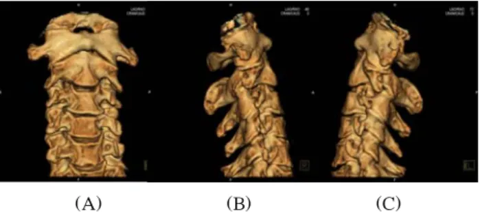
15)(A) (B) (C)
Fig. 1. A, B and C, Cervical spine computed tomography with 3-dimensional reformations showed anterior subluxation of the left lateral mass at C1 and posterior subluxation of the right lateral mass at C1.
Fig. 2. Neck CT angiogram showed hypoplastic change of right vertebral artery, but evidence of traumatic injury of both vertebral arteries was absent.
(A) (B) (C)
Fig. 3. A, B and C, After closed manual reduction under general anesthesia, CT scan showed good position of the C1–
C2 complex.
Fielding and Hawkins
1)described 4 types of AARF based on the extent of the shift of C1 on C2 and the integrity of the transverse ligament, which have been widely accepted. Type I is the most common pattern and is characterized by an intact transverse ligament. The fixed C1 C2 rotation is symmetrical and within the normal range – of rotation for the C1 C2 joints. Type II deformity is – identified by mild deficiency of the transverse ligament with an ADI of 3 to 5 mm. The intact joint acts as the pivot point for anterior unilateral displacement of the opposite side. Type III deformity is defined by a greater than 5 mm ADI, indicating disruption of both the transverse and alar ligaments. Both lateral masses are displaced anteriorly, and one side is rotated further forward than the other. Type IV lesion is described as a posterior shift of one or both lateral masses of the atlas. The importance of the classification scheme provided by Fielding and Hawkins is the increasing risk of spinal instability with potential neurologic compromise and a higher likelihood of recurrent deformity, with the more severe types. According to this classification, our case is type I AARF because of an intact transverse ligament : Measured ADI is 2 mm.
Stability of the upper cervical spine is almost entirely dependent on the surrounding soft tissue ligamentous constraints. The transverse ligament is the primary stabilizing force.
20)Therefore, the choice between conservative treatment and C1 C2 fusion is directly – dependent on whether or not the transverse ligament is torn or avulsed. With an intact transverse ligament, management is usually conservative.
21)The transverse ligament may be incompetent, leading to a widened ADI. Magnetic resonance imaging accurately depicts the anatomical integrity of the transverse ligament. The normal ligament has homogeneous low signal intensity on gradient-echo images, where as loss of continuity of the ligament with regions of high signal intensity characterize a tear.
22)The goals of treatment are to restore normal pain-free range of motion, prevent, or reverse neurologic compromise and restore spinal stability. Wise et al. noted that following a satisfactory closed reduction, a period of immobilization is adequate without the need for surgery. Axial traction
using Gardner-Wells tongs is recommended in the adult patient. Following satisfactory reduction in the acute setting, immobilization using a rigid orthosis is recommended for approximately 3 months. Levine and Edwards
23)recommended performing the manipulation with the patient awake using topical anesthetic in the posterior pharynx. The finger presses in counter rotation to the dislocation on the lateral mass of the atlas through the posterior wall of the pharynx. Gardner-Well tongs are used for control of rotation and gentle traction simultaneously.
They note that the reduction is often accompanied by an audible “pop” and can be confirmed by palpation of the ring of C1 through the mouth. If closed reduction is unsuccessful, open reduction must be performed.
21,24)The ring of C1 exposed, and a wire passed under the arch of C1. After the cranial traction with Gardner-Well tongs, the ring is then manually derotated using the C1 sublaminar wire at the same time. In our case, cervical traction was unsuccessful but reduction was ultimately achieved by closed manual reduction under general anesthesia.
Conclusion
We report one rare case of type I traumatic atlantoaxial rotatory fixation (AARF) developed in an elderly patient and underwent closed manual reduction under general anesthesia.
국문초록
환추 축추 회전성 고정증은 제 경추와 제 경추 관절 - 1 2
이 잠겨져 고정된 병적 상태로 대부분 환추 축추 관절의 -
여러 해부학적 취약성을 가진 유년시기에 상기도 감염과
관련하여 발생되는 드문 질병이다 본 저자들은 고령의 .
환자에서 외상후 발생한 type I 환추 축추 회전성 고정증 -
환자를 치험하였기에 문헌 고찰과 함께 보고하고자 한 다.
Key words 환추 축추 회전성 고정증 유년시기 해부학 : - , ,
적 취약성
고신대학교 의과대학 학술지 제 권 호 24 1 , 2009
References