148
Introduction
Double-chambered right ventricle (DCRV) is a cardiac dis- ease of the right ventricular outflow tract, where anomalous muscle bundles or a fibrous band cause obstruction.1-3)
A malaligned interventricular septal wall can develop in pa- tients with aneurismal changes of ventricular septal defect (VSD), the degree of which usually increases with time.4) Fur- thermore, when combined with DCRV, VSD with malalign- ment between the outlet and trabecular septa was associated with tetralogy of Fallot. A subaortic ridge in the malalign- ment-type VSD is caused by turbulent flow.5-7) We describe here in the unusual case of a 56-year-old man who had a spon- taneously healed membranous type VSD with malaligned in- terventricular septal wall, DCRV, subaortic ridge, and sur- vived with surgical treatment.
Case
A 56-year-old male presented with resting dyspnea and chest discomfort for several years. He had no history of any surgical or catheter-based cardiovascular interventions. Physi- cal examination showed a cardiac systolic murmur (grade 4/6) on the left upper sternal border, a systemic blood pressure of 140/75 mmHg, and a heart rate of 110 bpm. Electrocardiog- raphy showed sinus tachycardia, right axis deviation and fre- quent premature ventricular contraction with right ventricu- lar bundle branch block morphology.
Transthoracic and transesophageal echocardiography re- vealed a membranous type VSD with anteriorly malaligned interventricular septum, aneurysmal changes, subaortic ridge and DCRV with right ventricular hypertrophy (Fig. 1). Mea- surement of pressure gradients using a Doppler image was not
pISSN 1975-4612/ eISSN 2005-9655 Copyright © 2011 Korean Society of Echocardiography www.kse-jcu.org http://dx.doi.org/10.4250/jcu.2011.19.3.148
CASE REPORT J Cardiovasc Ultrasound 2011;19(3):148-151
Spontaneously Healed Membranous Type Ventricular Septal Defect with
Malaligned Interventricular Septal Wall and Double-Chambered Right Ventricle in a 56-Year-Old Patient
Jung Sun Cho, MD, PhD, Ho-Joong Youn, MD, PhD, Sung-Ho Her, MD, PhD, Soe Hee Ahn, MD, Mahn-Won Park, MD, Min Suk Choi, MD, Jae Bum Lee, MD, Jeong U Baeg, MD, Chan Seok Park, MD, PhD and Mi-Jeong Kim, MD, PhD
Department of Internal Medicine, School of Medicine, The Catholic University of Korea, Seoul, Korea
A 56-year-old male presented with resting dyspnea and chest discomfort for several years. During transthoracic and transesophageal echocardiography, a spontaneously healed membranous type ventricular septal defect (VSD) with malaligned interventricular septal wall, aneurysmal changes, a subaortic ridge and a double-chambered right ventricle (DCRV) was observed.
When combined with DCRV, VSD with malalignment between the outlet and trabecular septa was associated with tetralogy of Fallot. The subaortic ridge was due to turbulent flow caused by the malalignment-type VSD. The VSD with malaligned interventricular septal wall can be developed after aneurismal changes of a perimembranous VSD. We report here in the unusual case of a 56-year-old patient who had a pathology complex comprising DCRV, subaortic ridge, spontaneously healed membranous type VSD with malaligned interventricular septal wall, and survived with surgical treatment.
KEY WORDS: Membranous type ventricular septal defect · Double-chambered right ventricle · Subaortic ridge · Malaligned septal wall.
• Received: February 7, 2011 • Revised: June 3, 2011 • Accepted: August 17, 2011
• Address for Correspondence: Ho-Joong Youn, Department of Internal Medicine, School of Medicine, The Catholic University of Korea, 505 Banpo-dong, Seocho-gu, Seoul 137-701, Korea Tel: +82-2-3779-1066, Fax: +82-2-1588-1511, E-mail: [email protected]
• This is an Open Access article distributed under the terms of the Creative Commons Attribution Non-Commercial License (http://creativecommons.org/licenses/by-nc/3.0) which permits unrestricted non-commercial use, distribution, and reproduction in any medium, provided the original work is properly cited.
Ventricular Septal Defect with Double-Chambered Right Ventricle | Jung Sun Cho, et al.
149 Fig. 1. Preoperative transthoracic and transesophageal echocardiography. (A) Transthoracic echocardiography reveals a high-velocity systolic jet arising in the mid right ventricular outflow tract. (B) Right ventricular hypertrophy is seen on the parasternal long axis view. Transesophageal echocardiography reveals a subaortic ridge (arrowhead) (C) and aneurysmal changes (arrowhead) of the ventricular septal defect with anterior deviated interventricular septum (D). AV: aortic valve, RV: right ventricle, LV: left ventricle, IVS: interventricular septum.
C A
D B
Fig. 2. Preoperative right and left ventriculography. Right ventriculography at end-systole reveals an anomalous muscle bundle dividing the cavity into 2 chambers (A), and left ventriculography shows spontaneous closed malalignmented ventricular septal defect with aneurysmal changes (B). DCRV:
double-chambered right ventricle.
A B
Journal of Cardiovascular Ultrasound 19 | September 2011
150
made because the ultrasound beam could not be made parallel to the outflow tract jet. Right ventriculography confirmed an anomalous obstruction within the right ventricle. Preopera- tive cardiac catheterization demonstrated a right ventricle-to- pulmonary artery gradient of 89 mmHg (Fig. 2 and 3). Left ventriculography revealed a membranous VSD with anteriorly
malaligned interventricular septum, aneurysmal changes. The patient underwent open-heart surgery involving resection of an encircling infundibular fibrous band through right atrioto- my. Resection of the subaortic ridge was not carried out because it caused mild aortic regurgitation without pressure gradients in the left ventricular outflow tract. Dyspnea was improved at fol- low-up. Transthoracic echocardiography taken after surgery revealed that the high-velocity systolic jet in the mid right ventricular outflow tract disappeared and that right ventricu- lar outflow tract diameter was much improved (Fig. 4).
Discussion
Approximately two-thirds of patients have VSD that is di- minished in size by aneurysmal changes with apposition of tricuspid tissue over the VSD.4) In our case, transthoracic and transesophageal echocardiography revealed a membranous type VSD with anteriorly malaligned interventricular septum, aneurysmal changes. The parasternal long axis view is useful for determining the degree of aortic overriding (Fig. 1D).
DCRV is an infundibular pulmonary stenosis resulting from discrete fibromuscular narrowing or hypertrophied subvalvu- lar muscle bundles. In many cases, VSD is combined DCRV.1-3) Also, malaligned between the outlet and trabecular septa VSD with DCRV is associated with TOF.6)8)9) In our patient, DCRV was not associated TOF because anteriorly malaligned inter- venticular septum without malalignment of infudibular sep- tum was combined. VSD was located very close to the right aortic cusp and showed spontaneous closure with aneurysmal formation, which is rare without aortic valve prolapse.9-11)
The subaortic ridge is a fibrous structure from the crest of the ventricular septum to the left ventricular outflow tract.
This case had a subaortic ridge caused by increased turbulence
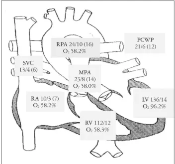
Fig. 3. Preoperative cardiac catheterization demonstrates a right ventricle-to-pulmonary artery gradient of 89 mmHg. There is no significant step-up of O2 saturation in the right ventricular sample that is higher than the highest right atrial sample because of spontaneously closed malalignmented ventricular septal defect. Systolic pressure/
diastolic pressure is expressed as a mean pressure and O2 saturation as a percentage. SVC: superior vena cava, RA: right atrium, RV: right ventricle, MPA: main pulmonary artery, RPA: right pulmonary artery, PCWP: pulmonary capillary wedge pressure, LV: left ventricle.
Fig. 4. Postoperative transthoracic and transesophageal echocardiography. A: Transthoracic echocardiography reveals that a high-velocity systolic jet arising in the mid right ventricular outflow tract disappeared after surgery. B: Transesophageal echocardiography reveals much improved right ventricular outflow tract diameter after surgery. AV: aortic valve, PA: pulmonary artery.
A B
13/4 (6)SVC MPA
23/8 (14) O2 58.0%
RPA 24/10 (16) O2 58.2%
RA 10/3 (7) O2 58.2%
PCWP 21/6 (12)
LV 136/14 O2 96.2%
RV 112/12 O2 58.3%
Ventricular Septal Defect with Double-Chambered Right Ventricle | Jung Sun Cho, et al.
151 adjacent to the area of VSD and enhanced by septal malalign-
ment.6)7) A subaortic ridge may result in progressive subaortic stenosis or aortic valve deformities, and resection is usually recommended. In our case, the patient underwent resection of an encircling infundibular fibrous band through right atrioto- my. However, resection of the subaortic ridge was not carried out because it caused mild aortic regurgitation without any pressure gradients in the left ventricular outflow tract.
In summary, the malaligned interventricular septum in membranous type VSD can develop secondary to aneurismal changes of VSD. We reported the unusual case of a patient who had a pathology complex comprising DCRV, subaortic ridge, and VSD with malaligned interventricular septal wall and survived with surgical treatment.
References
1. Maron BJ, Ferrans VJ, White RI Jr. Unusual evolution of acquired in- fundibular stenosis in patients with ventricular septal defect. Clinical and morphologic observations. Circulation 1973;48:1092-103.
2. Choi YJ, Park SW. Characteristics of double-chambered right ventricle in adult patients. Korean J Intern Med 2010;25:147-53.
3. McElhinney DB, Chatterjee KM, Reddy VM. Double-chambered right ventricle presenting in adulthood. Ann Thorac Surg 2000;70:124-7.
4. Wu MH, Wu JM, Chang CI, Wang JK, Wu YN, Chien SC, Lue
HC. Implication of aneurysmal transformation in isolated perimembranous ventricular septal defect. Am J Cardiol 1993;72:596-601.
5. Matina D, van Doesburg NH, Fouron JC, Guérin R, Davignon A.
Subxiphoid two-dimensional echocardiographic diagnosis of double-cham- bered right ventricle. Circulation 1983;67:885-8.
6. Wang JK, Wu MH, Chang CI, Chiu IS, Chu SH, Hung CR, Lue HC. Malalignment-type ventricular septal defect in double-chambered right ventricle. Am J Cardiol 1996;77:839-42.
7. Zielinsky P, Rossi M, Haertel JC, Vitola D, Lucchese FA, Rodrigues R. Subaortic fibrous ridge and ventricular septal defect: role of septal ma- lalignment. Circulation 1987;75:1124-9.
8. Wu MH, Wang JK, Chang CI, Chiu IS, Lue HC. Implication of ante- rior septal malalignment in isolated ventricular septal defect. Br Heart J 1995;74:180-5.
9. Oppenheimer-Dekker A, Gittenberger-de Groot AC, Bartelings MM, Wenink AC, Moene RJ, van der Harten JJ. Abnormal architec- ture of the ventricles in hearts with an overriding aortic valve and a peri- membranous ventricular septal defect (“Eisenmenger VSD”). Int J Cardiol 1985;9:341-55.
10. Vogel M, Smallhorn JF, Freedom RM, Coles J, Williams WG, Trusler GA. An echocardiographic study of the association of ventricular septal defect and right ventricular muscle bundles with a fixed subaortic ab- normality. Am J Cardiol 1988;61:857-60.
11. Kim CJ, Chai IH, Koh KK, Sohn DW, Lee MM, Park YB, Choi YS, Seo JD, Lee YW. Double chambered right ventricle (DCRV) in adult and adolescence. Korean Circ J 1990;20:248-55.