Pseudoaneurysm of the splenic artery - an uncommon cause of delayed hemorrhage after pancreaticoduodenectomy
Radu Dumitru1, Ana Carbunaru2, Mugur Grasu1, Mihai Toma1, Mihnea Ionescu2, and Traian Dumitrascu2
1Center of Interventional Radiology and Medical Imaging, 2Center of General Surgery and Liver Transplant, Fundeni Clinical Institute, Bucharest, Romania
Delayed post-pancreatectomy hemorrhage (PPH) is a relatively uncommon, but feared, complication after pan- creaticoduodenectomy (PD). A splenic artery pseudoaneurysm is a rare cause of delayed PPH after a PD. This paper describes the case of a patient with PD used to treat a distal bile duct cholangiocarcinoma complicated with a clinically significant pancreatic fistula and secondary intraabdominal abscess. Computed tomography-guided drainage of the ab- scess was performed with an apparently favourable outcome; the patient was discharged on postoperative day (POD) 35 and the abdominal drains were removed on POD 50. On POD 80, the patient was readmitted for a severe digestive hemorrhage. Computed tomography revealed a pseudoaneurysm of the splenic artery with a subsequent hematoma formation. Immediately, an angiography was performed and coils were successfully mounted. This case illustrates the rare possibility of the development of a splenic artery pseudoaneurysm with severe delayed PPH after PD complicated with a clinically significant pancreatic fistula, even after the patient was discharged from the hospital. An interventional radiology approach represents the first treatment option in hemodynamically stable patients with high success rates.
(Ann Hepatobiliary Pancreat Surg 2016;20:204-210)
Key Words: Pancreaticoduodenectomy; Pancreatic fistula; Delayed hemorrhage; Splenic artery pseudoaneurysm;
Interventional angiography
Received: May 10, 2016; Revised: August 22, 2016; Accepted: September 26, 2016 Corresponding author: Traian Dumitrascu
Center of General Surgery and Liver Transplant, Fundeni Clinical Institute, Fundeni Street no 258, Bucharest 022328, Romania Tel: +0040213180417, Fax: +0040213180417, E-mail: traian.dumitrascu76@gmail.com
Copyright Ⓒ 2016 by The Korean Association of Hepato-Biliary-Pancreatic Surgery
This is an Open Access article distributed under the terms of the Creative Commons Attribution Non-Commercial License (http://creativecommons.org/
licenses/by-nc/4.0) which permits unrestricted non-commercial use, distribution, and reproduction in any medium, provided the original work is properly cited.
Annals of Hepato-Biliary-Pancreatic Surgery ∙ pISSN: 2508-5778ㆍeISSN: 2508-5859
INTRODUCTION
Pancreaticoduodenectomy (PD) represents the standard approach for resectable periampullary malignancies.1 PD is a complex surgical procedure that is widely recognized to have increased rates of postoperative complications.1 Thus, morbidity rates around 40% and in-hospital mortal- ity rates around 5% should be expected after a PD.1 Associated venous resections appear to increase both mor- bidity and mortality rates after a PD.2
Surgeon experience and annual hospital volume are im- portant determinants of early outcomes after PD.3 However, a recent meta-analysis did not identify the sur- geon caseload as a significant determinant of the post- operative mortality after PD.4 Analysis performed at the national level in the United States has shown that mortal- ity rates reported at 90 days are double compared with
the 30 day mortalities rates after pancreatectomies.5 Furthermore, in hospitals with an average annual volume of more than 40 PDs, the 90 day mortality rate was 4.7%
while in hospitals with less than 5 PDs per year the 90 day mortality rate was significantly higher – 14.2%.5 Nevertheless, it appears that high-volume surgeons are as- sociated with significantly lower in-hospital mortality rates, compared with low-volume surgeons, no matter what the hospital annual volume of PDs is.6 The largest, single surgeon, Western experience with a PD published so far belongs to John Cameron from the Johns Hopkins Hospital (USA).7 Thus, in a consecutive series of 2,000 PDs, periampullary malignancies represented 68% of the indications, with a morbidity rate of 45% and in-hospital mortality rate of 1.6%.7
The main source of morbidity after PD is represented by a pancreatic fistula, a complication that can be encoun-
mainly related to the variability of definitions used.8 A pancreatic fistula remains an important issue after PD and can lead to other potentially life-threatening complications such as delayed hemorrhagic complications.8,9
A delayed hemorrhage is considered a relatively un- common complication after PD. Thus, the reported rates of delayed hemorrhage after a PD in recent published large series vary between 2% and 8.4%.7,10-18 Although the post-pancreatectomy hemorrhage (PPH) accounts for only 1% to 13.2% of the postoperative complications, it is con- sidered a severe complication because it is associated with very high mortality rates. PPH accounts for 10.5% to 38%
of overall mortality after a PD.9-13,15,17,18
This paper pres- ents the diagnosis and management of a patient with a delayed hemorrhage after a PD due to a pseudoaneurysm of the splenic artery.
CASE
A 57-year-old woman with arterial hypertension, was investigated in the Gastroenterology Department for jaun- dice and weight loss. Laboratory tests showed the follow- ing abnormalities:
ㆍCholestasis – total/direct bilirubin serum lev- el=16.6/11.5 mg/dl (normal values, 0.3-1.2/0-0.4 mg/dl) and gamaglutamiltranspeptidase (GGT) serum level=424 U/L (normal values, 7-99 U/L);
ㆍLiver cytolysis – aspartate aminotransferase (AST) serum level=112 U/L (normal values, 0-35 U/L) and alanine aminotransferase (ALT) serum level=87 U/L (normal values, 0-45 U/L);
ㆍHyperglycemia – glucose serum level=143 mg/dl (normal values 74-106 mg/dl).
The serum levels of the tumor markers were 287.5 U/ml for CA 19-9 (normal values, 0-39 U/ml) and 4.9 ng/ml for CEA (normal values, 0-3.4 ng/ml), respectively.
The abdominal ultrasound examination, magnetic reso- nance colangiopancreatography, and endoscopic ultra- sound examinations suspected the diagnosis of a resect- able distal bile duct cholangiocarcinoma. Thus, the patient was referred to the Department of Surgery for resection.
In February 2015, the patient underwent pylorus-pre- serving PD for a diagnosis of distal bile duct chol-
with origin from the superior mesenteric artery was also observed and was spared during the resection. The re- construction after PD was made using the modified Child’s technique. Thus, the distal pancreatic stump (soft pancreas, tiny Wirsung duct that could not be identified to be stented) was anastomosed to the jejunum in an end-to-side fashion, using single-layer, interrupted, non-absorbable sutures. Furthermore, the bile duct was anastomosed to the jejunum in an end-to-side single-layer, continuous, absorbable suture, as it was the case also for duodenojejunal anastomosis. Three drains were left in place at the end of the operation.
At approximately 1 h after the end of the operation, the patient presented tachycardia, severe hypotension and significant external bleeding in one abdominal drain, with a 2 g/dl decrease in the hemoglobin serum level.
Immediately the patient was readmitted into the operating room and, with the diagnosis of early severe PPH with secondary hemorrhagic shock, a re-laparotomy was performed. After the re-laparotomy, the diagnosis of early severe PPH with secondary hemorrhagic shock was changed to a diagnosis of hemoperitoneum due to a small arterial source branching from the accessory right hepatic artery, which was sutured to control the bleeding.
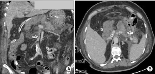
On POD 1, very high serum levels of amylase and li- pase were encountered: 1,025 U/L and 3,682 U/L, re- spectively (normal values, 25-100 U/L and 0-60 U/L, re- spectively). Furthermore, the clinical course of the patient was relatively well, with good intake tolerance, presence of the intestinal transit, normalisation of the amylase and lipase serum levels and no other significant abnormalities on laboratory tests, except for mild anemia. However, on POD 7, the patient developed an external pancreatic fistu- la (debit of approximately 500 ml/day), without any change of clinical status. The contrast-enhanced computed tomography performed on POD 7 revealed partial throm- bosis of the superior mesenteric vein (Fig. 1A, open white arrow), peripancreatic fluids and minor non-enhancing areas in the remnant pancreas (Fig. 1B, open white ar- row), but no signs of abscesses or infected necrosis. Thus, a conservative approach was considered, including anti- coagulant therapy and antibiotics. The contrast-enhanced computed tomography performed on POD 19 revealed no
Fig. 1. Abdominal contrast-en- hanced computed tomography in the venous phase performed on post-operative day (POD) 7 (A) frontal slice showing partial thrombosis of the superior mes- enteric vein (open white arrow);
(B) axial slice showing peri- pancreatic fluids and minor non-enhancing areas in the rem- nant pancreas (open white ar- row).
Fig. 2. Abdominal contrast-enhanced computed tomography axial slice in the venous phase performed on POD 19 showing thrombosis of the proximal splenic vein (open white arrow) and distal peripancreatic fluids (filled white arrow).
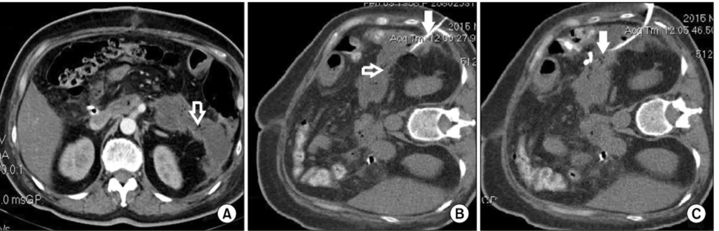
thrombosis of the superior mesenteric vein but thrombosis of the proximal splenic vein (Fig. 2, open white arrow) with distal peripancreatic fluids (Fig. 2, filled white ar- row). But again, there were no signs of abscesses or in- fected necrosis. The patient was in relatively good clinical condition and no significant abnormalities were detected in the laboratory tests. The pancreatic fistula had a favour- able outcome and, due to minimum drainage output, the drains were removed on POD 26. However, on POD 29 the patient presented with a fever and leukocytosis (a white cell count of 18,000 per cubic millimeter). The con- trast-enhanced computed tomography performed on POD 30 revealed a large, peripancreatic distal collection with gas inside suggesting the formation of a peripancreatic ab- scess (Fig. 3A, open white arrow). A successful percuta-
neous drainage of the abscess was performed (Fig. 3B and 3C, filled white arrow).
After that, the postoperative outcome was uneventful, and the patient was discharged on POD 35, with no fever, no abnormalities on the laboratory tests, and no collec- tions at the abdominal ultrasound examination, but with an abdominal drain left in place (a daily purulent outflow of around 50 ml). The abdominal drain was removed on POD 50 in an outpatient ward after a contrast-enhanced computed tomography examination which showed com- plete regression of the peripancreatic abscess (Fig. 4).
The final pathology examination of the operative speci- men revealed a 0.5 cm-sized, poorly differentiated distal bile duct cholangiocarcinoma (pT3), with perineural and microvascular invasion. None of the 26 harvested lymph nodes presented metastases.
The postoperative outcome was uneventful for a while however, on POD 80, the patient was readmitted for mele- na without significant clinical impact. The physical ex- amination was unremarkable except for the melena con- firmation during the rectal examination. The laboratory tests revealed only anemia – hemoglobin serum level=7.4 g/dl (normal values, 11.5-17 g/dl). The upper endoscopy did not show any abnormalities. The abdominal ultra- sound examination did not identify any abnormalities.
With conservative treatment and transfusions, the melena was remitted within two days and the patient was discharged.
On POD 84, the patient was readmitted with recurrent melena with severe clinical impact (tachycardia, hypo- tension). The laboratory tests showed severe anemia (hemoglobin 4.7 g/dl). After rapid transfusions and fluid
Fig. 3. Abdominal contrast-enhanced computed tomography axial slice in the venous phase performed on POD 30 showing (A) a large peripancreatic distal collection with gas inside suggesting the formation of a peripancreatic abscess (open white arrow);
(B) the percutaneous drainage catheter (filled white arrow) toward the peripancreatic abscess (open white arrow); (C) the drainage catheter (filled white arrow) successfully inserted in the peripancreatic abscess.
Fig. 4. Abdominal contrast-enhanced computed tomography axial slice in the arterial phase performed on POD 50 showing complete regression of the peripancreatic abscess (open white arrow - the drainage catheter).
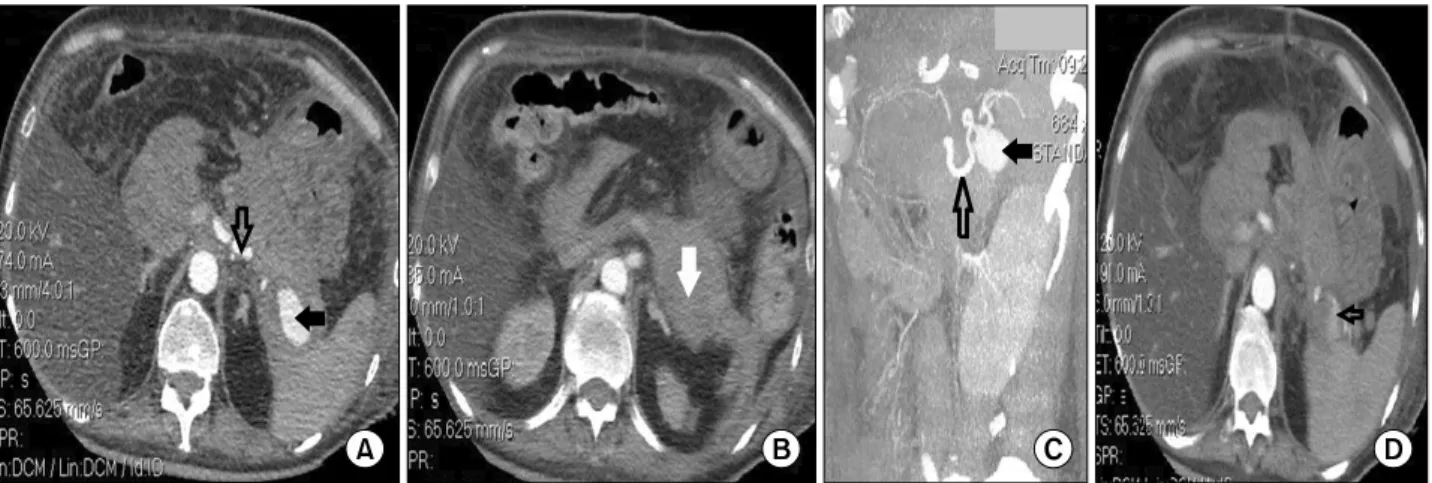
resuscitation, the patient underwent an enhanced-com- puted tomography that revealed a 3.2 cm×1.8 cm-sized pseudoaneurysm (Fig. 5A and 5C, filled black arrow) of the splenic artery (Fig. 5A and 5C, open black arrow) with a subsequent 9.5 cm×4.3 cm-sized hematoma for- mation (Fig. 5B, filled white arrow), and active bleeding in the distal part of the splenic artery (Fig. 5D, open black arrow). The patient was rapidly referred to the Interventional Radiology Department. A catheter was in- serted via the right femoral artery and the splenic artery (Fig. 6A, open black arrow) where the pseudoaneurysm was identified (Fig. 6A, filled black arrow). After that, several pushable coils (Fig. 6B, open black arrow) were successfully mounted with no bleeding and no
complications. The contrast-enhanced computed tomog- raphy control examinations, performed on day four and nine after the interventional procedure, showed no pseu- doaneurysm (Fig. 7A and 7B, open black arrow), com- plete remission of the hematoma (Fig. 7C, filled white ar- row), and several ischemic areas within the spleen, with- out a tendency of modification or abscesses at repeated examinations (Fig. 7A and 7B, filled black arrow). After the procedure, the clinical condition of the patient sig- nificantly improved with hemodynamic stability and no signs of hemorrhage.
Although the interventional radiology approach was a success for the splenic artery pseudoaneurysm, the patient died at a later time due to acute liver insufficiency not related to the pseudoaneurysm or interventional radiology procedure.
DISCUSSION
Delayed PPH is defined as bleeding that occurs more than 24 hours after the index operation, according to the International Study Group for Pancreatic Surgery.9 The median reported time between the pancreatic resection and appearance of a delayed PPH is around 12-39 days.11-13,16,17,19
A large part of these delayed PPHs oc- curred after the patient was discharged from the hospital.11,12 In the reported patient, the delayed PPH oc- curred on POD 80, after discharge. It appears that the mortality rate is higher in patients with a delayed PPH,
Fig. 5. Abdominal contrast-enhanced computed tomography in the arterial phase performed on POD 84 (A) axial slice showing a 3.2 cm×1.8 cm-sized pseudoaneurysm (filled black arrow) of the splenic artery (open black arrow); (B) axial slice showing a high-density lesion indicating a subsequent 9.5 cm×4.3 cm-sized hematoma formation (filled white arrow); (C) frontal slice with reconstruction showing the pseudoaneurysm (filled black arrow) of the splenic artery (open black arrow); (D) axial slice showing extravasations of contrast agent suggesting active bleeding in the distal part of the splenic artery (open black arrow).
Fig. 6. (A) Digital subtraction angiogram obtained through the celiac trunk showing the splenic artery (open black arrow) with the pseudoaneurysm (filled black arrow) (C - the catheter positioned at the celiac ostium); (B) angiography showing pushable coils (open black arrow) on either side of a false aneurysm of the splenic artery, with no extravasations of contrast agent (C - the catheter positioned at the proximal part of the splenic artery).
at more than 39 days after surgery.19 A severe PPH im- plies a large volume of blood loss (a drop of hemoglobin level ≥3 g/dl) with clinically significant impairment, need for transfusions and an invasive treatment approach,9 as was the case in the above presented patient.
As it was previously mentioned, a delayed PPH repre- sents a major concern after PD because it is an important source of postoperative mortality. Careful attention; a timely diagnosis of the source; and adequate, immediate therapy are of utmost importance for a successful outcome. A multidisciplinary approach created by experi- enced surgeons, interventional radiologists, endoscopists and intensivists is mandatory with these patients.
Delayed PPH is usually related to a local sepsis due to a pancreatic fistula, bile leak or intraabdominal abscess.9,15,17,20 Pathogenesis of a delayed PPH includes
enzymatic digestion, local infection with secondary vas- cular wall erosion, or vascular injury at the time of resection.9,20 In the reported patient, the pathogenesis of the pseudoaneurysm was most likely the enzymatic/in- fectious erosion of the splenic artery wall secondary to the postoperative clinically significant pancreatic fistula.
Clinically significant postoperative pancreatic fistula de- velopment after a PD was recently found to be an in- dependent risk factor for a delayed PPH.10,15 Furthermore, postoperative pancreatic fistula is reported in up to 80%
of the patients with a delayed PPH.11,13,17,19,21
The presence of bile in drainage and presence of clinical signs of in- fection (i.e., fever ≥38oC and leukocytosis >10,000 per cubic millimeter for more than 5 consecutive days) were found to be independent risk factors for a massive delayed PPH after a PD.22
Fig. 7. Check-up after embolization with an abdominal contrast-enhanced computed tomography axial slice in the arterial phase showing (A) on day 4 after the interventional radiology procedure the disappearance of the splenic artery pseudoaneurysm, pres- ence of tiny arciform images due to the metal artifacts created by coils (open black arrow), and several ischemic areas within the spleen (filled black arrow); (B) on day 9 after the interventional radiology procedure the presence of tiny arciform images due to the metal artifacts created by coils (open black arrow) and stationary ischemic areas within the spleen (filled black arrow), without tendency of modification or abscesses; (C) on day 9 after the interventional radiology procedure the complete remission of the peripancreatic hematoma (filled white arrow).
A splenic artery pseudoaneurysm is an uncommon pathology. Thus, as of 2007, no more than 200 patients with splenic artery pseudoaneurysms were described in the English literature.23 Furthermore, a splenic artery pseudoaneurysm represents an exceptional source for a delayed PPH after a PD.11-14,17,19,24,25 Most of the pseudoa- neurysms that occur after PD are located at the level of the gastroduodenal artery stump or hepatic ar- tery.9-11,13,14,16,17,19,20
Interestingly, a recent study suggested that the delayed PPH with origin from the splenic artery is more frequently encountered after pancreaticogas- trostomy reconstruction than after PD.25
Sentinel bleeding, defined as minor blood loss via sur- gical drains or the gastrointestinal tract, with an asympto- matic interval of at least 12 hours until the development of hemorrhagic shock,10 is sometimes the only clinical sign that announces a massive PPH, as was the case with the reported patient. Sentinel bleeding is encountered in up to 77.8% of the patients with severe PPH.10,13,16,17,21
Some studies suggest mandatory angiographic exploration in every patient with sentinel bleeding after PD because a pseudoaneurysm is thus discovered in 35% of the patients.26
An upper digestive hemorrhage can be a clinical sign in an important proportion of patients with a false aneur- ysm of the splenic artery.23 An upper endoscopy some- times does not identify the source of bleeding.
Contrast-enhanced computed tomography with multiplanar
reconstructions is mandatory in these situations to prompt- ly identify the origin of hemorrhage,24 as was the case in the above presented patient.
The management of delayed PPH after PD represents a challenging problem, particularly when a pancreatic fis- tula is associated. An endovascular approach represents the first treatment option in patients with delayed PPH of an arterial origin and in patients without hemodynamic instability.11,13,15,16,19-21,24,25
Arterial embolization consists of an occlusion on either side of a false aneurysm in order to prevent further bleed- ing via reverse flow or anastomosis,24 as was the case in the above presented patient. The success rate of an arterial embolization used to stop the bleeding is up to 100%.24 Recurrence of the bleeding is usually related to persis- tence of the favouring factors (i.e., pancreatic/bile leak, persistent abscess).19
Spleen infarction is a reported complication after splen- ic artery embolization but rarely is associated with clin- ically significant consequences,27 as was the case in the reported patient.
Surgery represents an alternative treatment approach in patients with delayed PPH with massive bleeding and he- modynamic instability, in patients where it is mandatory to solve the underlying source of hemorrhage to prevent further arterial erosion (i.e., completion of a pan- createctomy for a severe pancreatic fistula),13,15,20,21,25
when interventional radiology procedures have failed13 or
when immediate radiology interventions are not available.11,25 A recent meta-analysis associated surgery with significantly increased mortality rates compared with the interventional radiology approach for delayed PPH.17
In conclusion, this case illustrates the rare possibility of development of a splenic artery pseudoaneurysm with severe delayed PPH after PD complicated with a clinically significant pancreatic fistula, even after the patient was discharged from the hospital. Proper recognition of senti- nel bleeding might play a major role for timely therapy of delayed PPH after PD. An interventional radiology ap- proach represents the first treatment option in hemody- namically stable patients, with high success rates of stop- ping the bleeding.
REFERENCES
1. Popescu I, Dumitraşcu T. Pancreatoduodenectomy--past, present and future. Chirurgia (Bucur) 2011;106:287-296.
2. Dumitrascu T, Dima S, Brasoveanu V, Stroescu C, Herlea V, Moldovan S, et al. Impact of a portal/superior mesenteric vein resection during pancreatico-duodenectomy for pancreatic head adenocarcinoma. Minerva Chir 2014;69:301-313.
3. Schmidt CM, Turrini O, Parikh P, House MG, Zyromski NJ, Nakeeb A, et al. Effect of hospital volume, surgeon experience, and surgeon volume on patient outcomes after pancreati- coduodenectomy: a single-institution experience. Arch Surg 2010;145:634-640.
4. Gooiker GA, van Gijn W, Wouters MW, Post PN, van de Velde CJ, Tollenaar RA; Signalling Committee Cancer of the Dutch Cancer Society. Systematic review and meta-analysis of the vol- ume-outcome relationship in pancreatic surgery. Br J Surg 2011;98:485-494.
5. Swanson RS, Pezzi CM, Mallin K, Loomis AM, Winchester DP.
The 90-day mortality after pancreatectomy for cancer is double the 30-day mortality: more than 20,000 resections from the na- tional cancer data base. Ann Surg Oncol 2014;21:4059-4067.
6. Mathur A, Luberice K, Ross S, Choung E, Rosemurgy A. Pan- creaticoduodenectomy at high-volume centers: surgeon volume goes beyond the Leapfrog criteria. Ann Surg 2015;262:e37-e39.
7. Cameron JL, He J. Two thousand consecutive pancreati- coduodenectomies. J Am Coll Surg 2015;220:530-536.
8. Bassi C, Dervenis C, Butturini G, Fingerhut A, Yeo C, Izbicki J, et al. Postoperative pancreatic fistula: an international study group (ISGPF) definition. Surgery 2005;138:8-13.
9. Wente MN, Veit JA, Bassi C, Dervenis C, Fingerhut A, Gouma DJ, et al. Postpancreatectomy hemorrhage (PPH): an Internatio- nal Study Group of Pancreatic Surgery (ISGPS) definition.
Surgery 2007;142:20-25.
10. Ansari D, Tingstedt B, Lindell G, Keussen I, Ansari D, Andersson R. Hemorrhage after major pancreatic resection: in- cidence, risk factors, management, and outcome. Scand J Surg 2016. pii: 1457496916631854. [in press]
11. Asai K, Zaydfudim V, Truty M, Reid-Lombardo KM, Kendrick
M, Que F, et al. Management of a delayed post-pancreato- duodenectomy haemorrhage using endovascular techniques. HPB (Oxford) 2015;17:902-908.
12. Correa-Gallego C, Brennan MF, D'Angelica MI, DeMatteo RP, Fong Y, Kingham TP, et al. Contemporary experience with post- pancreatectomy hemorrhage: results of 1,122 patients resected between 2006 and 2011. J Am Coll Surg 2012;215:616-621.
13. Ding X, Zhu J, Zhu M, Li C, Jian W, Jiang J, et al. Therapeutic management of hemorrhage from visceral artery pseudoaneur- ysms after pancreatic surgery. J Gastrointest Surg 2011;15:
1417-1425.
14. Huo Y, Chi J, Zhang J, Liu W, Liu D, Li J, et al. Endovascular intervention for delayed post-pancreaticoduodenectomy hemor- rhage: clinical features and outcomes of transcatheter arterial em- bolization and covered stent placement. Int J Clin Exp Med 2015;8:7457-7466.
15. Jilesen AP, Tol JA, Busch OR, van Delden OM, van Gulik TM, Nieveen van Dijkum EJ, et al. Emergency management in pa- tients with late hemorrhage after pancreatoduodenectomy for a periampullary tumor. World J Surg 2014;38:2438-2447.
16. Lee HG, Heo JS, Choi SH, Choi DW. Management of bleeding from pseudoaneurysms following pancreaticoduodenectomy.
World J Gastroenterol 2010;16:1239-1244.
17. Roulin D, Cerantola Y, Demartines N, Schäfer M. Systematic review of delayed postoperative hemorrhage after pancreatic resection. J Gastrointest Surg 2011;15:1055-1062.
18. Wellner UF, Kulemann B, Lapshyn H, Hoeppner J, Sick O, Makowiec F, et al. Postpancreatectomy hemorrhage--incidence, treatment, and risk factors in over 1,000 pancreatic resections.
J Gastrointest Surg 2014;18:464-475.
19. Ching KC, Santos E, McCluskey KM, Orons PD, Bandi R, Friend CJ, et al. Covered stents and coil embolization for treat- ment of postpancreatectomy arterial hemorrhage. J Vasc Interv Radiol 2016;27:73-79.
20. Camerlo A, Turrini O, Marciano S, Sarran A, Berdah S, Delpero JR, et al. Delayed arterial hemorrhage after pancreatico- duodenectomy: is conservation of hepatic arterial flow vital?
Pancreas 2010;39:260-262.
21. Yekebas EF, Wolfram L, Cataldegirmen G, Habermann CR, Bogoevski D, Koenig AM, et al. Postpancreatectomy hemor- rhage: diagnosis and treatment: an analysis in 1669 consecutive pancreatic resections. Ann Surg 2007;246:269-280.
22. Tien YW, Lee PH, Yang CY, Ho MC, Chiu YF. Risk factors of massive bleeding related to pancreatic leak after pancrea- ticoduodenectomy. J Am Coll Surg 2005;201:554-559.
23. Agrawal GA, Johnson PT, Fishman EK. Splenic artery aneur- ysms and pseudoaneurysms: clinical distinctions and CT appearances. AJR Am J Roentgenol 2007;188:992-999.
24. Cazejust J, Raynal M, Bessoud B, Tubiana JM, Menu Y.
Diagnosis and radiological treatment of digestive haemorrhage following supramesocolic surgery. Diagn Interv Imaging 2012;93:e148-e158.
25. Darnis B, Lebeau R, Chopin-Laly X, Adham M. Postpancrea- tectomy hemorrhage (PPH): predictors and management from a prospective database. Langenbecks Arch Surg 2013;398:441-448.
26. Tien YW, Wu YM, Liu KL, Ho CM, Lee PH. Angiography is indicated for every sentinel bleed after pancreaticoduo- denectomy. Ann Surg Oncol 2008;15:1855-1861.
27. Piffaretti G, Tozzi M, Lomazzi C, Rivolta N, Riva F, Caronno R, et al. Splenic artery aneurysms: postembolization syndrome and surgical complications. Am J Surg 2007;193:166-170.