170
J. Appl. Biol. Chem. 52(4), 170-173 (2009) Article
Immunoblotting Reactivity of Vitellogenin Antibodies against Native Vitellogenins and a Vitellogenin Protein Fragment Produced in E. coli
Hyung-Seok Ryu, In-Young Jang, and Woo-Yeon Kim*
Department of Biotechnology, Chung-Ang University, Anseong 456-756, Korea Received November 30, 2009; Accepted December 4, 2009
Vitellogenin (Vtg) is found in the serum of both female and male fish that have been exposed to environmental endocrine disrupters or estrogen hormones, and is used as a biomarker for such contamination. In our current study antibodies raised against the purified flatfish Vtg were tested for their reactivity on immunoblots to flatfish and carp Vtg, and also to a Vtg protein fragment produced in E. coli. Polyclonal antibodies raised against purified flatfish Vtg reacted well with Vtg in the serum of flatfish and carp induced with 17β-estradiol, but not with the Vtg protein fragment produced in E. coli.
Key words: antibody, flatfish vitellogenin, reactivity
It has been reported previously that vitellogenin (Vtg), an estrogen-dependent egg yolk protein precursor and phospho- lipoglycoprotein [Tyler et al., 1996], is a potent fish biomarker.
Vtg is normally only present in the blood of female fish, but males also harbor Vtg genes and the estrogen receptors necessary for Vtg regulation [Tyler et al., 2002]. Hence, the exposure of both female and male fish to estrogen hormones or endocrine disrupters such as 17-ethinylestradiol, dioxins and polychlorinated biphenyls (PCBs) can trigger Vtg gene expression. As a consequence of these stimuli, Vtg accumulates abnormally in the blood [Chen, 1983; Tyler et al., 1996].
There has been growing concern in recent years regarding the possible adverse effects of man-made substances on the endocrine systems in humans and other animals [Colborn and Clement, 1992]. Estrogenic effects are usually evaluated through the measurement of Vtg in the blood plasma of male fish. Vtg is a yolk protein precursor and is normally secreted by the liver of female fish during the development of ovarian follicles (vitellogenesis). Following this secretion into the bloodstream, the protein is transported and incorporated into the oocytes as yolk. The synthesis of Vtg in the liver is under the control of the estrogen receptor and is induced by estrogens, most notably 17β-estradiol. Male fish do not normally have measurable quantities of Vtg in their blood, because of their relatively small plasma concentration of endogenous estrogen. However, the male fish liver is capable of synthesizing and secreting Vtg in
response to stimulation of exogenous estrogens such as endocrine disrupters [Wallace, 1985]. Importantly also, fish represents an important source of human contamination by endocrine disrupters because they are a component of the upper part of the food chain. Stringent surveillance is now required to detect possible contamination of edible fresh- and salt-water fish with endocrine disrupters due to the possible repercussion for human health [Kim et al., 2006b].
A rapid and sensitive screening tool that can efficiently measure the level of Vtg in fish serum is urgently needed for the effective assessment of endocrine disrupter contamination in fish. This is particularly important for species that are favored for human consumption, such as salt-water flatfish, in Korea, as it will help to ensure the safety of these commercially valuable stocks. A label free immunosensor that does not require a probe molecule for signal transduction and can measure changes in physical parameters, such as frequency or a refractive index, caused by an immune response [Oh et al., 2004; Nam et al., 2006; Park et al., 2006], would potentially be a very effective and desirable for on-site Vtg detection. This is due to the simplicity of the measurement procedures, the robustness of the technique against the background interference that is normally found in the case of a labeled immunosensor or ELISA procedure, and also the real-time nature of this methods [Kolosova et al., 2000; Kim et al., 2006a; 2007a]. To develop effective immunosensors for fish Vtg, a robust and potent antibody will be essential.
In current study, we employed the polyclonal antibodies previously raised against flatfish Vtg purified from the serum of flatfish exposed to estrogen [Kim et al., 2007b]. We then compared the reactivity of these antibodies to flatfish Vtg, carp
*Corresponding author
Phone: +82-31-675-3063; Fax: +82-31-675-0405 E-mail: [email protected]
doi:10.3839/jabc.2009.029
Reactivity of flatfish vitellogenin antibodies on immunoblots 171
Vtg, and a Vtg protein fragment produced in E. coli.
Materials and Methods
Reagents. Low range and high range protein molecular weight markers were purchased from Bio-Rad (Hercules, CA).
All other chemicals were obtained from Sigma (St. Louis, MO).
Preparation of flatfish Vtg antibodies and ELISA. Vtg was induced in the flatfish and purified according to the method of Moon et al. [2006] with slight modifications. Antibodies against this Vtg preparation were generated as described previously [Kim, et al., 2007b]. ELISA was then performed using these antibodies as follows. Fish control serum or serum induced with 17β-estradiol, was diluted with the ELISA coating buffer (0.032 M Na2CO3 and 0.068 M NaHCO3) to 250 ng/well, was added to a 96-well plate in 50µL aliquots, followed by incubation of the plate for 2 h at 37oC. After discarding the coating solution, 200
µL of 2% skim milk was dispensed into each well of the plate to block unoccupied surfaces for 30 min at 37oC. The wells were then washed once for 1 min with 150µL of PBST (PBS containing 1% Tween 20), and incubated with varying amounts of anti-flatfish Vtg antibodies [Kim et al., 2007b] in 100µL of PBS at 37oC for 1 h. After again washing the plate three times with PBST, and 100µL of horseradish peroxidase-conjugated goat anti-mouse IgG solution (1/5,000 dilution) was added to the wells for 1 h at 37oC. Finally, the plate was washed five times with PBST, followed by the addition of 100µL of TMB (3,3',5,5'-tetramethylbenzidine) substrate buffer (Sigma), a soluble colorimetric substrate for horseradish peroxidase (HRP) to each well. After a 30 min incubation at 25oC the reaction was stopped by addition of 100µL of 0.05 M H2SO4. Absorbance readings at 450 nm were then recorded using a Bio-Rad model 550 microplate reader.
RNA isolation, cDNA synthesis, and RT-PCR. Livers were collected from the 17β-estradiol-treated flatfish and total RNA was isolated from these tissues using RNeasy mini kit (Qiagen, Hilden, Germany) according to the manufacturer’s instructions.
The purified RNA was dissolved in RNase-free water and stored at −80oC until use. cDNA was synthesized using an RNA PCR kit Ver. 3.0 (Takara, Kyoto, Japan) using these total RNA.
Approximately 2µL of RNA sample was used for cDNA synthesis with oligo dT primers. Reverse-transcribed cDNA was amplified in a 20µL final reaction volume using AccuPower PCR premix (Bioneer, Daejeon, Korea). To obtain the fish Vtg cDNA fragment of 461 bp, a PCR amplification of 35 cycles [1 min at 94oC, 1 min at 55 to 65oC. (gradient), and 40 sec at 72oC.]
was carried out using Ex-Taq (Takara) and a flatfish Vtg-F primer containing EcoRI restriction enzyme site (5'- CACGAATTCGCATTACTCCTCAGTG-3') and a Vtg-B primer containing a SalI restriction enzyme site (5'-GTTGTCG
ACGACAGTTGTCCAGGT-3'). These primers were designed using known sequences in the NCBI database and the predicted sites of antigeneicity.
Cloning and expression of a fish Vtg cDNA fragment. The amplified fish Vtg cDNA fragments were digested using both
EcoRI and SalI restriction enzymes, isolated from agarose gel slices by using a Gene CleanII kit (BIO 101, Carlsbad, CA), and ligated into the pET28a vector pre-digested with EcoRI and SalI to generate the recombinant vector, pET28a-Vtg. This expression vector was then used to transform E. coli BL21(DE3) cells. E.
coli harboring pET28a-Vtg were grown to an optical density at 600 nm of 0.6, and then were induced with IPTG (isopropyl thiogalactopyranoside) at a final concentration of 1 mM. The culture was grown for a further 4 h and protein expression was determined using 12% SDS-PAGE as described by Laemmli [1970].
Western blotting. Proteins were equally loaded and separated on 7% or 12% SDS-PAGE and then electrotransferred onto polyvinylidene difluoride (PVDF) membranes at 14 V for 12 h in a cold room in accordance with the method of Burnette [1981] with some modifications. Immediately following electro- transfer, the PVDF membrane was incubated in blocking solution [non-fat dry milk to 5%(w/v)] in TBST [50 mM Tris, 150 mM NaCl, 1 mM HCl, and Tween 20 to 0.1%(v/v) in TBS], pH 7.5, at RT for 1 h on a rocking platform. The blocked membranes was then washed four times with TBST, pH 7.5, immersed in blocking solution containing Vtg antibody (1/
10,000 dilution), and then incubated for 30 min at RT on a rocking platform. The membrane was further rinsed four times with TBST, and incubated with 1/10,000-diluted alkaline phosphatase-conjugated goat anti-rabbit IgG (Sigma) in blocking solution for 30 min at RT. The membrane was rinsed again four times with TBST. Finally, a stock solution of NBT/BCIP (Promega, Madison, WI) was used to visualize the reactive bands.
Results and Discussion
Expression of the flatfish Vtg cDNA fragment. Total RNA was extracted from the 17β-estradiol-treated flatfish liver, as previous studies have revealed that Vtg is produced in the liver of fish [Smeets et al., 1999]. Vtg cDNA was RT-PCR amplified using oligonucleotide primers that were designed using Vtg sequences in the NCBI database. A 461 bp cDNA fragment band was thus obtained (data not shown). This PCR product was digested with EcoRI and SalI and ligated into the pET28a vector.
The resulting pET28a-Vtg plasmid was transformed into E. coli BL21(DE3) cells and used to produce a recombinant protein (see lane 5 of Fig. 1). The molecular weight of the recombinant Vtg fragment produced was estimated to be about 23 KDa,
172 Hyung-Seok Ryu et al.
which is slightly above that predicted by the size of the Vtg fragment and vector.
Comparison of Vtg antibodies in Western blotting. Anti- flatfish Vtg antibodies utilized in previous report [Kim et al., 2007b], were tested in ELISA and Western blotting experiments for their reactivity with the carp Vtg, flatfish Vtg, and the recombinant flatfish Vtg fragment that we produced (Fig. 1).
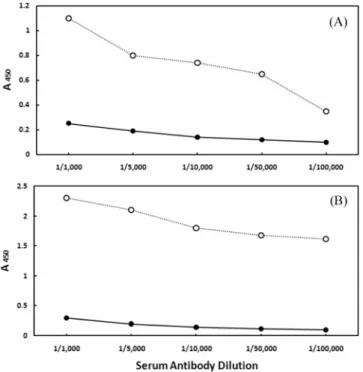
Fig. 2 shows the reactivity of these antibodies against control
and 17β-estradiol-treated serum from carp and flatfish at various dilutions, measured by ELISA at 450 nm. Compared with the control serum which showed conspicuously low values, the 17β- estradiol-treated serum from both fish had much higher reactivity to the anti-flatfish Vtg antibodies. As expected, this reactivity was two-fold higher in the flatfish serum compared with the carp serum [compare (b) to (a) of Fig. 2].
The antibody reactivity of the flatfish Vtg fragment, presumably unmodified due to its production in bacteria, was tested by immunoblotting analysis. The polyclonal antibodies raised against the purified flatfish Vtg, were previously shown to react well with purified flatfish Vtg and could be used successfully in immunosensor experiments [Kim et al., 2007b].
The flatfish Vtg induction pattern was also found to be similar (data not shown) to that reported in a previous carp Vtg experiment [Moon et al., 2006]. Fig. 3 shows the reactivity by western blotting of these of antibodies against both untreated and 17β-estradiol-treated carp and flatfish serum, and also in extracts of E. coli expressing the flatfish Vtg cDNA fragment. Positive Vtg signals were found in the serum of flatfish and carp treated with 17β-estradiol, but not in the corresponding untreated controls or E. coli extracts (Fig. 3). The flatfish Vtg protein (approximately 180 KDa), indicated by the arrow in Fig. 3, was located between the myosin (200 KDa) and β-galactosidase (116 KDa) of molecular weight markers (not shown on the blot).
These results suggest that covalent modifications of Vtg [Custodia-Lora et al., 2004] are required for antibody reactivity.
Fig. 1. SDS-PAGE (12%) analysis of a flatfish Vtg protein fragment produced in E. coli. Lane 1, Bio-Rad low range protein molecular weight markers [phosphorylase b (97,000), serum albumin (67,000), ovalbumin (45,000), carbonic anhydrase (31,000), trypsin inhibitor (21,000), lysozyme (14,000)]; lane 2, culture broth of BL21(DE3) cells harboring pET28a prior to induction with IPTG; lane 3, culture broth of BL21(DE3) cells harboring pET28a after induction with IPTG; lane 4, culture broth of BL21(DE3) cells harboring pET28a-Vtg prior to induction with IPTG; lane 5, culture broth of BL21(DE3) cells harboring pET28a-Vtg after induction with IPTG. Arrow indicates the flatfish Vtg protein fragment (~23 KDa) produced in E.coli.
Fig. 2. ELISA binding graph of rabbit anti-flatfish Vtg polyclonal antibodies. (a) Carp control serum and serum induced with 17β- estradiol. (b) Flatfish control serum and serum induced with 17β- estradiol. ○, 17β-estradiol induced serum; ●, untreated control serum.
Fig. 3. Western blotting analysis using rabbit anti-flatfish Vtg polyclonal antibodies of carp serum, flatfish serum, and a recombinant flatfish Vtg protein fragment produced in E. coli. Lanes 1-6, 7% SDS- polyacrylamide gel; lanes 7-11, 12% SDS-polyacrylamide gel; lanes 1, 6 and 7, Bio-Rad high range protein molecular weight markers; lane 2, male carp serum without induction; lane 3, male carp serum induced with 17β-estradiol dissolved in peanut oil; lane 4, male flatfish serum without induction; lane 5, male flatfish serum induced with 17β- estradiol dissolved in peanut oil; lane 8, culture broth of BL21(DE3) cells harboring pET28a prior to induction with IPTG; lane 9, culture broth of BL21(DE3) cells harboring pET28a after induction with IPTG; lane 10, culture broth of BL21(DE3) cells harboring pET28a- Vtg prior to induction with IPTG; lane 11, culture broth of BL21(DE3) cells harboring pET28a-Vtg after induction with IPTG. Arrow indicates the flatfish Vtg protein (180 KDa) located between the myosin (200 KDa) and β-galactosidase (116 KDa) of high range protein molecular weight markers (not shown on the blot).
Reactivity of flatfish vitellogenin antibodies on immunoblots 173
The polyclonal antibodies we tested in this study were found to be very sensitive for the detection of Vtg in carp and flatfish, and could therefore be used in an immunosensor screening tool for endocrine disrupters monitoring Vtg quantity.
Acknowledgments
This research was supported by a Chung-Ang University Research Grant in 2006.
References
Burnette WN (1981) Western Blotting: Electrophoretic transfer of proteins from sodium dodecyl sulfate-polyacrylamide gels to unmodified nitrocellulose and radiographic detection with antibody and radioiodinated protein A. Anal Biochem112, 195- Chen TT (1983) Identification and characterization of estrogen-203.
responsive gene products in the liver of rainbow trout. Can J Biochem Cell Biol 61, 802-810.
Colborn T and Clement C (1992) Chemically induced alterations in sexual and functional development. In The Wildlife/Human Connection, Princeton Scientific Publishing, Princeton, NJ, Custodia-Lora N, Novillo A, and Callard IP (2004) Effect ofUSA.
gonadal steroids on progesterone receptor, estrogen receptor, and vitellogenin expression in male turtles (Chrysemys picta). J Exp Zoology 301, 15-25.
Kim N, Park IS, and Kim DK (2006a) Optimization of quartz crystal microbalance-precipitation sensor measuring acetylcholinesterase activity. J Microbiol Biotechnol 16, 1523-1528.
Kim N, Park IS, and Kim WY (2006b) Detection of carp vitellogenin with piezoelectric immunosensor. J Korean Soc Appl Biol Chem49, 254-258.
Kim N, Park IS, and Kim WY (2007a) Salmonella detection with a direct-binding optical grating coupler immunosensor. Sens Actuators B Chem121, 606-615.
Kim N, Ryu HS, and Kim WY (2007b) Flatfish vitellogenin detection using optical waveguide lightmode spectroscopy-
based immunosensor. J Microbiol Biotechnol17, 1445-1451.
Kolosova AY, Samsonova YV, and Egorov AM (2000) Competitive ELISA of chloramphenicol: Influence of immunoreagent structure and application of the method for the inspection of food of animal origin. Food Agric Immunol 12, 115-125.
Laemmli UK (1970) Cleavage of structural proteins during assembly of the head of bacteriophage T4. Nature 227, 680- Moon DK, Kim N, and Kim WY (2006) Reactivity of the685.
antibodies against purified carp vitellogenin and a synthetic vitellogenin peptide. J Korean Soc Appl Biol Chem 49, 196- Nam YS and Choi JW (2006) Fabrication and electrical characteristics201.
of ferredoxin self-assembled layer for biomolecular electronic device application. J Microbiol Biotechnol 16, 15-19.
Oh BK, Kim YK, Park KW, Lee WH, and Choi JW (2004) Surface plasmon resonance immunosensor for the detection of Salmonella typhimurium. Biosens Bioelectron 19, 1497-1504.
Park JS, Lim SH, Sim SJ, Chae H, Yoon HC, Yang SS, and Kim BW (2006) Enhancement of sensitivity in interferometric biosensing by using a new biolinker and prebinding antibody. J Microbiol Biotechnol16, 1968-1976.
Smeets JMW, Rankouhi TR, Nichols KM, Komen H, Kaminski NE, Giesy JP, and Martin van den Berg M (1999) In vitro vitellogenin production by carp (Cyprinus carpio) hepatocytesas a screening method for determining (anti)estrogenic activity of xenobiotics. Toxico Appl Pharmacol157, 68-76.
Tyler CR, Van der Eerden B, Sunpter JP, Jobling S, and Painter G (1996) Measurement of vitellogenin, a biomarker for exposure to oestrogen, in a wide variety of cyprinids. J Comp Physiol
166, 418-426.
Tyler CR, Van Aerle R, Nilsen MV, Blackwell R, Maddix S, Nilsen BM, Berg K, Hutchinson TH, and Goksoyr A (2002) Monoclonal antibody enzyme-linked immunosorbent assay to quantify vitellogenin for studies on environmental estrogens in the rainbow trout (Oncorhynchus mykiss). Environ Toxicol Chem 21, 47-54.
Wallace RA (1985) Vitellogenesis and oocyte growth in nonmammalian vertebrates. In Developmental Biology, Browder LW (ed.), pp. 127-177. Plenum Press, New York, USA.