서론
Albrektsson1등은 1986년에 성공기준(success criteria)을 언급할 때, 임플란트 보철 이후 1년 동안 변연골 흡수가 1 - 2 mm 이내 이며, 경과관찰 기간 동안 매년 0.05 - 0.2 mm 이내의 변연골 흡수 가 있으면 임플란트가 성공적인 것으로 제시하였다.
International Congress of Oral Implantologists (ICOI)2는 2007년에 구 강임플란트의 건강지표(Health Scale for Dental implants)에 대해 논 의하였고, 임플란트 성공에 대해 4가지 그룹으로 I. Success; II.
Satisfactory survival; III. Compromised survival; IV. Failure (clinical or absolute failure)로 나눴으며, success 에 대해 a) 기능 중 통증이 없 어야 하며 (No pain or tenderness upon function), b) 동요도가 없고 (0 mobility), c) 초기수술 이후 방사선적으로 2 mm 이하의 골소실이 있어야 하며(< 2 mm radiographic bone loss from initial surgery), d) No exudates history로 규정하였다. 또한 심미적 부분을 포함한 환자 의 만족이 이러한 성공기준에 고려되어야 한다고 제안하였다.
임플란트는 골유착을 통해 골과 직접적으로 결합하게 되는
데 지속적인 골흡수와 골형성이 동일 위치에서 순차적으로 일 어나며 골개조가 일어난다.3특히 골에 응력이 집중되는 곳인 implant neck 부위에서 많이 일어나며, 골개조 과정 중 기능이나 부하가 시작되면서 implant neck 주위에서 변연골 흡수가 일어나 게 된다. 이러한 변연골 흡수가 계속적으로 진행될 경우, 골에 서 발생되는 strain은 불리해지며, bone crest에 대한 응력이 극적 으로 증가할 수 있다.4또한 해면골에 응력이 증가하게 되며 측 방력 등에 의해 disintegration이 일어나 임플란트 실패가 일어날 수 있다.5그렇기 때문에 초기의 변연 골 흡수를 조절하는 것이 임플란트 성공과 관련하여 중요하다. 부하가 시작되고 나서 초기 1년 정도의 기간 동안 marginal bone loss가 많이 일어나게 되 는데 이에 대한 원인은 명확히 밝혀지지는 않았으나, occlusal overload,5,6presence of microgap,7reformation of biologic width8,9에 의 한 것으로 생각되고 있다. 여러 가지 원인들로 인해 발생 가능 한 초기의 marginal bone loss에 따라 임플란트 주위 연조직이 remodeling 되며, 이에 따라 치료의 심미성에 영향을 주게 된다.
따라서 여러 가지 변연골 흡수 유발 원인에 대처하여 implant http://dx.doi.org/10.4047/jkap.2011.49.4.346 REVIEW ARTICLE
*교신저자: 박박영영범범
120-752 서울 서대문구 신촌동 연세의료원 치과대학병원 02-2228-3164: e-mail, [email protected] 원고접수일: 2011년 7월 9일 / 원고최종수정일: 2011년 10월 17일 / 원고채택일: 2011년 10월 24일
임플란트 주위 조직 보존을 위한 임플란트 경부의 디자인에 관한 고찰
김홍준∙김지환∙김성태∙이재훈∙박영범*
연세대학교 치과대학 치과보철학교실
연구 목적: 임플란트 식립 후 변연골 흡수에 따라 임플란트 주위 연조직이 재구성되며, 이에 따라 치료의 예후 및 심미성 등에 영향을 주게 된다. 그러므로 임플란트 경부 주위 골조직 보존을 위한 임플란트 경부에 다양한 디자인이 연구되고 있다. 본 고찰의 목적은 초기 변연골 흡수의 원인과 이에 따른 임플란트 주위의 연조직 변화에 대해 고찰하고, 어떠한 임플란트 경부 디자인이 임플란트 주위 조직의 보존에 유리한 지 알아보고자 한다.
연구 재료 및 방법: Pubmed database에서 임플란트 초기 변연골 흡수의 원인과 관련된 논문과 임플란트 경부의 여러 디자인에 관한 논문을 검색하여 분석하였다. 임플란트 경부 디자인은 one piece implant, two piece implant, internal hex abutment, external hex abutment, taper joint connection, butt joint connection, scalloped design abutment, platform switching concept에 관해 검토하였다.
결과:초기의 임플란트 주위 조직 보존에 대하여 one piece implant가 two piece implant보다 유리한 것으로 여러 임상적, 실험적 연구가 있다. Two piece implant에서는 internal hex abutment가 external hex abutment보다, taper joint connection가 butt joint connection보다 유리할 것으로 보여진다. Scalloped design abutment에 대해서는 논쟁의 여지가 있어 더 많은 연구가 필요할 것으로 판단된다. Platform switching concept은 그 원인이 명확히 밝혀지지는 않았으나 임상적, 실험적으로 초기 임플란트 주위 조직 보존에 대해 유리한 것으로 판단된다.
결론: 임플란트 경부의 디자인마다 각각의 장단점이 있고 추가적인 연구가 더 필요한 제한이 있지만 현재까지의 선행 연구들을 분석 종합해 보면 초기 임플란트 주위 조 직 보존을 고려한다면 가능한 경우 one piece implant가 유리할 것으로 판단되며, 보철적인 문제나 다른 이유로 인하여 two piece implant를 고려할 경우 platform switching concept, internal connection abutment, taper joint connection을 이용하는 것이 임플란트 주위 조직 보존에 좀더 유리할 것으로 사료된다. (대한치과보철학회지 2011;49:346-53)
주요단어: 임플란트 경부 디자인, One piece implant, Internal hex abutment, Taper joint connection, Platform switching concept
주변 골조직을 보존하기 위한 있는 임플란트 경부 디자인이 연구되어 왔다. 본 고찰의 목적은 이러한 marginal bone loss의 원인과 implant 주위의 연조직 반응에 대해 고찰하고, 어떠한 implant neck design이 임플란트 주위 조직의 보존에 유리한 지 알 아보는 것이다.
본론
Etiology of early marginal bone loss
Occlusal overload
임플란트는 자연치와는 달리 치주인대가 없이 골과 직접 결 합하므로 과도한 교합력에 주위 골 흡수가 일어나고 골 유착 실패의 원인이 된다. 임플란트는 수직력보다는 측방력에 더 취약하다.10Wiskott 등4은 bone과 implant 계면에서 발생하는 stress 에 의해 strain이 발생하게 되며, 그 양에 따라 골이 생성되기도 하고, 흡수되기도 한다고 하였으며, 그 양이 증가하게 될 경우 bone에 fatigue & creep을 일으킨다고 하였다. Isidor 등11은 원숭이 를 대상으로 하여 overload에 대한 임플란트 주위 조직의 반응을 살펴보았고, 측방력으로써 overload를 가한 결과, overload에 의해 osseointegration이 부분적 혹은 전부 상실되었다고 하였다.
Quirynen 등6은 전치부 contact이 없는 환자, parafuntion이 있는 환자, 전악에 임플란트 보철을 한 환자 등에서 overload가 발생 하게 되며, 이런 환자들에게서 marginal bone loss가 더 증가한다고 하였다.
Reformation of biologic width
임플란트에도 자연치의 biologic width 와 유사한 개념이 존재 하여 이 biologic width를 침범할 경우 bone loss가 일어나게 된다.
Berglundh 등8은 이러한 biologic width에 의한 bone loss는 implant와 abutment 사이의 interface로부터 일정한 거리를 유지한다고 하였 다. Hermann9의 개를 대상으로 한 연구에서 1-piece implant에서는 rough/smooth surface의 경계가 bone loss에 영향을 미치며, 2-piece implant에서는 crestal bone level이 임플란트와 abutment의 inter- face에 위치한 microgap의 위치에 따라 결정되며, 대략적으로 microgap의 2 mm 하방에 위치하게 된다고 하였다. 또한 이는 submerged 나 non-submerged의 수술방법과 무관하게 직접적으로 crestal bone loss에 영향을 미친다고 하였다. 또한 1-piece implant의 rough/smooth surface가 alveolar crest에 위치되었을 때 2-piece의 모든 경우보다 bone loss가 적게 일어났다. 즉 biologic width의 재형 성으로 인한 골흡수가 발생하며, 이는 microgap으로 부터 일정 한 거리를 유지하게 되고, 1-piece impalnt에서는 이러한 microgap 이 존재하지 않기 때문에 biologic width 측면에서 상대적으로 bone loss에 유리하다.
Presence of microgap
① Micro gap에의 bacterial colonization
Quirynen12과 Persson13은 internal surfaces of submerged implants or their restorative component parts에 microbial species가 cultivation 된 다고 하였다. 또한 Lindhe14과 Berglundh 등15은 peri-implant mucosa 의 특징을 분석하였고, 그 결과 plaque accumulation 과 상관없이 microgap을 기준으로 apical& coronal 부위 0.5 - 0.75 mm에 염증세 포가 모여있는 abutment infiltrated connective tissue area가 존재한다 고 하였다(Fig. 1). 이 부위에 bacterial colonization이 일어나게 되며 이로 인해 implant와 abutment가 연결된 이후 marginal bone loss가 일어난다고 하였다.
② Micro gap에의 micromovement
Crestal bone loss를 줄이기 위해 alveolar crest보다 1 - 2 mm 정도 상방에 microgap이 위치하도록 식립하게 되었다. 그러나 여전히 crestal bone loss는 존재하였으며, 이를 설명하기 위한 가설 중 하 나가‘movements between components’에 의한 marginal bone loss이 다. 이에 대한 영향을 알아보기 위해 Hermann등9은 ① microgap size에 따른 marginal bone loss의 변화와, ② micromovement에 의한 marginal bone loss의 영향을 알아보기 위해 two-piece implant에서 fixture와 abutment를 welding을 시행한 group과 fixture와 abutment가 screw에 의해 연결된 group에서의 marginal bone loss 변화를 비교 하였다. 그 결과 micromovement를 제한한 경우, marginal bone loss가 적게 나타났으며, microgap size와는 관계가 없었다. 즉 microgap이 있더라도 그 micromovement를 제한할 수 있다면 bone loss를 줄일 수 있는 것으로 예상된다.
Soft tissue sealing 임
임플플란란트트 주주위위 biologic width의의 구구성성
Biologic width는 sulcular epithelium, junctional epithelium, underly- Fig. 1. Infiltrated connective tissue zone around the implant.
ing connective tissue zone 으로 구성되어있다. Sulcular epitheliem의 apical 부위는 매우 얇으며, 임플란트 표면에 hemi desmosome 과 유사한 구조로 접착이 된다. Connective tissue는 scar like tissue와 유 사하며, 임플란트 표면에 다른 attachment 없이 직접 접촉한다.
Underlying connective tissue zone에는 염증세포가 모여 있는 부분 인 abutment infiltrated connective tissue가 존재한다.16Implant 에서의 biologic width에 대한 연구논문은 동물을 대상으로 한 경우가 대부분이며, 어느 정도의 distance를 가졌는지에 대하여 명확한 정의를 내리기는 어렵다. 그러나 치아에서의 biologic width보다 조금 더 큰 값을 갖는 것으로 생각되며, Cochran, Berglundh, Abrahamsson 등은 동물실험에서 각각 biologic width가 3.08 mm17/ 3.80 mm15/ 3.42 mm16로 언급하였다.
임
임플플란란트트 주주위위 biologic width의의 기기능능
Lindhe14와 Berglundh15등은 1992년, 동물실험을 하여 biologic width 를 이루는 연조직은 leukocyte의 migration을 일으키며, infiltrated con- nective tissue zone을 형성하여 underlying bone에의 보호기능을 한다고 하였다. Zitzmann 등19은 사람에서 plaque accumulation에 대 한 peri -implant mucosa와 gingiva의 반응을 연구하기 위해 12명의 부분무치악 환자를 대상으로 임상 검사 및 연조직 biopsy를 하 여 immunohistochemical analysis를 시행하였다. Peri-implant mucosa 와 gingiva 두 부위의 infiltrated connective tissue에서 염증세포가 모 이게 되며 그 중에 B cell과 T cell의 비율이 높아진다고 하였다.
또한 Bullon 등20은 2004년, healthy gingiva, peri implantitis mucosa, aggres- sive periodontitis gingiva 그룹으로 나눠 각각의 그룹에서 Biopsy spec- imen을 제작하여 histological and immunohistochemical analysis가 시 행했다. T cell에 대한 항체인 CD3과 B cell에 대한 항체인 CD20의 양이 healthy gingival 에서보다 peri-implantitis mucosa와 aggressive periodontitis gingiva에서 증가되었으며, 특히 T cell의 양이 많이 증 가된다고 하였다.
임
임플플란란트트 주주위위 biologic width에에서서의의 mucosal thickness의의 영영향향 Peri-implant mucosa의 두께는 적절한 epithelial connective tissue attach- ment를 이루기 위해 필요하며, 이러한 soft tissue dimension이 만족 되지 않을 경우 골흡수가 일어나서 새로운 적절한 biologic width 를 이룬다고 한다.21Berglundh 등15은 동물실험을 통해 2nd surgery 시에 test group에서 mucosa를 2 mm 정도로 얇게 만들었고, control group에서는 조작하지 않았다. 두 group 에서 total biologic width는 유의한 차이를 보이지 않았으며, test group에서 연조직 치유 기 간 동안 점진적인 골흡수가 관찰되었다. Control group을 가진 사 람에 대한 연구는 많지 않으나 peri-impalnt mucosa가 적절한 두께 를 유지해야 좋은 임상적 결과를 얻을 수 있다고 사료된다.22
Implant neck design for prevention of marginal bone loss and soft tissue sealing
Marginal bone loss를 줄이고 적절한 soft tissue sealing을 얻기
위해 implant neck 부위에 다양한 노력이 있어왔다. Smooth surface 를 이용하던 것에 대해 stress distribution과 osseointegration을 위해 rough surface 나 rough surface with microthread 등을 이용하는 노력 에서 형태, 연결부위의 변화 등 다양한 노력이 있어왔다. 여기서 는 표면의 texture와 관련된 부분은 방대한 자료 관계로 제외하고 neck 부위 형태와 연결 형태와 관련하여 알아보고자 하였다.
One-piece or two piece implant
Hermann23은 동물실험을 통해 같은 crest bone level에 임플란트 를 식립한 경우 One piece 임플란트에서 two piece 임플란트 보다 marginal bone loss의 양이 적다고 하였다. Heijdenrijk24등은 Implant- abutment의 microgap 이 존재하지 않는 one piece implant 와 two piece implant non-submerged, two piece implant submerged 간의 5년간 bone loss를 관찰하였다. 5년간의 관찰결과 one piece와 two piece 간 의 유의한 차이가 관찰되지 않았다고 하였다. 그러나 저자는 보철물에 implant overdenture를 사용한 것이었는데, two piece implant에서는 5 mm 높이의 bar type을 사용하여 splint하였고, one piece implant에서는 bar type을 사용하지 않았다. 그렇기 때문 에 각 implant의 stress distribution에 차이를 일으켰을 것으로 생각 되며, T0 - T12 의 1년간의 기간에서는 one piece implant의 marginal bone loss가 다른 두 방식에 비해 적게 나타났다. 보철물이 적용 되고 4주가 지난 기간을 T0로 설정하였으며, 이후의 개월 수에 따라 T12, T24, T36, T48, T60으로 설정하였다. One piece implant의 경우 abutment와 fixture가 일체형으로서 microgap이 없는 형태이 며, 여기서는 보철물과 implant사이의 경계가 가장 apical 부위에 존재하는 microgap이다. Two piece implant에서는 abutment와 fixture 간의 microgap이 존재하는 형태이다. One-piece의 경우 일체형이 기 때문에 microgap에서의 bacterial colonization이 방지될 수 있으 며,25implant-abutment interface에서 일어날 수 있는 micromove- ment를 방지할 수 있다. 그러나 abutment의 taper 조절이 힘들다는 등 보철과 관련한 단점이 있다. 즉 marginal bone loss와 관련하였 을 때 one piece implant가 two piece implant 보다 더 유리할 것으로 사료된다.
Internal hex abutment와와 external hex abutment
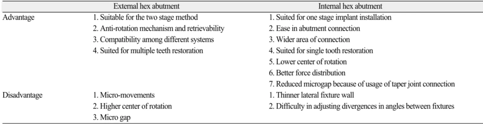
Maeda 등26은 2006년 실험에서 implant with external hex와 implant with internal hex에 vertical & horizontal load를 가하였는데 vertical load 를 가하였을 때는 유의 차가 없는 범위에서 상대적으로 external hex에서 cervical 부위에 strain값이 컸고, internal hex에서는 apex에 서 strain 값이 크게 나왔다. 특히 horizontal load를 가하였을 때 Internal hex system에서 cervical area에서의 상대적으로 적은 stress 가 발생하였다. 이는 bone preservation에 긍정적으로 기여할 것이 라고 하였다. 다만 tip 부위에서의 큰 stress가 fixture fracture에의 위 험요소가 될 수 있다고 하였다. Table 1과 같이 external hex abutment 와 internal hex abutment는 각각의 아래와 같은 장단점을 가진다.
Implant-abutment connection에에서서 Taper joint와와 butt joint Norton 등은 taper joint connection이 banding force에 저항하는 능 력을 증가 시킨다고 하였다. Levine 등27은 임상적으로 보철수복 과 관련한 문제가 taper joint connection의 경우가 butt joint보다 적게 일어난다고 하였다. 그 원인으로는 sutter 등이 언급한 내용 으로 fixture와 abutment와의 gap이 10 um 보다 적은 경우 thread부 위에 functional load가 줄어들고, taper 부위에서 vibration을 완화시 켜 thread 부위에 micromovement가 없는 것을 들었다. Merz 등28은 2000년에 taper joint와 butt joint에 대해 유한요소분석을 시행하여 axis에 대한 load의 방향에 따른 응력분포를 알아보고자 하였 다. Taper joint는 axis에 따라 작용하는 힘을 받았을 때 thread 대한 load가 완전히 상쇄되어 abutment loosening을 일으키지 않는다.
Taper에서의 friction에 의해 implant와 abutment간의 안정적이고 회 전이 없는 연결이 가능해진다. 이에 반해, butt joint는 load에 대해 부분적 혹은 완전히 상쇄시키게 되는데, 이는 시간이 지남에 따라 abutment loosening을 일으킬 것으로 여겨진다. Axis에서 15도 정도 벗어난 방향에서의 힘까지도 taper joint의 경우 thread는 거 의 영향을 받지 않아 유리하다고 하였다. Axis에서 30도 벗어난 힘에 대해서는 yield point 보다 높은 압력을 보이게 되었으나, 매 우 한정된 부분이었으며, stress의 경사가 높아 지지효과가 작용 될 수 있다. Pieri 등29은 2011년 abutment with taper joint와 abutment with conventional internal connection를 대상으로 12개월의 follow up 을 시행한 결과, marginal bone loss가 abutment with taper joint에서 적 게 일어났다고 하였다. 비록 pieri의 연구에서는 taper joint에서 platform switching의 개념이 함께 사용되었기 때문에 taper joint만 의 영향으로 말할 수는 없다. 다만 taper joint가 butt joint에 비해 micromovement가 상대적으로 적고, 응력분포에 우수한 결과를 가지기 때문에 marginal bone loss에 유리할 것으로 사료된다.
Scalloped design abutment
Flat-top implant가 통상적으로 사용되었으나 연조직 치유 및 치 조골형태를 심미적으로 하기 위한 design으로 scalloped design abutment이 나왔다. Interproximal bone의 보존에 도움이 될 것으로
사료되었다. Biologic width가 임플란트의 가장 apical 부위의 microgap부터 형성되게 되며, 그에 맞게 bone remodeling 된다고 하 여 이러한 형태를 이용한 것이다.30Bradley31는 Scalloped design에 대한 interproximal bone level에 대한 영향을 평가하였고, scalloped implant는 수복 후 interproximal bone level이 first thread보다 coronal 에 위치했다고 하였다. 유의미한 차이가 있는지 비교하지는 않았으나 scalloped implant가 interproximal bone level을 유지하는데 유리하다고 하였다. 그러나 Nowzari32의 연구에서는 scalloped design의 marginal bone loss가 conventional 의 경우보다 더 많게 나 타나는 상반된 결과가 보고되기도 하였다. 현재 scalloped design abutment에 대한 연구는 case report가 대부분이며, 초기에는 implant shoulder 부위에서 2 mm 정도 하방에서 bone remodeling되 지만 결국에는 평균적인 bone level은 임플란트의 first thread 수준 이 된다고 하였다. 또한 임플란트 사이의 거리가 bone level과 soft tissue attachment에 중요한 요인인데 이것에 대한 고려가 함께 고려하여 연구가 진행되지 않았다. Scalloped design abutment이 inter proximal bone level 에 어떠한 영향을 끼칠지에 대해서는 더 많은 연구가 필요할 것으로 사료된다.
Platform switching
Platform switching의의 기기원원
역사적으로 two-piece dental implant systems은 outer edge of the implant platform에서 prosthetic components이 위치하는 형태로 수복되어 왔 다. 1991년에 wide-diameter implants with matching wide-diameter platforms이 소개되었다. 그러나 처음 소개되었을 때 matching- diameter prosthetic components 가 사용할 수 가 없었기 때문에 5.0 mm 나 6.0 mm의 wide diameter implant에 standard diameter (4.1 mm)의 healing abutments 와 prosthetic components가 이용되었다. 장 기간의 방사선 사진을 통한 follow up에서“platform-switched”된 임플란트가 conventional 하게 사용된 platform matched prosthesis에 서 보다 골의 수직적 변화가 더 적었다고 보고하였다.
Table 1. Advantages and disadvantages of external hex abutment and internal hex abutment
External hex abutment Internal hex abutment
Advantage 1. Suitable for the two stage method 1. Suited for one stage implant installation 2. Anti-rotation mechanism and retrievability 2. Ease in abutment connection 3. Compatibility among different systems 3. Wider area of connection 4. Suited for multiple teeth restoration 4. Suited for single tooth restoration
5. Lower center of rotation 6. Better force distribution
7. Reduced microgap because of usage of taper joint connection
Disadvantage 1. Micro-movements 1. Thinner lateral fixture wall
2. Higher center of rotation 2. Difficulty in adjusting divergences in angles between fixtures 3. Micro gap
Platform switching의의 Rationale
첫번째로 biologic width와 관련되어있는 것으로 사료된다.
Berglundh 등15은 1991년 implant-abutment의 interface 즉, microgap을 기준으로 apical, coronal로 각각 0.5 - 0.75 mm 의 범위로 abutment infil- trated connective tissue 가 존재하여 bacterial colonization을 이루게 된 다고 하였다. 이를 기준으로 하방 1 mm의 부위에 bacterial colo- nization이 없는 CT가 존재하며, crestal bone이 위치한다고 하였다.
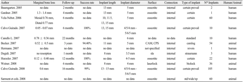
그렇기 때문에 two-stage implant에서는 biologic seal을 형성하기 위 해 3 mm의 최소 두께가 필요하며, 그렇지 않으면 crestal bone resorption이 올 것이라고 하였다. Platform switching은 implant- abutment의 microgap을 안쪽으로 이동시키게 된다. Infiltrated con- nective tissue를 안쪽으로 이동시켜 microgap으로 부터의 microbac- terial leakage를 crestal bone으로부터 멀리 위치시키게 하여 bone resorptsion을 줄여준다.33두 번째는 잔류응력과 관련한 역학적인 원인이다. 2007년 Maeda34는 3차원 유한요소 분석을 통해 Platform switching의 생역학적 분석을 하였다. Abutment의 stress distribution 을 보면 Normal model에서는 implant top의 periphery에서 lateral sur- face를 따라 stress가 집중되었으나, platform switching model에서는 stress가 implant의 center로 이동하였다. Platform switching은 stress concentration area를 cervical bone implant interface에서 멀어지게 하 는 장점이 있어 bone loss에 유리할 것으로 사료된다. 그러나 abutment or abutment screw로 증가된 load는 탄성한계를 넘어서게 될 경우 abutment screw의 변형을 일으킬 가능성이 있다고 하였 다. 이러한 platform switching concept은 다양한 임상연구에서 marginal bone preservation에 유리한 결과를 보이고 있다. Table 2에 서는 비록 단기간 연구들이지만 marginal bone preservation에 plat- form switching concept이 임상적으로 적합한 결과를 보여주고 있음을 알 수 있다. 또한 success rate와 관련하여서는 기존의 임 플란트와 유사한 수준을 나타내고 있다.35
Longitudinal study는 일반적으로 5년을 기준으로 삼고 있다.
Platform switching 에 대하여 5년 이상의 논문은 많지 않으나, Vigolgo 등36은 2009년에 발표한 논문에서 platform switching의 5년 동안의 crestal bone loss를 platform matching 과 비교하고 임상적 평 가를 하기 위해, 2000 - 2002년에 식립된 구치부 임플란트를 대 상으로 Platform matching (85개)와 platform-switching (97개)를 방사 선사진을 통해 5년간 crestal bone loss를 관찰하였다. 모두 External hex type 을 사용하였고, 5 mm 지름의 fixture를 이용하였다.
Platform switching concept이 platform matching에 비해 골 소실량이 초기 1년 동안 0.3 mm정도 적게 나타났으며 두 그룹 모두 1년이 지난 이후의 bone level은 안정적인 모습을 보였다. Wagenberg와 Froum37은 2010년, platform switching concept에서 implant survival과 crest bone level을 평가하기 위해 1992년에서 2006년까지 76명 의 환자에 대해 106개의 platform switched implant를 follow up 한 결 과, marginal bone loss는 mesial 에서 84%가 0.8 mm 이하, 98 % 가 2.0 mm 이하로 나타났으며, distal에서는 88%가 0.8 mm, 99%가 2.0 mm 이하로 나타났다. 비록 이 연구에서 control group이 없으나, plat- form switching concept이 interproximal crestal bone levels을 보존하는 것에 대해 효과적이라고 하였다. 여기서 사용된 것은 모두 external connections, no collar, thread가 fixture 최상부까지 있으며, 4 mm 지름의 abutments를 사용한 것이었다.
비록 아직 longitudinal study는 많지 않으나 현재까지 나와 있는 연구를 바탕으로 보았을 때 platform switching concept은 단기간뿐 아니라 장기간의 예후도 임상적으로 적합할 것으로 사료된다.
Soft tissue sealing & esthetic
Tarnow 등38,39은 전치부에서 gingival papilla를 유지하는 중요 한 것은 cervical bone around the neck of the implant를 가능한 보존하 는 것이라고 하였다. 그러나 abutment가 implant에 연결될 때 fixture
Table 2. Clinical outcome of platform switching concept
Author Marginal bone loss Follow-up Success rate Implant length Implant diameter Surface Connection Type of implant N* Implants Human/Animal
Baumgarten. 2005 no data 2 months no data 13 mm 5 mm osseotite internal certain prevail 2 human
Gardner. 2005 1.3 - 1.4 mm 4 months no data 13 mm 5 mm osseotite internal certain 1 human
Vela-Nebot. 2006 Mesial 0.76 mm, 6 months no data 10, 11.5, 5 mm osseotite internal certain 60 human
Distal 0.77 mm 13, 15 mm
Calvo Guirado. 2007 0.05 - 0.07 mm 6 months 100% 13, 15 mm 4/5/4 mm - osseotite internal certain prevail 10 human 5/6/5 mm
Canullo L. 2007 0.78 ± 0.36 mm 22 months no data no data 6 mm no data no data standard 10 human
Becker. 2007 0.52 ± 0.5 mm 3 years 94.40% 11 mm 5 mm CAM, CPS internal camlog 54 animal
Hermann. 2007 no data no data no data no data no data not specified internal revois 1 human
Degidi. 2007 no resorption 1 month no data 8 mm 3.5 mm sla internal straumann 3 human
Hurzeler. 2007 0.12 ± 0.40 mm 12 months 100% no data 4-5 mm osseotite internal certain 22 human
Weiner. 2008 no data 6 months no data 8 mm 4 mm laserlock internal biolock 36 animal
Calvo Guirado. 2008 0.6 mm 16 months 99% 13 - 15 mm 4/5/4 mm - osseotite internal certain prevail 105 human 5/6/5 mm
Sarment et cols. 2008 no data no data no data no data no data osseotite internal std/wide/xp 60 animal Calvo Guirado et al. Med Oral Patol Oral Cir Bucal. 2009
주위로 전형적인 골소실이 일어난다. 이 때의 골 소실은 micro- gap에서 골의 방향으로 수평적으로 1.3 - 1.4 mm일어난다고 한 다. 그러한 bone remodeling에 의해 연조직 역시 영향을 받는다고 하였다.
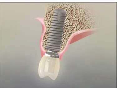
또한 biologic width를 위해 연조직의 적절한 두께가 유지 되어 야 하는데, Hermann 등40은 2007년에 발표한 연구에서 Fig. 2에서 보듯 platform switching에서는 적절한 연조직 두께를 얻기에 용이 하여 biologic width의 유지에 유리하다고 하였다. 2007년 Calvo41의 논문에서는 10명의 환자를 대상으로 platform switched implant를 식립하여 6개월간 follow up을 시행하였다. 심미적인 결과를 보 였으며, platform switching concept이 crestal bone loss를 줄이고 gin- giva papilla를 유지하는 것에 유리하다고 하였다(Fig. 2).
결론
현재까지 여러 연구 결과들을 분석 고찰해 본 결과 marginal bone loss를 유발하는 원인들은 명확하게 밝혀진 것은 없으나 occlusal overload와 biologic width 등이 중요한 원인들로 판단된다.
본 고찰에서 여러 가지 가능한 변연골 흡수 유발 원인들에 대 처하기 위한 임플란트 경부 형태 및 특징들을 살펴 보았고 이 런 임플란트 경부의 형태나 특성이 임플란트 주변 조직의 보 존에 관련 있음을 판단할 수 있었다. One-piece implant가 two- piece implant와 비교하여 fixture-abutment microgap이 없어서 biologic width 측면이나 microgap으로 인한 micromovement, bacterial colo- nization에 유리하여 marginal bone preservation에 우수할 것으로 사 료된다. 그러나 abutment taper를 조절하기 힘들고 자연스러운 emergency profile을 형성하기 힘들며, 전치부와 같은 부위에서 심 미성을 고려할 때 사용하기 어려운 경우가 있다. Two-piece implant의 사용하는 경우 internal hex abutment가 external hex abutment
에 비하여 응력이 상대적으로 implant neck 부위에 적게 분포되 기 때문에 상대적으로 유리할 것으로 사료된다. 또한 internal hex abutment에서 taper joint connection이 butt joint connection 보다 응 력을 보다 균등하게 배분하며, micromovement를 적게 발생시 키며, 측방력에 대해서도 thread 부위에 load가 적게 가해지게 된다. 그렇기 때문에 보다 marginal bone preservation에 우수할 것 으로 사료된다. 그리고 scalloped design abutment는 Marginal bone preservation에 유리할지에 대해서는 아직 연구가 부족하기 때문 에 근거는 충분하지 않다고 사료된다. Platform switching concept 은 응력분포 및 microgap의 수평적 이동을 통한 biologic width &
bacterial colonization 등에 관련하여 platform matching concept보 다 유리한 것으로 사료된다. 또한 soft tissue의 적절한 두께를 유 지하기 유리한 구조이기 때문에 soft tissue seal이 우수하며 심미 적인 결과를 얻을 수 있을 것으로 보인다. 단기간 연구에서는 임상적으로 우수한 결과를 보이고 있으며, 장기간의 연구는 아직 많지 않으나 긍정적인 결과를 보여주고 있다. 본 고찰에 서 밝힌 바와 같이 여러 선행 연구들이 확정적인 결과를 제시 하고 있지는 못하지만 제한적인 결론은 one piece implant를 사용 하기 적절한 부위에는 one-piece implant를 사용하고, 만약 심미나, 보철적인 문제와 관련하여 two piece implant를 사용한다면, inter- nal hex abutment, taper joint connection, platform switching concept이 포함된 임플란트를 사용하는 것이 임플란트 주위 조직의 보존 관점에서는 유리할 수 있을 것으로 사료된다.
참고문헌
1. Albrektsson T, Zarb G, Worthington P, Eriksson AR. The long- term efficacy of currently used dental implants: a review and pro- posed criteria of success. Int J Oral Maxillofac Implants 1986;1:11-25.
2. Misch CE, Perel ML, Wang HL, Sammartino G, Galindo- Moreno P, Trisi P, Steigmann M, Rebaudi A, Palti A, Pikos MA, Schwartz-Arad D, Choukroun J, Gutierrez-Perez JL, Marenzi G, Valavanis DK. Implant success, survival, and failure: the International Congress of Oral Implantologists (ICOI) Pisa Consensus Conference. Implant Dent 2008;17:5-15.
3. Davies JE. Mechanisms of endosseous integration. Int J Prosthodont 1998;11:391-401.
4. Wiskott HW, Belser UC. Lack of integration of smooth titanium surfaces: a working hypothesis based on strains generated in the surrounding bone. Clin Oral Implants Res 1999;10:429-44.
5. Kitamura E, Stegaroiu R, Nomura S, Miyakawa O. Biomechanical aspects of marginal bone resorption around osseointegrated implants: considerations based on a three-dimensional finite element analysis. Clin Oral Implants Res 2004;15:401-12.
6. Quirynen M, Naert I, van Steenberghe D. Fixture design and overload influence marginal bone loss and fixture success in the Bra�nemark system. Clin Oral Implants Res 1992;3:104-11.
7. Ericsson I, Persson LG, Berglundh T, Marinello CP, Lindhe J, Klinge B. Different types of inflammatory reactions in peri-implant soft tissues. J Clin Periodontol 1995;22:255-61.
Fig. 2. Sealing around the soft tissue is excellent and adequate thickness could be maintained at the fixture-abutment interface in platform switching concept.
8. Berglundh T, Lindhe J. Dimension of the periimplant mucosa.
Biological width revisited. J Clin Periodontol 1996;23:971-3.
9. Hermann JS, Schoolfield JD, Schenk RK, Buser D, Cochran DL.
Influence of the size of the microgap on crestal bone changes around titanium implants. A histometric evaluation of unloaded non-sub- merged implants in the canine mandible. J Periodontol 2001;72:
1372-83.
10. Misch CE, Dietsh-Misch F, Hoar J, Beck G, Hazen R, Misch CM.
A bone quality-based implant system: first year of prosthetic load- ing. J Oral Implantol 1999;25:185-97.
11. Isidor F. Histological evaluation of peri-implant bone at implants subjected to occlusal overload or plaque accumulation. Clin Oral Implants Res 1997;8:1-9.
12. Quirynen M, van Steenberghe D. Bacterial colonization of the internal part of two-stage implants. An in vivo study. Clin Oral Implants Res 1993;4:158-61.
13. Persson LG, Lekholm U, Leonhardt A, Dahlen G, Lindhe J.
Bacterial colonization on internal surfaces of Bra�nemark system implant components. Clin Oral Implants Res 1996;7:90-5.
14. Lindhe J, Berglundh T, Ericsson I, Liljenberg B, Marinello C.
Experimental breakdown of peri-implant and periodontal tissues.
A study in the beagle dog. Clin Oral Implants Res 1992;3:9-16.
15. Berglundh T, Lindhe J, Ericsson I, Marinello CP, Liljenberg B, Thomsen P. The soft tissue barrier at implants and teeth. Clin Oral Implants Res 1991;2:81-90.
16. Abrahamsson I, Berglundh T, Wennstro¨m J, Lindhe J. The peri-implant hard and soft tissues at different implant systems.
A comparative study in the dog. Clin Oral Implants Res 1996;
7:212-9.
17. Cochran DL, Hermann JS, Schenk RK, Higginbottom FL, Buser D. Biologic width around titanium implants. A histometric analysis of the implanto-gingival junction around unloaded and loaded nonsubmerged implants in the canine mandible. J Periodontol 1997;68:186-98.
18. Berglundh T, Lindhe J, Marinello C, Ericsson I, Liljenberg B. Soft tissue reaction to de novo plaque formation on implants and teeth.
An experimental study in the dog. Clin Oral Implants Res 1992;3:1-8.
19. Zitzmann NU, Berglundh T, Marinello CP, Lindhe J. Experimental peri-implant mucositis in man. J Clin Periodontol 2001;28:517-23.
20. Bullon P, Fioroni M, Goteri G, Rubini C, Battino M.
Immunohistochemical analysis of soft tissues in implants with healthy and peri-implantitis condition, and aggressive peri- odontitis. Clin Oral Implants Res 2004;15:553-9.
21. Iacono VJ; Committee on Research, Science and Therapy, the American Academy of Periodontology. Dental implants in pe- riodontal therapy. J Periodontol 2000;71:1934-42.
22. Berglundh T, Abrahamsson I, Welander M, Lang NP, Lindhe J.
Morphogenesis of the peri-implant mucosa: an experimental study in dogs. Clin Oral Implants Res 2007;18:1-8.
23. Hermann JS, Buser D, Schenk RK, Cochran DL. Crestal bone changes around titanium implants. A histometric evaluation of unloaded non-submerged and submerged implants in the canine mandible. J Periodontol 2000;71:1412-24.
24. Heijdenrijk K, Raghoebar GM, Meijer HJ, Stegenga B, van der Reijden WA. Feasibility and influence of the microgap of two im- plants placed in a non-submerged procedure: a five-year follow-
up clinical trial. J Periodontol 2006;77:1051-60.
25. Broggini N, McManus LM, Hermann JS, Medina RU, Oates TW, Schenk RK, Buser D, Mellonig JT, Cochran DL. Persistent acute inflammation at the implant-abutment interface. J Dent Res 2003;82:232-7.
26. Maeda Y, Satoh T, Sogo M. In vitro differences of stress con- centrations for internal and external hex implant-abutment con- nections: a short communication. J Oral Rehabil 2006;33:75-8.
27. Levine RA, Clem DS 3rd, Wilson TG Jr, Higginbottom F, Solnit G. Multicenter retrospective analysis of the ITI implant sys- tem used for single-tooth replacements: results of loading for 2 or more years. Int J Oral Maxillofac Implants 1999;14:516-20.
28. Merz BR, Hunenbart S, Belser UC. Mechanics of the implant- abutment connection: an 8-degree taper compared to a butt joint connection. Int J Oral Maxillofac Implants 2000;15:519-26.
29. Pieri F, Aldini NN, Marchetti C, Corinaldesi G. Influence of im- plant-abutment interface design on bone and soft tissue levels around immediately placed and restored single-tooth implants: a randomized controlled clinical trial. Int J Oral Maxillofac Implants 2011;26:
169-78.
30. Wo¨hrle PS. Nobel Perfect esthetic scalloped implant: rationale for a new design. Clin Implant Dent Relat Res 2003;5:64-73.
31. McAllister BS. Scalloped implant designs enhance interproximal bone levels. Int J Periodontics Restorative Dent 2007;27:9-15.
32. Nowzari H, Chee W, Yi K, Pak M, Chung WH, Rich S.
Scalloped dental implants: a retrospective analysis of radi- ographic and clinical outcomes of 17 NobelPerfect implants in 6 patients. Clin Implant Dent Relat Res 2006;8:1-10.
33. Jansen VK, Conrads G, Richter EJ. Microbial leakage and marginal fit of the implant-abutment interface. Int J Oral Maxillofac Implants 1997;12:527-40.
34. Maeda Y, Miura J, Taki I, Sogo M. Biomechanical analysis on platform switching: is there any biomechanical rationale? Clin Oral Implants Res 2007;18:581-4.
35. Lo′pez-Marl′L, Calvo-Guirado JL, Martl′n-Castellote B, Gomez- Moreno G, Lo′pez-Marl′M. Implant platform switching concept:
an updated review. Med Oral Patol Oral Cir Bucal 2009;14:
e450-4.
36. Vigolo P, Givani A. Platform-switched restorations on wide-di- ameter implants: a 5-year clinical prospective study. Int J Oral Maxillofac Implants 2009;24:103-9.
37. Wagenberg B, Froum SJ. Prospective study of 94 platform-switched implants observed from 1992 to 2006. Int J Periodontics Restorative Dent 2010;30:9-17.
38. Tarnow DP, Cho SC, Wallace SS. The effect of inter-implant dis- tance on the height of inter-implant bone crest. J Periodontol 2000;71:546-9.
39. Tarnow D, Elian N, Fletcher P, Froum S, Magner A, Cho SC, Salama M, Salama H, Garber DA. Vertical distance from the crest of bone to the height of the interproximal papilla between adjacent implants. J Periodontol 2003;74:1785-8.
40. Hermann F, Lerner H, Palti A. Factors influencing the preservation of the periimplant marginal bone. Implant Dent 2007;16:165-75.
41. Calvo Guirado JL, Saez Yuguero MR, Pardo Zamora G, Muñoz Barrio E. Immediate provisionalization on a new implant design for esthetic restoration and preserving crestal bone. Implant Dent 2007;16:155-64.
Considerations in implant crestal module to preserve peri-implant tissue
Hong- Jun Kim, DDS, Jee-Hwan Kim, DDS, PhD, Sung-Tae Kim, DDS, PhD, Jae-Hoon Lee, DDS, PhD, Young-Bum Park*, DDS, MS, PhD Department of Prodthodontics, Yonsei University College of Dentistry, Seoul, Korea
Purpose: The peri-implant soft tissue is remodeled by the initial marginal bone resorption affecting the prognosis and esthetic result of treatment. Thus various designs on implant neck design are studied to preserve peri-implant bone. The purpose of this study is to review on the causes of initial marginal bone resorption, the configuration of peri-implant soft tissue, and the implant crestal module favorable in preserving peri-implant tissue. Materials and methods: The studies on the causes of initial marginal bone resorption and the implant crestal modules are researched and reviewed using Pubmed database. The implant crestal modules including one piece and two-piece implant, internal and exter- nal hex abutment, taper and butt joint connection, scalloped design abutment, and platform switching concept are reviewed. Results: A number of clinical and experimental studies preferred one piece implant to two-piece in preserving initial peri-implant tissue. For two piece implants, internal hex abutment and taper joint connection appear more favorable than external hex abutment and butt joint connection relatively. Controversial issues still exist on scalloped design requiring more studies on it. Although the ratio- nale is not certain, the concept of platform switching seems favorable in preserving initial peri-implant tissue based on clinical and experimental studies. Conclusion: Each implant crestal module contains its own advantages and disadvantages with various controversial issues. In the aspect of preservation of initial peri-implant tissue, however, one-piece implant seems beneficial. In cases when two-piece implant is more appropriate due to prosthodontic concerns or any other problems, the application of platform switching con- cept, internal connection abutment, and taper joint connection may be favorable for the preservation of peri-implant tissues. (J Korean Acad Prosthodont 2011;49:346-53) Key words: Implant crestal module, Implant neck design, One piece implant, Internal hex abutment, Taper joint connection, Platform switching concept
*Corresponding Author: Young-Bum Park
Department of Prosthodontics, College of Dentistry, Yonsei University, 250 Seongsanno, Seodaemun-gu, Seoul, 120-752, Korea +82 2 2228 3164: e-mail,[email protected]
Article history
Received July 9, 2011 / Last Revision October 17, 2011 / Accepted October 24, 2011