INTRODUCTION
The liver has a unique anatomy and physiology which allows for liver procurement from a living donor. Because of recent ad- vances in surgical techniques, living-donor liver transplantation (LDLT) in adults is being performed with increasing frequency and is considered as the treatment of choice for end-stage liver disease (1, 2). Living donor hepatectomy offers an exclusive in- sight into liver regeneration in normal livers (3-6). In healthy donors, up to 75% of the whole liver volume can be donated (1, 2). However, donation of the partial liver is not without risk for donors. The risk for donors increases with increase of the graft volume. Because donor safety is paramount in LDLT, knowl-
edge about the postoperative changes in the donor’s remnant liver and spleen is important. Because of the ability of the liver to regenerate, both the transplanted graft and the remaining liv- er are expected to regenerate and compensate for the loss of he- patic volume in the recipient and donor, respectively. It has been well documented that remnant liver enlargement occurs in the postoperative period following partial liver donation (7). The spleen also enlarges following major hepatic resection and after liver donation (7-9). To explain this phenomenon, relative por- tal hypertension (increase of the portal pressure after donor hepatectomy as compared with the preoperative state) and ele- vation in the levels of growth factors have been postulated as causes (10). However, the exact mechanism of the increase in
J Korean Soc Radiol 2013;69(5):377-384 http://dx.doi.org/10.3348/jksr.2013.69.5.377
Received June 26, 2013; Accepted September 2, 2013 Corresponding author: Hunkyu Ryeom, MD Department of Radiology, Kyungpook National University Hospital, 130 Dongdeok-ro, Jung-gu, Daegu 700-721, Korea.
Tel. 82-53-420-5390 Fax. 82-53-422-2677 E-mail: hkryeom@knu.ac.kr
This is an Open Access article distributed under the terms of the Creative Commons Attribution Non-Commercial License (http://creativecommons.org/licenses/by-nc/3.0) which permits unrestricted non-commercial use, distri- bution, and reproduction in any medium, provided the original work is properly cited.
Purpose: To define the changes in liver and spleen volumes in the early postopera- tive period after partial liver donation for living-donor liver transplantation (LDLT) and to determine factors that influence liver and spleen volume changes.
Materials and Methods: 27 donors who underwent partial hepatectomy for LDLT were included in this study. The rates of liver and spleen volume change, measured with CT volumetry, were correlated with several factors. The analyzed factors in- cluded the indocyanine green (ICG) retention rate at 15 minutes after ICG adminis- tration, preoperative platelet count, preoperative liver and splenic volumes, resected liver volume, resected-to-whole liver volume ratio (LVR/LVW), resected liver volume to the sum of whole liver and spleen volume ratio [LVR/(LVW + SV0)], and pre and post hepatectomy portal venous pressures.
Results: In all hepatectomy donors, the volumes of the remnant liver and spleen were increased (increased rates, 59.5 ± 50.5%, 47.9 ± 22.6%). The increment rate of the remnant liver volume revealed a positive correlation with LVR/LVW (r = 0.759, p <
0.01). The other analyzed factors showed no correlation with changes in liver and spleen volumes.
Conclusion: The spleen and remnant liver volumes were increased at CT volumetry performed 2 weeks after partial liver donation. Among the various analyzed factors, LVR/LVW influences the increment rate of the remnant liver volume.
Index terms Hepatectomy Transplantation Liver
Spleen
Computed Tomography CT Volumetry
Factors Influencing Liver and Spleen Volume Changes after Donor Hepatectomy for Living Donor Liver Transplantation
1생체 간이식을 위한 부분 간이식 절제술 후 간과 비장의 부피 변화에 영향을 주는 요인들1
Ji Hea Bae, MD
1, Hunkyu Ryeom, MD
1, Jung Hup Song, MD
2Departments of 1Radiology, 2Occupational Medicine, Kyungpook National University Hospital, Daegu, Korea
Non-enhanced CT scans were performed first. For dynamic con- trast-enhanced CT, 2 mg/kg of a non-ionic contrast agent (Om- nipaque 300; GE Healthcare, Milwaukee, WI, USA) was adminis- trated intravenously at a flow rate of 3 mL/sec using a power injector. Imaging was conducted at three different phases (he- patic arterial dominant, portal venous dominant, and late equi- librium phases) determined by bolus tracking and automated triggering technology. Non-enhanced scans and portal venous dominant phase imaging were obtained from the dome of the diaphragm to the pubic symphysis. The hepatic arterial domi- nant phase was obtained from the dome of the diaphragm to the iliac crest, to cover the entire liver. The protocol was as fol- lows: 140 kVp; 350 mA; section thickness, 0.625 mm; pitch, 1.75; table speed, 35 mm/sec (17.5 mm per rotation with two rotations); and gantry speed, 0.5 second per rotation. The trans- verse section data were reconstructed with 5-mm-thick sections at 5-mm intervals in the transverse plane. Coronal reformatted images were reconstructed with 3-mm sections at 3-mm inter- vals. The 5-mm transverse and 3-mm coronal images were re- constructed at the operator’s console and transferred to a picture archiving and communication system (PACS) workstation (IN- FINITT PACS, Infinitt Co., Ltd, Seoul, Korea) as a separate se- ries of scans for interpretation.
CT volumetry for the whole liver and spleen was performed at PACS workstations by one radiologist with six years of abdomi- nal CT imaging experience (J. B.). Transverse plane images of the portal venous dominant phase were used for the CT volumetry.
A manual trace of the contours of all liver and all spleen sections was performed on a PACS viewer with an electronic cursor. The manufacturer’s software automatically calculated the number of pixels included within the traced contours for each section and provided the cross-sectional area of each organ on a section-by- section basis. The circumscribed areas were then multiplied by the CT section thickness, yielding an approximate volume for the liver or spleen section, and the volumes of all sections were summed to give the total volume of the liver or spleen.
Statistical Analysis
The remnant liver volume in the immediate postoperative pe- riod (LV0) was calculated by subtracting the resected liver vol- ume (LVR) from the preoperative whole liver volume (LVW).
The rates of liver volume change and the rate of spleen volume the liver and spleen volume has not been completely elucidated.
A direct correlation between the portal pressure and volume changes in the liver and spleen has not been reported. Therefore, we attempted to delineate liver and spleen volume changes dur- ing the early postoperative period 2 weeks after surgery and tried to identify possible influencing factors, including portal venous pressure changes, of the liver and spleen volume changes.
MATERIALS AND METHODS
Patients and Surgical Record Review
Institutional review board approval was obtained and the re- quirement for informed patient consent was waived for this ret- rospective study. Review of the electronic medical record data- base and surgical record database of our institution revealed 27 LDLT between September 2001 and November 2005. During this period, intraoperative measurement of the portal venous pressure before and after donor hepatectomy was routinely per- formed. A total of 27 donor hepatectomies were performed in 27 patients during this period. There were 23 men and 4 wom- en. The mean age of the patients was 32.7 years old (age range, 17-54 years old). Right hemihepatectomy was performed in 24 patients. In 7 patients with right hemihepatectomy, the middle hepatic vein was included in the transplant. Extended right hemihepatectomy was performed in 3 patients.
The patients’ electronic medical records and surgical records were reviewed to find relevant data including the plasma indocy- anine green (ICG) retention rate at 15 minutes after ICG admin- istration (ICG R15), preoperative platelet count, resected liver vol- ume, and the portal venous pressure measured before (PV0) and after (PV1) hepatectomy. The percent change in the portal ve- nous pressure was defined as (PV1 - PV0) × 100 / PV0. The re- sected liver volume was calculated based on the assumption that the mean density of healthy liver tissue was 1.00 g/mL (11).
CT Scans and CT Volumetry
At our institution, multi-detector row helical computed to- mography (MDCT) is routinely used for preoperative imaging evaluation of LDLT donors. After donor hepatectomy, donors are regularly followed-up at 2 weeks after hepatectomy with an MDCT. All CT scans were performed with a 16-channel MDCT scanner (Lightspeed 16; GE Healthcare, Milwaukee, WI, USA).
(Fig. 1A, B). The mean liver volume two weeks after hepatecto- my in this series was 58.99 ± 9.51% of the original liver volume.
The mean liver volume at two weeks after hepatectomy was sig- nificantly lower than that of the original liver volume (p < 0.01) (Fig. 2). In all hepatectomy donors, the remnant liver showed an increment of the volume two weeks after hepatectomy (p <
0.01) (Fig. 3). The increment rate of the remnant liver volume was 59.45 ± 50.52% (Table 1). The average spleen volume at do- nation (SV0) was 168.23 ± 54.31 cm3 and the average spleen vol- ume two weeks after donation (SV2wks) was 247.65 ± 86.33 cm3 (Fig. 1C, D). There was a statistically significant increment in the splenic volume in donors two weeks post-donation (p <
0.01), with a mean increment of splenic size of 47.94 ± 22.61%
change were defined as [(LV2wks - LV0) / LV0] and [(SV2wks - SV0) / SV0], respectively, where LV2wks is the liver volume at 2 weeks af- ter hepatectomy, LV0 is the remnant liver volume after hepatec- tomy, SV2wks is the spleen volume at 2 weeks after hepatectomy, and SV0 is the preoperative spleen volume. Volume changes were correlated with several relevant and measurable factors that could influence volume changes. The resected-to-whole liver vol- ume ratio was defined as LVR/LVW, and the resected liver to the sum of the liver and spleen volume ratio was defined as LVR/ (LVW + SV0), where LVR is the resected liver volume, LVW is the preoperative whole liver volume, and SV0 is the preoperative spleen volume.
Measured volumetric data and relevant clinical parameters, including ICG R15, preoperative platelet count, measured portal venous pressures and changes in the portal venous pressure, were statistically analyzed to look for correlations. The paired t- test was used to compare the liver and spleen volume before and after donation. In all cases, a p value < 0.05 was considered sig- nificant. For statistical analysis, Pearson’s correlation analysis was performed by using software (SPSS for Window, version 11.0; SPSS Inc., Chicago, IL, USA).
RESULTS
The average whole liver volume (LVW) at donation was 1331.57
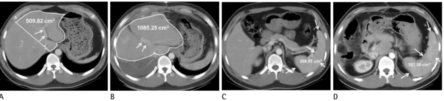
± 225.59 cm3 and the average resected liver volume (LVR) was 801.15 ± 138.29 cm3; the average remnant liver volume immedi- ately after donation (LV0) was 527.25 ± 166.85 cm3, and the liver volume 2 weeks after donation (LV2wks) was 782.48 ± 168.90 cm3 Fig. 1. CT scans of a 25-year-old male donor.
A, B. CT scans obtained preoperatively (A) and 14 days after the right hepatectomy (B) at the level of the left portal vein (arrows) show postop- erative enlargement of the remnant left hepatic lobe. The immediate postoperative remnant left hepatic lobe volume calculated by CT volumetry and intraoperative measurement was 509.82 cm3 (area encircled by a white line in A) and the left lobe volume measured 2 weeks after right hep- atectomy was increased to 1085.25 cm3 (area encircled by a white line in B).
C, D. CT images obtained preoperatively (C) and 14 days after right hepatectomy (D) show postoperative enlargement of the spleen (arrows). By CT volumetry, the preoperative splenic volume was 255.97 cm3 and the splenic volume 2 weeks after right hepatectomy was increased to 397.30 cm3.
A B C D
Fig. 2. The original liver volume versus the liver volume at 2 weeks af- ter donor hepatectomy.
Note.-LVW =whole liver volume at donation, LV2wks =liver volume at 2 weeks after hepatectomy
0 500 1000 1500 2000
Volume (cm3 )
LVw LV2wks
Mean, 1331.57 cm3
Mean, 782.48 cm3
cluding portal venous pressure, showed no correlation with the changes of the liver and spleen volumes (Table 2).
DISCUSSION
The liver has the ability to self-regenerate when there is loss of functional liver tissue. This property of the liver and recent ad- vances in surgical techniques have made LDLT feasible (1, 2).
LDLT can alleviate the problem of organ shortage that is due to a markedly limited cadaveric donor supply, and it can reduce mortality while patients are on the waiting list. In adult patients, a right lobe graft is mainly used to cope with the volume re- quirement for the recipient. The capability of the liver to regen- erate provides great advantages for living donor liver transplan- tation compared with living related kidney transplantation (12).
However, the process of regeneration is a complex one and it is still not completely understood (7). There have been many stud- ies on the regeneration of the liver after hepatectomy, but these have been done in diseased livers, and their results cannot be applied directly to living donor hepatectomy (13). The liver after liver resection for benign diseases of the liver can be a model for liver regeneration in normal human livers (14). Greene et al.
(15) demonstrated with an animal model experiment that endo- thelial cells are involved in the regulation of the regenerating adult liver, and they suggested that angiogenesis controls the re- (Table 1) (Fig. 4). The mean platelet count of donors at donation
(PLT0) was 255.54 × 10³/mm3 (normal range 150-400 × 103/ mm3), and the mean platelet count at 2 weeks (PLT2wks) was 268.12 × 10³/mm3. There was no statistical significance differ- ence in platelet levels pre-donation and at 2 weeks post-dona- tion (Table 1).
The remnant liver volume increment rate revealed a positive correlation with the resected-to-whole liver volume ratio (LVR/ LVW) (r = 0.759, p < 0.01) (Fig. 5). The other analyzed factors, in-
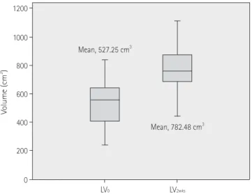
Fig. 3. Volume changes of the remnant liver. The remnant liver showed increment of volumes two weeks after hepatectomy (p < 0.01).
Note.-LV0 = remnant liver volume immediately after donation, LV2wks
=liver volume at 2 weeks after hepatectomy 0
400
200 600 800 1000 1200
Volume (cm3)
LV0 LV2wks
Mean, 527.25 cm3
Mean, 782.48 cm3
Table 1. Liver and Spleen Volume and Platelet Count Changes after Donor Hepatectomy
At Donation 2 Weeks after Donation Increment Rate (%) Paired t-Test Remnant liver volume 527.25 ± 166.85 cm3 782.48 ± 168.90 cm3 59.45 ± 50.52 p < 0.01
Spleen volume 168.23 ± 54.31 cm3 247.65 ± 86.33 cm3 47.94 ± 22.61 p < 0.01
Platelet count 255.54 × 10³/mm3 268.12 × 10³/mm3 0.41 ± 20.11 p > 0.05
Table 2. Correlation Analysis between Volume Changes and Assessed Factors
LVW LVR LVR/LVW SV0 LV0 IGC-R15 PLT0 PP0 PP1 %PP
Increase Lw + SV0 LVR/ (LVw + SV0)
%LV0 -0.451 0.155 0.759* 0.120 -0.718 -0.350 0.287 0.122 0.172 0.038 -0.385 0.478
increase 0.018 0.440 0.000 0.550 0.000 0.979 0.147 0.543 0.390 0.852 0.047 0.155
%SV -0.094 0.247 0.442 -0.077 0.442 0.152 -0.169 0.286 0.270 -0.003 -0.101 0.475
increase 0.641 0.215 0.021 0.704 0.021 0.458 0.399 0.759 0.172 0.989 0.616 0.012 Note.-Numbers are correlation coefficient and p-values.
*The remnant liver volume increment rate (%LV0 increase) revealed positive correlation with the resected-to-whole liver volume ratio (LVR/LVW) by Pearson's correlation analysis.
ICG R15 = indocyanine green retention rate at 15 minutes, LVR = resected liver volume, LVW = whole liver volume at donation, LV0 = remnant liver volume immediately after donation, LV2wks = liver volume at 2 weeks after hepatectomy, PLT0 = platelet count before hepatectomy, PLT2wks = platelet count at 2 weeks after hepatectomy, PP0 = portal vein pressure before hepatectomy, PP1 = portal vein pressure after hepatectomy, SV0 = spleen volume at donation, SV2wks = spleen volume at 2 weeks after hepatectomy, %LV0 increase = [(LV2wks - LV0) / LV0], %SV increase = [(SV2wks - SV0) / SV0]
tion of splenic engorgement (17). The results of several recent animal studies indicate that both the liver and spleen respond to the same growth factors, epidermal growth factor, transforming growth factor-a, or perhaps a combination of these factors (18- 21). In humans, the relationship between splenic enlargement and post-hepatectomy is rather complex and controversial.
Ando et al. (10) recently found that the increment in the splenic volume correlated well with the increment in the remnant liver generative process. After a partial hepactectomy, massive hepa-
tocyte proliferation is observed to begin immediately, peaking at 48 hours postoperatively. Endothelial cells, on the other hand, lag behind and peak only 4 days after the operation proper (15).
In our series, the mean liver volume as measured 2 weeks after hepatectomy was 58.99 ± 9.41% of the original liver volume.
This percentage was significantly lower when compared to the pre-donation volumes overall in all donors. Ibrahim et al. (7) re- ported that full liver regeneration was not attained at 6 months after donation in most donors. Some authors believe that the liver will enter into a slower pace of regeneration once it reaches an adequate hepatocyte mass to support normal metabolism (4- 6, 16). In this study, the increment rate of the remnant liver vol- ume showed a wide range (59.45 ± 50.52%). One of the patients in this study more than doubled their liver volume (222%) by 2 weeks after donor hepatectomy. In this patient, the resected-to- whole liver volume ratio (LVR/LVW) was the highest among the included patients (78.2%). Therefore, we presume this excep- tionally high resected-to-whole liver volume is the cause of the extremely high rate of volume increase.
Our study showed that the volume increment rate of the rem- nant liver 2 weeks after partial liver donation correlated not with the absolute resected liver volume (LVR) but with the relative re- sected liver volume compared to the whole liver (LVR/LVW).
These results suggest that the trigger for liver regeneration may be the relative shortage of liver tissue for supporting normal me- tabolism. However, our study showed that there was no correla- tion between portal venous pressure and the volume increment of the remnant liver. This result suggests that relative portal hy- pertension is not a major factor in triggering liver regeneration.
According to recent other studies, we presume that some other complex mechanism including humoral factors such as growth factors, including epidermal growth factor, and transforming growth factor-a, are involved in the process.
It has been known that there is splenic enlargement in pa- tients who undergo liver resection (7-9). This enlargement was most marked in patients with liver cirrhosis in whom hyper- splenism can be detrimental (9). Ando et al. (10) showed that the spleen enlarges as much as 155 ± 40% within 14 days after hepatectomy for biliary cancer. It has been conventionally con- jectured that the spleen enlarges as a consequence of relative portal hypertension, and that the enlargement is simply a reflec-
Fig. 4. Splenic volume change. There was a statistically significant in- crement in splenic volume at 2 weeks after liver donation (p < 0.01).
Note.-SV0 = spleen volume at donation, SV2wks = spleen volume at 2 weeks after hepatectomy
Fig. 5. The correlation between the increment rate of the remnant liv- er and relative volume of the resected liver to the whole liver. The per- cent remnant liver volume increase (%LV0 increase) revealed positive correlation with the resected-to-whole liver volume ratio (LVR/LVW) (r
= 0.759, p < 0.01).
Note.-LVR = resected liver volume, LVW = whole liver volume at donation, LV0 = remnant liver volume immediately after donation
0
0 200
50 100
100 300
150 400
200 500
250 Volume (cm3)%LV0 increase
SV0
40.0 50.0 60.0 70.0 80.0
SV2wks
%LVR/LVw Mean, 168.23 cm3
R = 0.759, p < 0.01
Mean, 247.65 cm3
useful both for candidates involved in liver donation as well as for patients undergoing other forms of hepatic surgery.
REFERENCES
1. Nadalin S, Bockhorn M, Malagó M, Valentin-Gamazo C, Frilling A, Broelsch CE. Living donor liver transplantation.
HPB (Oxford) 2006;8:10-21
2. Hardy KJ. Liver surgery: the past 2000 years. Aust N Z J Surg 1990;60:811-817
3. Bucher NL. Experimental aspects of hepatic regeneration.
N Engl J Med 1967;277:686-696 contd
4. Fausto N. Liver regeneration: from laboratory to clinic.
Liver Transpl 2001;7:835-844
5. Court FG, Wemyss-Holden SA, Dennison AR, Maddern GJ.
The mystery of liver regeneration. Br J Surg 2002;89:1089- 1095
6. Olthoff KM. Molecular pathways of regeneration and re- pair after liver transplantation. World J Surg 2002;26:831- 837
7. Ibrahim S, Chen CL, Wang CC, Wang SH, Lin CC, Liu YW, et al. Liver regeneration and splenic enlargement in donors after living-donor liver transplantation. World J Surg 2005;29:1658-1666
8. Zollinger RM, Zollinger RM Jr. Left hepatic lobectomy. In Zollinger RM, Zollinger RM Jr. Atlas of Surgical Operations, 4th ed. New York: Macmillian, 1990:172-175
9. Akimaru K, Onda M, Tajiri T, Yoshida H, Yokomuro S, Mama- da Y, et al. Hypersplenism induced by hepatectomy. Hepa- togastroenterology 2001;48:1170-1175
10. Ando H, Nagino M, Arai T, Nishio H, Nimura Y. Changes in splenic volume during liver regeneration. World J Surg 2004;28:977-981
11. Lemke AJ, Hosten N, Neumann K, Müller B, Neuhaus P, Fe- lix R, et al. [CT volumetry of the liver before transplanta- tion]. Rofo 1997;166:18-23
12. Marcos A, Fisher RA, Ham JM, Shiffman ML, Sanyal AJ, Luketic VA, et al. Liver regeneration and function in donor and recipient after right lobe adult to adult living donor liver transplantation. Transplantation 2000;69:1375-1379 13. Tanaka W, Yamanaka N, Oriyama T, Katoh T, Kuroda N, Oka-
moto E. Multivariate analysis of liver regenerative capacity volume and suggested the presence of a common growth factor.
Sato et al. (22), on the other hand, suggested that the percent in- crease in liver volume was inversely related to the spleen vol- ume. Both of the above studies were done in diseased livers with underlying parenchymal disease. Ibrahim et al. (7) reported that the spleen enlarges as the liver regenerates after donor hepatec- tomy. Ibrahim et al. (7) also reported that there was a strong correlation between the amount of the resected liver tissue and the size of the splenic enlargement. This favors the hypothesis that both organs are stimulated by the same growth factor, and in some donors, this process seems to be more active leading to greater enlargement of both the spleen and liver. However, our study results showed no correlation between the volume incre- ment rate of the remnant liver and that of the spleen. Further- more, the volume increment ratio of the spleen did not show any correlation with the portal venous pressures measured dur- ing the surgery, before clamping and after declamping of the main portal vein. We believe that these discrepancies could be explained by the multifactorial nature of liver and spleen regen- eration. Liver and spleen regeneration is not affected by a single factor but by many different physiological and anatomical ones that together form a web of complex relationships (7).
Our study has some limitations. Our study data was small, and only 27 cases were reviewed. Because this was a retrospec- tive analysis, there could be other factors such as growth factors acting on the liver and spleen causing volume enlargement or humoral factors that were not assessed in this series. One possi- ble relevant factor is the change in the donors’ body weight be- fore donation and 2 weeks after. This factor cannot be analyzed because there was no data on the donors’ body weight at 2 weeks post-donation in the electronic medical record database.
In conclusion, the spleen and remnant liver volumes were in- creased at CT volumetry performed 2 weeks after partial liver donation. Remnant liver regeneration after liver donation is cor- related with the relative resected liver volume as compared to the whole liver volume. The other analyzed factors, including the portal venous pressure, were not correlated with the liver or spleen volume changes. Liver and spleen regeneration is a high- ly complex process in humans. Many factors play a part in the physiological regeneration of the liver and spleen. More studies are warranted to define the possible presence of any other fac- tors that affect the rate of splenic regeneration. This would be
Nagasue N. Transforming growth factor-beta1 released from the spleen exerts a growth inhibitory effect on liver regeneration in rats. Lab Invest 2003;83:1595-1603 19. Kaido T, Oe H, Yoshikawa A, Okajima A, Imamura M. Ex-
pressions of molecules associated with hepatocyte growth factor activation after hepatectomy in liver cirrhosis.
Hepatogastroenterology 2004;51:547-551
20. Rosenkranz E, Charters AC 3rd, Orloff MJ. Regeneration in rat liver injured by carbon tetrachloride. Surg Forum 1975;
26:411-412
21. Tomiya T, Tani M, Yamada S, Hayashi S, Umeda N, Fujiwara K. Serum hepatocyte growth factor levels in hepatecto- mized and nonhepatectomized surgical patients. Gastro- enterology 1992;103:1621-1624
22. Sato K, Tanaka M, Tanikawa K. The effect of spleen volume on liver regeneration after hepatectomy--a clinical study of liver and spleen volumes by computed tomography.
Hepatogastroenterology 1995;42:961-965 after hepactectomy in humans. J Hep Bil Pancr Surg 1997;
4:78-82
14. Yamanaka N, Okamoto E, Kawamura E, Kato T, Oriyama T, Fujimoto J, et al. Dynamics of normal and injured human liver regeneration after hepatectomy as assessed on the basis of computed tomography and liver function. Hepa- tology 1993;18:79-85
15. Greene AK, Wiener S, Puder M, Yoshida A, Shi B, Perez- Atayde AR, et al. Endothelial-directed hepatic regeneration after partial hepatectomy. Ann Surg 2003;237:530-535 16. Nakagami M, Morimoto T, Itoh K, Arima Y, Yamamoto Y,
Ikai I, et al. Patterns of restoration of remnant liver vol- ume after graft harvesting in donors for living related liv- er transplantation. Transplant Proc 1998;30:195-199 17. Nagasue N, Yukaya H, Ogawa Y, Higashi T. Portal pressure
following partial to extensive hepatic resection in patients with and without cirrhosis of the liver. Ann Chir Gynaecol 1983;72:18-22
18. Ueda S, Yamanoi A, Hishikawa Y, Dhar DK, Tachibana M,
생체 간이식을 위한 부분 간이식 절제술 후 간과 비장의 부피 변화에 영향을 주는 요인들1
배지혜
1· 염헌규
1· 송정흡
2목적: 생체 기증 간이식을 위해 부분 간 절제 후 수술 후 초기에 간과 비장의 변화에 대해 알아보고 간과 비장의 부피 변 화에 영향을 주는 인자를 알아보고자 하였다.
대상과 방법: 부분 간 절제술을 시행한 총 27명의 기증자가 이 연구에 포함되었다. 간과 비장의 부피 변화의 비율은 CT 볼륨메트리를 이용해 측정하였고 여러 가지 요인들과의 상관관계를 알아보았다. 분석된 여러 요인들로는 indocyanine green (이하 ICG) 시약 주입 후 15분 후 ICG 시약 정체 비율, 수술 전 혈소판 수치, 수술 전 간과 비장의 부피, 절제된 간 의 부피, 전체 간의 부피 중 절제된 간의 부피 비율, 전체 간과 비장의 부피 중 절제된 간의 부피 비율과 부분 간 절제 수술 전 그리고 수술 후 문맥압의 변화에 대해 연구하였다.
결과: 모든 부분 간 기증자에게서 간 절제 후 남아있는 간과 비장의 부피는 증가되었다(간과 비장의 부피 증가 비율은 각 각 59.5 ± 50.5% 그리고 47.9 ± 22.6%). 남아있는 간의 부피 증가 비율은 전체 간의 부피 중 절제된 간의 부피 비율 과 양성 상관관계를 나타내었다(r = 0.759, p < 0.01). 그러나 분석된 다른 요인들은 간 절제 후 남아있는 간과 비장의 부피변화와 상관관계가 없었다.
결론: 부분 간 기증 후에 잔존 간과 비장의 부피는 2주 후 측정에서 증가하였다. 여러 분석된 요인들 중 전체 간의 부피 중 절제된 간의 부피 비율이 남아있는 간 부피 증가 비율에 영향을 주었다.
경북대학교병원 1영상의학과, 2산업의학과