- 61 -
KISEP Case Reports J Rhinol 4((((1)))), 1997
A Case of Oncocytic Schneiderian Papilloma in the Maxillary Sinus
Hun-Jong Dhong, M.D.
1, Dong-Soo Han, M.D.
1, Kwang-Chol Chu, M.D.
1and Young-Hyeh Ko, M.D.
2 ABSTRACTOncocytic Schneiderian papilloma (OSP) is a rare neoplasm of the nose and paranasal sinuses, and is often mistaken histo- logically for papillary adenocarcinoma or even rhinosporidiosis. Recently, we experienced a case of oncocytic Schneiderian papilloma of the left maxillary sinus developed in 53-year-old female patient. She complained of unilateral nasal obstruction, bloody rhinorrhea and frontal headache. The tumor mass was resected surgically through intranasal endoscopic and Caldwell- Luc’s approach. We report its clinical manifestations and histologic characteristics.
KEY WORDS:Oncocytic schneiderian papilloma·Maxillary sinus.
INTRODUCTION
Papillomas which occur in the nasal cavity and the para- nasal sinuses are classified into everted squamous papilloma, inverted papilloma, and oncocytic Schneiderian papilloma (OSP) according to their pathophysiologic manifestation.1)2) OSP is a rarely found benign tumor occurring in the nasal cavity and the paranasal sinus with main symptoms such as unilateral nasal obstruction, epistaxis and pain. Its clinical ch- aracteristics include a high recurrence rate, a tendency for destroying peripheral tissue and transforming into malignancy, which are similar to those of a malignant tumor.3)4)5) The aut- hors report a recent case of OSP in the maxillary sinus which was admitted to the hospital complaining of unilateral epista- xis and headache as main symptoms and completely recovered after surgery.
CASE REPORT
A 53-year-old woman presented with a 2-year history of left-sided rhinorrhea and nasal obstruction. She had previously been diagnosed as chronic paranasal sinusitis by the general practitioner that was refractory to antibiotic therapy. Frontal
headache and occasional left-sided epistaxis developed over the previous several months.
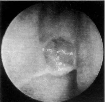
Physical examination revealed the presence of a polypoid mass filling the middle meatus of the left nasal cavity. Nasal endoscopic examination revealed that the left middle meatus was filled with a papillomatous mass and muco-sanguineous secretion (Fig. 1). The uncinate process was displaced in an- terior and medial directions by the tumor, and the protruding tumor showed a tendency of easy bleeding. The left middle turbinate was also partially displaced to the nasal septum. The inferior turbinate and the choana seemed to be intact. Tissue biopsy conducted at the first examination revealed chronic inflammation.
A coronal computed tomography scan revealed the homo- geneous soft tissue mass which filled the left middle meatus and the maxillary sinus. The left uncinate process and the medial wall of the left maxillary sinus showed a partial bony destruction by the tumor together with expansion of the adj- acent bone tissue (Fig. 2).
At operation, the mass originated from the natural ostium of the left maxillary sinus and the medial wall of the maxillary sinus showed a partial bony erosion. There was no specific finding in the infraorbial wall and the inferior wall of maxi- llary sinus. After the mass in the left middle meatus and the bone around the natural ostium was removed the mass in the maxillary sinus was removed. The mass contained abscess, and looked like granulation tissue with a fragile and easily ble- eding tendency. The frozen biopsy revealed co-existence of everted squamous and inverted papillomas. To remove the mass in the maxillary sinus which could not be approached by endoscopy, we employed Caldwell-Luc’s approach which allowed access to the anterolateral wall of the maxillary sinus Department of 1Otorhinolaryngology-Head and Neck Surgery,
2Diagnostic Pathology, College of Medicine, Sungkyunkawn University, Seoul, Korea
Address correspondence and reprint requests to Hun-Jong Dh- ong, M.D., Department of Otorhinolaryngology-Head and Neck Surgery, Samsung Medical Center, 50 Il won-dong Seoul 135- 230, Korea
Tel:82-3410-3573, Fax:82-3410-3879 Accepted for publication February 12, 1997
62 / J Rhinol 4(1), 1997
and completely removed the mass in the left maxillary sinus.
The mass looked soft with a relatively smooth surface of weak pinkish and yellow-white colors. The masses in the na- sal cavity and the maxillary sinus examined by microsopy showed everted squamous and inverted papilloma (Fig. 3).
The mass was lined with pseudostratified columnar epithelium consisting of small, thick and circular nuclei without dysplasia and granular eosinophilic cytoplasm. Mucous secretions were scattered on the mass (Fig. 4).
On the follow-up examinations, the patient recovered wit- hout recurrence of the intranasal and intramaxillary mass for 12 months after the surgery.
DISCUSSION
The papilloma occurring in the nasal cavity and the para- nasal sinuses is a neoplasm found in the epithelium and can
Fig. 2. A coronal CT scan showing large expansile soft-tissue density in the left nasal cavity and maxillary sinus, and medially displaced medial wall of the maxillary sinus.
Fig. 1. Preoperative endoscopic finding showing inflammatory polypoid masses in the middle meatus.
Fig. 4. Light micrograph showing epithelium composed of irre- gularly distributed strips of eosinophilic cells with interspersed mucous cells (H & E stain, ×200).
Fig. 3. Light micrograph showing characteristic exophytic (A) and inverted (B) growth pattern of oncocytic Schneiderian papilloma (H & E stain, ×100).
Dhong et al.:Oncocytic Schneiderian Papilloma / 63
be classified into the following three types:(1) everted sq- uamous papilloma, exophytic, septal, squamous, or fungiform papilloma (2) inverted papilloma, Schneiderian, transitional cell, or Ringertz papilloma, and (3) oncocytic Schneiderian papilloma, cylindrical cell papilloma, or microcystic papillary adenoma.2)3)4)6)7) Oncocytic Schneiderian papilloma (OSP) had been called ‘cylindrical cell papilloma’ but was later named ‘oncocytic Schneiderian papilloma’ since Barnes and Bedetti3) proved that the epithelia of cylindrical cell papillo- ma are true oncocytes which originate in the nasal cavity and paranasal sinus mucosae in terms of their immunohistoche- mical, histochemical and ultrastructural aspects.
The incidence of papilloma in the nasal cavity and para- nasal sinuses is 4-4.7%. Among them, everted papilloma and inverted papilloma account for 45-50% each, while OSP is rarely reported at a rate of 3-5%.1)3)
The papilloma can occur at all age, but everted papilloma and inverted papilloma are reportedly found frequently in males of 40-50 years of age.1)8)9) Compared with inverted papilloma, OSP shows no sexual difference in occurrence, but has frequent occurrence after the fifth decade.1)3)7)10)
Like other papillomas, the cause of OSP has not been clar- ified, but viral infection, allergy, chronic inflammation, smo- king and environmental carcinogens are suggested as possible causes.3)4)
The most common early symptom of OSP is unilateral na- sal obstruction with intermittent epistaxis and pain which may last from several months to years, while rhinorrhea, paranasal sinustis and allergic symptoms may manifest.4)7)10) Our case showed unilateral nasal obstruction and rhinorrhea at the ea- rly stage, later accompanied by intermittent epistaxis and he- adache.
The mass originating in the maxillary sinus or the lateral nasal wall shows smooth, edematous and polypoid manifest- ations on physical examination, which can be confused with an inflammatory polyp, and exhibit varied colors of red, brown, pink, grey and yellow.3)4)5)6)7) In our case, the tumor was obs- erved as a pinkish polypoid mass with mucoid, bloody sec- retions in the left middle meatus. Bony destruction was not observed during surgery.
More than one paranasal sinus in the unilateral paranasal sinuses showed haziness and soft tissue density through a si- mple X-ray examination.3) Most CT and MRI scans reveal masses originating from the unilateral nasal cavity side wall or paranasal sinus which showes enhancement. If the tumors expanded, displacement of structures adjacent to the tumor, thinning of the medial wall of the paranasal sinus and bony destruction are observed.5)7) In our case, displacement of the uncinate process, and medial wall of the maxillary sinus and partial bony destruction in the posterior wall of the maxillary
sinus were suspected. Chronic maxillary sinusitis, mucocele, and lymphoma should be considered as possible radiological diagnosis. Fungal sinusitis can be differentiated by presence of calcification.
Microscopically, under low power, OSP has a characteristic everted and inverted growth pattern. In our case, histological findings showed highly increased vascularity, surrounding str- oma and everted and inverted growth patterns. The tumors are usually lined with pseudostratified columnar cells of 2-8 layers and consists of single, small, thick circular, or oval nuclei, and granular cytoplasm. On the epithelium, cilia may exist and intraepithelial microabscess and mucin-filled micr- ocysts are commonly observed.3)4)5)6)7) Differential diagnosis from adenocarcinoma and rhinosporidiosis is also important.1) Discrimination is possible because adenocarcinoma consists of well-differentiated epithelial cells in a monolayer, while OSP consists of multi-layered epithelia.4) OSP should be hi- stologically differentiated from rhinosporidiosis because of a pathologic finding due to the microcystic structure of the ep- ithelium.1)4)
In principle, the treatment should be complete removal of the tumor by exposing its entire mass since may be associated with or develop into malignant tumors. Medial maxillectomy through lateral rhinotomy or radical excision through midfa- cial degloving approach have been attempted.4)7)10) In our case, we employed intranasal endoscopic and Caldwell-Luc’s app- roaches to completely resect the tumors. Radical maxillectomy or postoperative radiotherapy are planned according to the extension of the tumor.4) Radiation therapy can be employed in case it is accompanied by malignant tumor, but is not eff- ective for OSP.1)
Hyams1) reported postoperative recurrence in 40% of 10 cases, and Barnes and Bedetti3) reported 25-35% recurrence rate after an incomplete resection through an intranasal ap- proach and 0-15% after exposing the lesion though lateral rhinotomy and resecting the medial wall of the maxillary sinus en bloc. Clinical characteristics of malignant transfor- mation and recurrence of OSP is similar to inverted papill- oma.4)5)7)11) In our case, the tumor was completely resected through intranasal endoscopic and Caldwell-Luc’s approaches.
The maxilliary sinus mucosa restored to normal without evi- dence of recurrence after 12 months of follow-up.
CONCLUSION
The authors have recently experienced a case of oncocytic Schneiderian papilloma which occurred in the nasal cavity and paranasal sinuses and obtained favorable surgical outcome by removing the tumor through intranasal and Caldwell-Luc’s approaches.
64 / J Rhinol 4(1), 1997
REFERENCES
1) Hyams VJ. Papillomas of the nasal cavity and paranasal sinuses:
A clinicopathologic study of 315 cases. Ann Otol Rhinol Laryngol 1971;180:192-206.
2) Michaels L, Young M. Histogenesis of papillomas of the nose and paranasal sknuses. Arch Pathol Lab Med 1995;119:821-6.
3) Barnes L, Bedetti C. Oncocytic Schneiderian papilloma: A reapp- raisal of cylinderical cell papilloma of the sinonasal tract. Hum Pathol 1984;15:344-51.
4) Cunningham MJ, Brantley S, Barnes L, Schramm VL. Oncocytic Schneiderian papilloma in a young adult: A rare diagnosis. Oto- laryngol Head Neck Surg 1987;97:47-51.
5) Kapadia SB, Barnes L, Pelzman K, Mirani N, Heffner DK, Bedetti C. Carcinoma ex oncocytic Schneiderian (cylindrical cell) papil-
loma. Am J Otolaryngol 1993;14:332-8.
6) Batsakis TG. Squamous cell Papillomas of the oral cavity, sinon- asal tract and larynx. In: Tumors of the head and neck. 2nd ed, Baltimore, Williams, 1979:130-7.
7) Frantz TD, Rasgon BM, Rumore GJ. Pathologic quiz case: Onco- cytic schneiderian papilloma. Arch Otolaryngol Head Neck Surg 1994;120:102-6.
8) Ahn HY, Yeo SK, Seok SY, Hong NP, Cho JS, Cha CI. Inverted papilloma and malignant transformation. Korean J Otolaryngol 1994;37:306-15.
9) Min YG, Hong SH, Kim HJ, Rhee CS. A clinical study on inverted papilloma of the nose and paranasal sinuses. Korean J Otolaryngol 1991;34:962-7.
10) Kennedy KS. Cylindrical cell papilloma of the maxillary sinus. Am J Rhinol 1990;4:185-8.
11) Ward BE, Fechner RE, Mills SE. Carcinoma arising in oncocytic Schneiderian papilloma. Am J Surg Pathol 1990;14:364-9.