GSTM1 Tissue Genotype as a Recurrence Predictor in Non- muscle Invasive Bladder Cancer
Tissue genotyping is more useful approach than using blood genomic DNA, which can reflect the effects of the somatic mutations in cancer. Although polymorphisms in glutathione S-transferase (GST ) have been associated with the risk of bladder cancer (BC) development, few reports provide information about the prognosis of BC. We investigated glutathione S-transferase mu (GSTM1) and glutathione S-transferase theta (GSTT1) genotypes using genomic DNA from primary 165 BC tissue samples to assess the association with disease prognosis. DNA samples from tumor were analyzed by multiplex polymerase chain reaction (PCR). The results were compared with clinicopathological parameters. The prognostic significance of the GSTs was evaluated by Kaplan-Meier and multivariate Cox regression model. Kaplan-Meier estimates revealed significant differences in time to tumor recurrence according to the GSTM1 tissue genotype (P = 0.038) in non- muscle invasive bladder cancer (NMIBC). Multivariate Cox regression analysis also revealed that the tissue GSTM1 genotype (hazards ratio [HR]: 0.377, P = 0.031) was an
independent predictor of bladder tumor recurrence in NMIBC. This identification of GSTM1 tissue genotype as a prognosticator for determining recurrence in NMIBC should prove highly useful in a clinical setting.
Key Words: Urinary Bladder Neoplasms; Tumor Markers, Biological; GSTM1; Glutathione S-Transferase T1
Yun-Sok Ha1, Chunri Yan1,2, Pildu Jeong1, Won Tae Kim1, Seok-Joong Yun1, Isaac Yi Kim3, Sung-Kwon Moon4, and Wun-Jae Kim1,2
1Department of Urology, College of Medicine, Chungbuk National University, Cheongju; 2BK21 Chungbuk Biomedical Science Center, School of Medicine, Chungbuk National University, Cheongju, Korea; 3Section of Urologic Oncology, The Cancer Institute of New Jersey, Robert Wood Johnson Medical School, New Brunswick, New Jersey, USA;
4Department of Food and Biotechnology, Chungju National University, Cheongju, Korea
Received: 3 November 2010 Accepted: 24 December 2010 Address for Correspondence:
Wun-Jae Kim, MD
Department of Urology, Chungbuk National University Hospital, 410 Seongbong-ro, Heungdeok-gu, Cheongju 361-711, Korea Tel: +82.43-269-6371, Fax: +82.43-269-6144
E-mail: [email protected]
This study was supported by a grant of Korea Healthcare technology R & D Project, Ministry of Health & Welfare, Republic of Korea (A100651-1011-0000100).
DOI: 10.3346/jkms.2011.26.2.231 • J Korean Med Sci 2011; 26: 231-236 Oncology & Hematology
INTRODUCTION
Bladder cancer (BC) involves a heterogeneous cell population, and numerous factors are likely to be involved in tumorigenesis (1). These factors result in uncontrolled growth of the cell popu- lation, decreased cell death, invasion and metastasis, and may influence the patient’s prognosis. Identification of the aggres- sive features of the cancer in patients with BC is very important for adequate management of this disease (2). While several mo- lecular markers for the development, recurrence and progres- sion of BC have been identified (3, 4), the limited value of these established prognostic markers has emphasized the need for novel molecular indicators of BC outcomes.
Previous molecular epidemiologic studies have revealed that polymorphisms in glutathione S-transferase-mu (GSTM1) and glutathione S-transferase-theta (GSTT1) have been implicated as risk factors in BC (5, 6). Furthermore, a small number of re- ports have added valuable information towards our understand- ing of the clinicopathologic characteristics of BC (7, 8).
Several environmental factors, as well as the activities of de-
toxification enzymes, have been associated with tumor devel- opment in BC, and could also affect the clinical course of this disease. To date, there has been no published report describing the prognostic value of homozygous deletions of GSTM1 and GSTT1 in tumor tissues as markers of disease prognosis in pri- mary BC patients with long-term follow-up. This study is the first and the only study that used tumor tissue to analyze the GSTM1 and GSTT1 genotypes to evaluate the prognosis of BC instead of blood genomic DNA. In the current study, we identi- fied the GSTM1 tissue genotype as a prognosticator of recurrence in patients with primary non-muscle invasive bladder cancer (NMIBC).
MATERIALS AND METHODS Patients and tissue samples
We used primary BC tissue samples taken all from the initial primary tumor resection (no prior intravesical therapy or sys- temic adjuvant chemotherapy) from 165 consecutive cases of patients with histologically-verified transitional cell carcinoma,
which were obtained from Chungbuk National University Hos- pital between 1995 and 2007. To reduce confounding factors that may affect the analyses, any patients diagnosed with con- comitant carcinoma in situ (CIS) lesion or CIS lesion alone were excluded. All tumors were macro-dissected, typically within 15 min of surgical resection. Each BC specimen was confirmed by pathological analysis of a part of the tissue sample in fresh-fro- zen sections from cystectomy and transurethral resection (TUR) specimens, and then frozen in liquid nitrogen and stored at -80°C until use.
Tumors were staged according to the 2002 TNM classifica- tion (9) and the 1973 WHO grading system (10). In the case of NMIBC, TUR of the tumor was performed. A second TUR was performed 2-4 weeks after the initial resection when a BC spec- imen did not include proper muscle or when a high-grade was detected (11). Patients who had multiple tumors, large tumors (≥ 3 cm in diameter), or high grade NMIBC received one cycle of intravesical treatment (Bacillus Calmette–Guérin [BCG] or mitomycin-C) (11, 12). Response to treatment was assessed by cystoscopy and urinary cytology. Patients who were free of dis- ease within 3 months of commencement of treatment were as- sessed every 3 months for the first 2 yr and every 6 months there- after (11, 12). We defined recurrence as the relapse of primary NMIBC with a lower or equivalent pathologic stage, and pro- gression as disease with a higher TNM stage when relapsed. In cases of muscle-invasive bladder cancer (MIBC), radical cys- tectomy and complete pelvic lymph node dissection were per- formed. Patients with pT3, pT4, or node-positive disease, based on the analysis of radical cystectomy specimens, received at least four cycles of cisplatin-based chemotherapy. Either clini- cally metastatic disease, or non-cystectomy cases were exclud- ed in this study. Each patient has been followed and managed according to the standard recommendation (13). Among MIBC cases, we defined progression as local regional recurrence or metastasis after radical cystectomy and adequate adjuvant che- motherapy.
DNA extraction and multiplex PCR
Genomic DNA was isolated from frozen tumor tissue specimens using the Wizard Genomic DNA Purification System Kit (Pro- mega, Maddison, WI, USA) in accordance with the manufac- turer’s instructions. A multiplex PCR method was used to detect the presence or absence of the GSTM1 and GSTT1 polymor- phisms in genomic DNA samples, as described previously (14).
The primers used in the PCR were: 1) GSTM1 (219 bp) sense (5´- GAA CTC CCT GAA AAG CTA AAG C-3´) and anti-sense (5´-GTT GGG CTC AAA TAT ACG GTG G-3´); 2) GSTT1 (372 bp) sense (5´-TTA GCT GAC CTC GTA GCC AT-3´) and anti-sense (5´-GAA GTC CTT GGC CTT CAG AA-3´); and 3) β-globin (268 bp) sense (5´-GAA GAG CCA AGG ACA GGT AC-3´) and anti-sense (5´-CAA CTT CAT CCA CGT TCA CC-3´). DNA (200 ng) was amplified in
a total volume of 20 μL, containing 10 pM of each primer, 0.5 unit of Taq polymerase, 2.5 mM dNTP, and 10 × PCR buffer. Follow- ing an initial denaturation step at 94°C for 5 min, 40 cycles of am- plification were performed at: 1) 94°C for 60 sec; 2) 63°C for 60 sec; and 3) 72°C for 60 sec. A final extension step was then per- formed at 72°C for 10 min. Confirmation of PCR reactions was performed by electrophoresis of the amplified products on a 2%
agarose gel. Positive and negative control samples were analyzed in each experiment. GSTM1 and GSTT1 genotypes were only scored in the presence of the internal reference gene (β-globin) product.
Statistical analysis
The chi-square test and Fisher’s exact test were used to analyze the frequencies of the genotypes according to clinicopathologi- cal parameters. The Kaplan-Meier method was used to esti- mate time to recurrence and progression, and differences were assessed using log-rank statistics. The prognostic value of the GSTM1 and GSTT1 genotypes for recurrence and progression of BC was analyzed with multivariate Cox proportional hazard regression models. Statistical analysis was performed using SPSS 12.0 software (SPSS Inc., Chicago, IL, USA), and a P value of
< 0.05 was considered statistically significant.
Ethics statement
The collection and analysis of all samples was approved by the institutional review board of Chungbuk National University (IRB approved number 2006-01-001), and informed consent was obtained from each subject.
RESULTS
Baseline characteristics
Table 1 lists the baseline characteristics of the 165 patients en- rolled in this study. The median follow-up period of the primary
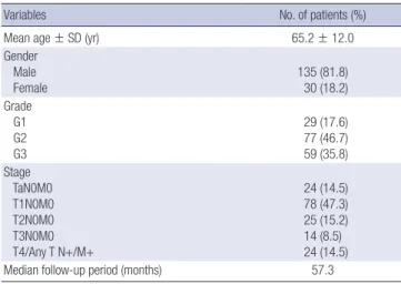
Table 1. Baseline characteristics
Variables No. of patients (%)
Mean age ± SD (yr) 65.2 ± 12.0
Gender Male Female
135 (81.8) 30 (18.2) Grade
G1 G2 G3
29 (17.6) 77 (46.7) 59 (35.8) Stage
TaN0M0 T1N0M0 T2N0M0 T3N0M0 T4/Any T N+/M+
24 (14.5) 78 (47.3) 25 (15.2) 14 (8.5) 24 (14.5)
Median follow-up period (months) 57.3
SD, standard deviation.
BC patients was 57.3 months (range, 1.0-161.4). Total 20 of 102 patients (19.6%) received the repeat TUR for the adequate tu- mor staging. Intravesical therapy was performed in 50 patients (49.0%) after primary TUR including 39 patients treated with BCG, and 11 with mitomycin-C. In our study, patients who com- pleted 6 times of intravesical therapy were counted in number.
Fig. 1 shows 2% agarose gel demonstrating multiplex PCR ge- notyping of genomic DNA samples for the detection of GSTM1 and GSTT1 gene deletion. The frequencies of the GSTM1-null and GSTT1-null types were 69.1% and 47.9%, respectively.
Relationship between GSTM1 and GSTT1 genotype, and clinicopathological parameters
Table 2 summarizes the correlations between genotype and clinicopathological parameters in patients with NMIBC. The GSTM1-null genotype was observed in 30 of 36 NMIBCs (83.3%) with recurrence and in 41 of 66 (62.1%) without recurrence, which represented a statistically significant difference (P = 0.026).
No significant correlations were observed between GSTM1 gen-
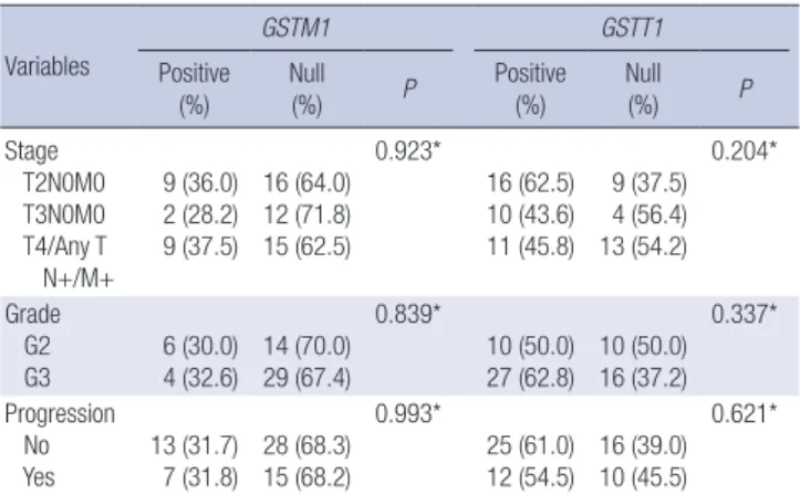
otype and the other clinicopathological parameters such as tu- mor stage, grade, tumor size, multiplicity and progression. The GSTT1-positive genotype was more frequently detected in large- sized (≥ 3 cm) than in small-sized NMIBC (< 3 cm) (28 of 46 or 60.9% vs 21 of 56 or 37.5%, P = 0.019). The other parameters were not significantly associated with GSTT1 genotype. Associations between the genotype of GSTM1 or GSTT1 and clinicopatho- logical features of MIBC were not found (Table 3).
Association between genotype of GSTM1 with recurrence of NMIBC
Kaplan-Meier estimates revealed a significant difference in time to tumor recurrence according to the GSTM1 genotype (log-rank test, P = 0.038, Fig. 2). However, recurrence-free survival was not related to the GSTT1 genotype. Multivariate Cox regression analysis revealed that the GSTM1 genotype was an independent predictor of bladder tumor recurrence (hazard ratio, 0.377; 95%
372 bp
M 1 2 3 4
268 bp 219 bp Fig. 1. Electrophoretic findings of GSTM1 and GSTT1, Lane M: molecular size marker (100 bp DNA ladder); Lane 1: GSTM1-null/GSTT1-null type; Lane 2: GSTM1-null/
GSTT1-positive type; Lane 3: GSTM1-positive/GSTT1-null type; Lane 4: GSTM1-posi- tive/GSTT1-positive type.
Table 2. Relationship between GSTM1 and GSTT1 genotypes, and clinicopathological parameters in non-muscle-invasive bladder cancer
Variables
GSTM1 GSTT1
Positive (%) Null
(%) P Positive
(%) Null
(%) P
Stage Ta T1
9 (37.5) 22 (28.2)
15 (62.5) 56 (71.8)
0.387*
15 (62.5) 34 (43.6)
9 (37.5) 44 (56.4)
0.105*
Grade G1 G2 G3
9 (31.0) 19 (33.3) 3 (30.0)
20 (69.0) 38 (66.7) 13 (70.0)
0.532*
16 (55.2) 25 (43.9) 8 (50.0)
13 (44.8) 32 (56.1) 8 (50.0)
0.602*
Size < 3 cm
≥ 3 cm 16 (28.6) 15 (32.6) 40 (71.4)
31 (67.4) 0.659*
21 (37.5)
28 (60.9) 35 (62.5) 18 (39.1)
0.019*
Number Single
Multiple 18 (29.5) 13 (31.7) 43 (70.5)
28 (68.3) 0.813*
30 (49.2)
19 (46.3) 31 (50.8) 22 (53.7)
0.778*
Recurrence No
Yes 25 (37.9) 6 (16.7) 41 (62.1)
30 (83.3) 0.026*
29 (43.9)
20 (55.6) 37 (56.1) 16 (44.4)
0.262*
Progression No
Yes 29 (32.2) 2 (16.7) 61 (67.8)
10 (83.3) 0.337†
42 (46.7)
7 (58.3) 48 (53.3) 5 (41.7)
0.447*
*Chi-Square test; †Fisher’s Exact Test. GSTM1, Glutathione S-transferase-mu 1; GSTT1, Glutathione S-transferase-theta 1.
Table 3. Relationship between GSTM1 and GSTT1 genotypes, and clinicopathological parameters in muscle invasive bladder cancer
Variables
GSTM1 GSTT1
Positive (%) Null
(%) P Positive
(%) Null
(%) P
Stage T2N0M0 T3N0M0 T4/Any T N+/M+
9 (36.0) 2 (28.2) 9 (37.5)
16 (64.0) 12 (71.8) 15 (62.5)
0.923*
16 (62.5) 10 (43.6) 11 (45.8)
9 (37.5) 4 (56.4) 13 (54.2)
0.204*
Grade G2
G3 6 (30.0)
4 (32.6) 14 (70.0) 29 (67.4)
0.839*
10 (50.0)
27 (62.8) 10 (50.0) 16 (37.2)
0.337*
Progression No
Yes 13 (31.7)
7 (31.8) 28 (68.3) 15 (68.2)
0.993*
25 (61.0)
12 (54.5) 16 (39.0) 10 (45.5)
0.621*
*Chi-Square test. GSTM1, Glutathione S-transferase mu 1; GSTT1, Glutathione S- transferase theta 1.
Fig. 2. Kaplan-Meier estimate curves predict the probability of recurrence according to the GSTM1 genotype in patients with non-muscle invasive bladder cancer.
Recurrence free survival
Time (Months)
Log-rank test, P = 0.038
GSTM1 positive
GSTM1 null
0.0 50.0 100.0 150.0
1.0
0.8
0.6
0.4
0.2
0.0
confidence interval, 0.156-0.914; P = 0.031, Table 4).
DISCUSSION
The activities of specific enzymes can change with genotype.
GSTM1 and GSTT1 are involved in cellular metabolism and detoxification of carcinogenic products, and these detoxifica- tion related-enzymes are associated with tumorigenesis of BC (5, 6, 15, 16). Specific enzymes that are known to be important in carcinogenesis can also play a critical role in disease recur- rence and progression after initial treatment.
In this study, we investigated the GSTM1 and GSTT1 geno- types in human primary BC tissues. The results demonstrated that the GSTM1 tissue genotype was a strong indicator for pre- dicting recurrence in patients with primary NMIBC. However, the GSTM1 genotype was not associated with tumor progres- sion. Previous blood genotype studies revealed that individuals with the GSTM1-null genotype were at great risk of developing BC (5, 6, 16). These results imply that the GSTM1-null genotype may contribute to the initiation of tumorigenesis in BC simply by promoting cell proliferation, rather than by the generation of certain aspects of the aggressive phenotype such as progression and invasion. There have been several studies aimed at identi- fying the biologic potential of bladder tumors, which may help to better predict the clinical outcome of BC, including recurrence and progression (17-20). Since the molecular genetic aspects are different between recurrence and progression in NMIBC (21-23), it seems necessary to discriminate these two pathogen- eses. Grossman et al. reported that p53 and retinoblastoma (RB) expression in T1 NMIBC can be used to predict progression only, and not recurrence without progression (19). Kim et al. (20) suggested that RUNX3 methylation was correlated with disease progression but did not detect a statistically significant associa- tion between RUNX3 methylation and recurrence in NMIBC. A small number of reports have described tumor markers related to recurrence without progression in NMIBC (17, 18), however, cell cycle markers were reported to provide no added prognos- tic information on tumor recurrence in NMIBC (17). Van Rhijn et al. (18) suggested that FGFR3 mutation was a strong indica-
tor for predicting recurrence in NMIBC. However, the design of that study carried limitations in terms of a relatively small sam- ple size and in including both primary and recurrence cases. In the present study, we used only 165 primary BC tissues with long-term follow-up. In our data, the conventional prognostic factors such as multiplicity, tumor size, stage and grade did not related to the recurrence in NMIBC on multivariate analysis.
Although we do not know the exact reason, one possibility is that the patients at high risk of recurrence or progression un- derwent second TUR or intravesical therapy, and these adju- vant treatments might act as the confounding effects against the conventional prognostic factors.
Most of the single nucleotide polymorphism (SNP) studies of GSTM1 and GSTT1 have been case control studies, or cross-sec- tional studies based on epidemiology. There are several distinc- tive aspects of the current study compared with previous SNP studies. First, we used genomic DNA from 165 primary BC tissue samples for the analysis. To date, there has been no study using tumor tissues to identify the genotype of GSTM1 and GSTT1.
DNA from tumor tissues represents the effects of the somatic mutations, alterations occurring in tumor DNA could affect the gene expression. We have the expression analysis data for GSTM1 and GSTT1 using tumor DNA. The mRNA expression of GSTM1 and GSTT1 was higher in BC tissues with positive genotypes than in those with null genotypes (24). It also revealed that tu- mor genotype might affect the expression of the specific gene.
In these regards, we suggest that using genomic DNA from tu- mor tissues to the genotypes of GSTM1 and GSTT1 is a more important and useful approach than using genomic DNA from blood which could also be affected by changes in the patient’s health status originating from other diseases unrelated to the cancer that investigators are attempting to analyze. Second, we performed definitive subgroup analysis and survival analyses such as Kaplan-Meier and Cox regression analysis with long- term follow-up. Only a small number of studies have reported GSTM1 and GSTT1 genotype according to the stage and grade of the BC (7, 8, 25), and the results were inconsistent among the different ethnic groups. We analyzed all the factors affecting BC prognosis, including stage, grade, multiplicity and size, in con- junction with GSTM1 and GSTT1 tissue genotypes. We found that the GSTT1-positive genotype was more frequently detected in large-sized (≥ 3 cm) than in small-sized NMIBC (< 3 cm). This finding supports our previous data showing that a GSTT1-null genotype is not a risk factor, but a protective factor, for BC (15).
The GSTT1-positive genotype may reflect the aggressiveness of NMIBC, but further investigation to confirm this proposal will be needed.
Frequent recurrence and progression are devastating events for both urologists and BC patients. The high incidence of recur- rence results in considerable costs that make NMIBC one of the most expensive diseases to treat (26). Progression from NMIBC Table 4. Multivariate Cox regression analysis for prediction of recurrence in non-muscle
invasive bladder cancer
Variables HR (95% CI) P
Age 0.991 (0.965-1.017) 0.476
Sex (male vs female) 1.099 (0.449-2.686) 0.836
Stage (Ta vs T1) 1.190 (0.481-2.949) 0.707
Grade (G1 and G2 vs G3) 0.851 (0.313-2.311) 0.751 Number (single vs multiple) 1.458 (0.693-3.069) 0.320 Size (< 3 cm vs ≥ 3 cm) 1.438 (0.734-2.815) 0.289 Intravesical therapy (No vs Yes) 1.384 (0.647-2.960) 0.403 GSTM1 (null vs positive) 0.377 (0.156-0.914) 0.031 HR, hazards ratio; CI, confidence interval; GSTM1, Glutathione S-transferase mu 1.
to MIBC or metastasis is not unusual and is often life-threaten- ing to the patient. Efforts towards preventing these events are ongoing. The traditional methods include a second TUR, intra- vesical drug instillation treatment, and early cystectomy (27, 28).
Some reports have revealed that early cystectomy results in a superior 5-yr survival rate in comparison with bladder-sparing surgery (2). Cystectomy may lead to severe complications or morbidity (29), and there has been much controversy as to wheth- er cystectomy represents over-treatment (30). If patients with NMIBC are likely to suffer from recurrence of the disease, but will never experience progression, the need for early cystecto- my is diminished. Therefore, the usefulness of GSTM1 tissue genotype as a recurrence prognosticator demonstrated in the current study must be emphasized to urologists. However, in- troducing this prognostic test for NMIBC into routine clinical practice requires further external validation in a prospective manner using a large number of samples.
In conclusion, we showed that the GSTM1 tissue genotype has a predictive value for determining recurrence in NMIBC. It is suggested that the GSTM1 tissue genotype may play an im- portant role in the prognosis of NMIBC in the clinical setting in the future.
REFERENCES
1. Hirao Y, Kim WJ, Fujimoto K. Environmental factors promoting bladder cancer. Curr Opin Urol 2009; 19: 494-9.
2. Borden LS Jr, Clark PE, Hall MC. Bladder cancer. Curr Opin Oncol 2003;
15: 227-33.
3. Kim TH, Jo SW, Lee YS, Kim YJ, Lee SC, Kim WJ, Yun SJ. Forkhead box O-class 1 and forkhead box G1 as prognostic markers for bladder cancer.
J Korean Med Sci 2009; 24: 468-73.
4. Ha YS, Kim MJ, Yoon HY, Kang HW, Kim YJ, Yun SJ, Lee SC, Kim WJ.
mRNA Expression of S100A8 as a prognostic marker for progression of non-muscle-invasive bladder cancer. Korean J Urol 2010; 51: 15-20.
5. Kim WJ, Lee HL, Lee SC, Kim YT, Kim H. Polymorphisms of N-acetyltrans- ferase 2, glutathione S-transferase mu and theta genes as risk factors of bladder cancer in relation to asthma and tuberculosis. J Urol 2000; 164:
209-13.
6. Brockmöller J, Cascorbi I, Kerb R, Roots I. Combined analysis of inherit- ed polymorphisms in arylamine N-acetyltransferase 2, glutathione S- transferases M1 and T1, microsomal epoxide hydrolase, and cytochrome P450 enzymes as modulators of bladder cancer risk. Cancer Res 1996;
56: 3915-25.
7. Song DK, Xing DL, Zhang LR, Li ZX, Liu J, Qiao BP. Association of NAT2, GSTM1, GSTT1, CYP2A6, and CYP2A13 gene polymorphisms with sus- ceptibility and clinicopathologic characteristics of bladder cancer in Cen- tral China. Cancer Detect Prev 2009; 32: 416-23.
8. Guey LT, García-Closas M, Murta-Nascimento C, Lloreta J, Palencia L, Kogevinas M, Rothman N, Vellalta G, Calle ML, Marenne G, Tardón A, Carrato A, García-Closas R, Serra C, Silverman DT, Chanock S, Real FX, Malats N; EPICURO/Spanish Bladder Cancer Study investigators. Ge- netic susceptibility to distinct bladder cancer subphenotypes. Eur Urol
2010; 57: 283-92.
9. Greene FL. The American Joint Committee on Cancer: updating the strat- egies in cancer staging. Bull Am Coll Surg 2002; 87: 13-5.
10. Mostofi FK, Sobin LH, Torloni H. Histological typing of urinary bladder tumors. International Histologic Classification of Tumors. Geneva:
World Health Organzation; 1973.
11. Babjuk M, Oosterlinck W, Sylvester R, Kaasinen E, Böhle A, Palou-Re- dorta J; European Association of Urology (EAU). EAU guidelines on non- muscle-invasive urothelial carcinoma of the bladder. Eur Urol 2008; 54:
303-14.
12. Hall MC, Chang SS, Dalbagni G, Pruthi RS, Seigne JD, Skinner EC, Wolf JS Jr, Schellhammer PF. Guideline for the management of nonmuscle in- vasive bladder cancer (stages Ta, T1, and Tis): 2007 update. J Urol 2007;
178: 2314-30.
13. Stenzl A, Cowan NC, De Santis M, Jakse G, Kuczyk MA, Merseburger AS, Ribal MJ, Sherif A, Witjes JA. The updated EAU guidelines on mus- cle-invasive and metastatic bladder cancer. Eur Urol 2009; 55: 815-25.
14. Chen H, Sandler DP, Taylor JA, Shore DL, Liu E, Bloomfield CD, Bell DA. Increased risk for myelodysplastic syndromes in individuals with glutathione transferase theta 1 (GSTT1) gene defect. Lancet 1996; 347:
295-7.
15. Kim WJ, Kim H, Kim CH, Lee MS, Oh BR, Lee HM, Katoh T. GSTT1-null genotype is a protective factor against bladder cancer. Urology 2002; 60:
913-8.
16. Bell DA, Taylor JA, Paulson DF, Robertson CN, Mohler JL, Lucier GW.
Genetic risk and carcinogen exposure: a common inherited defect of the carcinogen-metabolism gene glutathione S-transferase M1 (GSTM1) that increases susceptibility to bladder cancer. J Natl Cancer Inst 1993; 85:
1159-64.
17. Pfister C, Moore L, Allard P, Larue H, Lacombe L, Têtu B, Meyer F, Fradet Y. Predictive value of cell cycle markers p53, MDM2, p21, and Ki-67 in superficial bladder tumor recurrence. Clin Cancer Res 1999; 5: 4079-84.
18. van Rhijn BW, Lurkin I, Radvanyi F, Kirkels WJ, van der Kwast TH, Zwar- thoff EC. The fibroblast growth factor receptor 3 (FGFR3) mutation is a strong indicator of superficial bladder cancer with low recurrence rate.
Cancer Res 2001; 61: 1265-8.
19. Grossman HB, Liebert M, Antelo M, Dinney CP, Hu SX, Palmer JL, Bene- dict WF. p53 and RB expression predict progression in T1 bladder cancer.
Clin Cancer Res 1998; 4: 829-34.
20. Kim EJ, Kim YJ, Jeong P, Ha YS, Bae SC, Kim WJ. Methylation of the RUNX3 promoter as a potential prognostic marker for bladder tumor. J Urol 2008;
180: 1141-5.
21. Kim WJ, Kim EJ, Kim SK, Kim YJ, Ha YS, Jeong P, Kim MJ, Yun SJ, Lee KM, Moon SK, Lee SC, Cha EJ, Bae SC. Predictive value of progression- related gene classifier in primary non-muscle invasive bladder cancer.
Mol Cancer 2010; 9: 3.
22. Wu XR. Urothelial tumorigenesis: a tale of divergent pathways. Nat Rev Cancer 2005; 5: 713-25.
23. Kim YJ, Ha YS, Kim SK, Yoon HY, Lym MS, Kim MJ, Moon SK, Choi YH, Kim WJ. Gene signatures for the prediction of response to Bacillus Calme- tte-Guerin immunotherapy in primary pT1 bladder cancers. Clin Can- cer Res 2010; 16: 2131-7.
24. Ha YS, Yan C, Park C, Yun SJ, Moon SK, Choi YH, Kim WJ. GSTT1: A mark- er of the aggressiveness of bladder cancer. Urol Int 2010. doi: 10.1159/
000321689.
25. Srivastava DS, Mishra DK, Mandhani A, Mittal B, Kumar A, Mittal RD.
Association of genetic polymorphism of glutathione S-transferase M1, T1, P1 and susceptibility to bladder cancer. Eur Urol 2005; 48: 339-44.
26. Botteman MF, Pashos CL, Redaelli A, Laskin B, Hauser R. The health economics of bladder cancer: a comprehensive review of the published literature. Pharmacoeconomics 2003; 21: 1315-30.
27. Divrik RT, Yildirim U, Zorlu F, Ozen H. The effect of repeat transurethral resection on recurrence and progression rates in patients with T1 tumors of the bladder who received intravesical mitomycin: a prospective, ran- domized clinical trial. J Urol 2006; 175: 1641-4.
28. Thalmann GN, Markwalder R, Shahin O, Burkhard FC, Hochreiter WW, Studer UE. Primary T1G3 bladder cancer: organ preserving approach or immediate cystectomy? J Urol 2004; 172: 70-5.
29. Ramani VA, Bromage SJ, Clarke NW. A contemporary standard for mor- bidity and outcome after radical cystectomy. BJU Int 2009; 104: 628-32.
30. Shariat SF, Palapattu GS, Karakiewicz PI, Rogers CG, Vazina A, Bastian PJ, Schoenberg MP, Lerner SP, Sagalowsky AI, Lotan Y. Concomitant car- cinoma in situ is a feature of aggressive disease in patients with organ- confined TCC at radical cystectomy. Eur Urol 2007; 51: 152-60.
AUTHOR SUMMARY
GSTM1 Tissue Genotype as a Recurrence Predictor in Non-muscle Invasive Bladder Cancer
Yun-Sok Ha, Chunri Yan, Pildu Jeong, Won Tae Kim, Seok-Joong Yun, Isaac Yi Kim, Sung-Kwon Moon, and Wun-Jae Kim
Tissue genotyping is more useful approach than using blood genomic DNA, which can reflect the effects of the somatic mutations in cancer. We investigated the genotype of GSTM1 and GSTT1 in bladder cancer (BC) using genomic DNA from 165 BC tissue samples, to assess the association with disease prognosis in BC. Kaplan-Meier estimates revealed significant differences in time to tumor recurrence according to the GSTM1 tissue genotype (P = 0.038) in non-muscle invasive bladder cancer (NMIBC).
Multivariate Cox regression analysis also revealed that the tissue GSTM1 genotype (hazards ratio [HR]: 0.377, P = 0.031) was an independent predictor of bladder tumor recurrence in NMIBC. This identification of GSTM1 tissue genotype as a prognosticator for determining recurrence in NMIBC might prove highly useful in a clinical setting.