1. Introduction 1.1 Introduction
The common peroneal nerve contains L4-5 and
S1-2 nerve fibers and is located on the lateral side of the sciatic nerve [1, 2]. The common peroneal nerve is often damaged by dislocation of the knee joint or
Effect of kinesio taping on ankle strength, movement and function in patients with common
peroneal nerve paralysis
Si Eun Park1, Kyun-Hee Cho2, Shin Jun Park3*
1Professor, Department of Physical Therapy, Woosong University
2Physiotherapist., AVENS Hospital
3Professor, Department of Physical Therapy, Gangdong University
키네지오 테이핑이 온종아리신경 마비를 가진 환자의 발목관절의 근력, 움직임 및 기능에 미치는 영향
박시은1, 조균희2, 박신준3*
1우송대학교 물리치료학과 교수, 2아벤스병원 물리치료사, 3강동대학교 물리치료과 교수 Abstract The effect of kinesio taping in patients with common peroneal nerve paralysis (PNP) have not been investigated. This purpose of this study was to evaluate the effects of kinesio taping on patients with common PNP. Ten subjects who had common PNP were included in this study. Kinesio taping was applied to the ankle joint (direction of dorsiflexion and eversion). The measurements were by manual muscle test (MMT; tibialis anterior, peroneus longus), active ROM (ankle dorsiflexion, eversion), pain (visual analogue scale (VAS), pressure pain threshold (PPT)), and balance (one leg standing). Subjects were assessed at baseline and 8 weeks of intervention.
In the results, all subjects showed improvements in MMT, active ROM, Pain and balance at the 8-week. These findings are considered to be effective in applying kinesio taping on ankle joint in common PNP patients.
Key Words : Kinesio Taping, Peripheral Nerve Paralysis, Ankle Joint, Common PNP, Ankle Strength
요 약 온종아리신경 마비를 가진 환자에 대한 키네지오 테이핑의 효과는 연구되지 않고 있다. 따라서 본 연구의 목적
은 온종아리신경 마비로 인한 발목 관절 부위의 기능적인 움직임이 제한된 환자를 대상으로 키네지오 테이핑의 효과를 알아보고자 한다. 본 연구는 온종아리신경 마비를 가진 10명을 대상으로 연구를 실시하였다. 키네시오 테이핑 적용은 발 목관절에 적용하였다(발등굽힘, 바깥들림 방향). 키네지오 테이핑의 효과를 알아보기 위해 발목 관절의 도수근력검사(앞 정강근, 긴종아리근), 능동 관절가동범위(발등굽힘, 바깥들림), 통증(시각사상척도, 통증 압력의 역치) 및 균형(한 발 서기 동작)을 실시하였다. 측정은 중재 전, 중재 8주에 측정을 실시하였다. 연구결과 온종아리신경 마비 환자의 발목 관절에 키네지오테이핑을 중재 후 도수근력검사, 능동 관절가동범위, 통증, 균형을 모두 향상시켰다. 이러한 연구결과를 바탕으 로, 발목관절에 키네지오 테이핑 적용은 온종아리신경 마비 환자의 발목기능 및 통증과 균형에 효과적인 것을 알 수 있 었다.
키워드 :키네시오 테이핑, 말초신경마비, 발목관절, 온종아리신경, 발목근력
*Corresponding Author : Shin-Jun Park([email protected]) Received January 17, 2020
Accepted February 20, 2020
Revised January 31, 2020 Published February 29, 2020
fracture of the fibula or tibia, and is often paralyzed by nerve compression by cast splints or edema [3]. It is especially vulnerable to damage around the fibular head [4]. Common peroneal nerve paralysis (PNP) is a common mono-neuropathy in the lower limb [5].
Common PNP patients present with loss of ankle dorsiflexion and loss of eversion from the peroneus muscles, as well as dorsal foot sensory loss [6,7,8]. It isthe foot drop that is caused by the loss of ankle dorsiflexion.
Foot drop patients usually use an ankle-foot orthosis (AFO) that provides mediolateral stability at the ankle during the gait [9]. However, an AFO results in limitation of ankle joint mobility during gait, and patients often find the AFO to be uncomfortable [10].
Kinesio taping of the ankle joint provides support for stability of the joint and improvement of balance [11].
In patients with chronic ankle instability, kinesio taping provides a neutral ankle position during gait and jogging on a treadmill [12]. Kinesio taping is inexpensive and easy to apply.
Kinesio taping, invented by KenzoKaze, can be stretched up to 120-140% of its original length [13].
Kinesio taping, an organized wrapping technique with kinesio tape, can be used as a modality to correct joint position, decrease swelling, and increase proprioception as well as to prevent sport injuries [13,14]. The purpose of ankle taping is to restrict ankle inversion and plantar flexion motion [12]. Kinesio taping can be applied to PNP patients with foot drop. In previous studies, taping of the ankle joint was used in patients with ankle instability [12,13]. Because ankle instability patients are musculoskeletal injuries and PNP patients with foot drop are peripheral nerve injury, it is necessary to elucidate the effects of kinesiotaping in general PNP patients.
Therefore, the purpose of this study was to determine the effects of kinesio taping in patients with common PNP. We measured the effects of kinesio taping on ankle strength, active ROM, pain, and balance. These results may be relevant for clinical practice.
2. METHODS 2.1 SUBJECTS
The subjects were selected from S rehabilitation hospital (Pohang, South Korea). Ten patients who had common peroneal nerve paralysis (PNP) with foot drop were included in this study. Sample size was calculated using G * power.
The pilot study obtained an effect size of 0.90 and a total of ten subjects were calculated using alpha 0.05 and power 0.80. All subjects agreed to participate in the study after receiving explanations regarding the purpose and procedures of the intervention, and signed an informed consent statement before participation. This study was approved by the Institutional Review Board of Yongin University (2-1040966-AB-N-01-20-1903-HSR-136-2).
2.2 Methods
The intervention period was a total of 8 weeks, and subjects were assessed at baseline and 8 weeks of intervention [15]. The patients were evaluated for ankle strength, active ROM, pain, and balance. The ankle strength was assessed using a manual muscle test (MMT), which measures the strength of tibialis anterior peroneus longus (MMT was recorded as normal=5, good=4, fair=3, poor=2, trace=1, zero=0).
Active ROM of ankle joint was assessed using a universal goniometer, which measures the ankle movement angle (dorsiflexion and eversion). Each measurement was conducted two times, and an average value was used for data. To measure pain, pressure pain threshold (PPT) and visual analogue scale (VAS) were used. The PPT measured over the fibular head by algometry (Fabrication Enterprises, USA). This measures the amount of pressure applied to produce pain when the tissue (fibular head) is pressed. To measure balance, the one-leg standing test was conducted. The one-leg standing test was carried out in a standing position. In a standing position, the arms were folded and placed on the chest.
The patient was asked to support their weight on the
affected side while the other leg (the unaffected side) was flexed at the hip and knee joint by 90°. We recorded the length of time that the unaffected leg was elevated.
The physiotherapist attached kinesio taping to the ankle joint of subjects. In this study, standard 2-inch Kinesio taping (Benefact, Nippon Sigmax Co., Ltd., Japan) was applied to the ankle with the joint in dorsiflexion and eversion. The first tape was placed over the medial malleolus, wrapped around the plantar surface, and fixed on the lateral malleolus. The second and third tapes were overlapped on each other in the same manner by about 2 to 3 cm. In this study, the tape was stretched while a rubber band was used to hold eversion and dorsiflexion during the tape application [11]. The taping was performed with the subjects in the long sitting position, with knee joint in full extension. If skin problems appeared, the researcher removed the tape. But there was no particular problem. To suggest the validity of the study, kinesiotaping was directly attached by the investigator and the evaluation was evaluated by one uninformed physiotherapist.
2.3 Statistical analysis
Data analysis of this study was statistically treated with SPSS 21.0. The general characteristics of the subjects were calculated using descriptive statistics. Before and after the application of kinesiotapping, the comparison of MMT, ROM, PPT, VAS, and one-leg standing was analyzed by paired t-test. Significance was evaluated at the levels of p <.05.
3. RESULTS
The general characteristics of subjects are shown in Table 1.
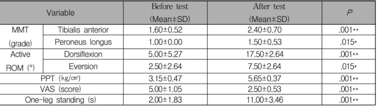
After kinesiotaping in PNP patients for 8 weeks, MMT (tibialis anterior, peroneus longus muscles) Active ROM (dorsiflexion, eversion), PPT (fibular head), and one-leg standing significantly increased.
VAS decreased significantly (Table 2).
4. DISCUSSION
Ankle taping is the most common method for supporting the ankle instability and preventing ankle injury in sports. However, effect of ankle taping in patients with common PNP have not been investigated. Patients with common PNP commonly present with loss of ankle dorsiflexion (weakness of the tibialis anterior) and eversion (weakness of the tibialis anterior, peroneus longus, and peroneus brevis), leading to foot drop with the characteristic foot slap during heel strike and a steppage gait [16]. In this study, subjects had unilateral foot drop due to damage to the common peroneal nerve. The most frequent cause of unilateral foot drop is common PNP of a spontaneous, traumatic, or pressure type [3].
Taping of the ankle joint is used to restrict ankle joint movement as well as to reduce the risk of ankle sprain [17]. In this study, we used kinesio taping on the ankle joint to assess its effectiveness on MMT, active ROM, pain, and balance in PNP patients.
Kinesiotaping has been shown to be effective in many studies [18,19].
In this study, the subjects showed increased muscle strength at 8 weeks (MMT of tibialis anterior and peroneus longus). A previous study reported that
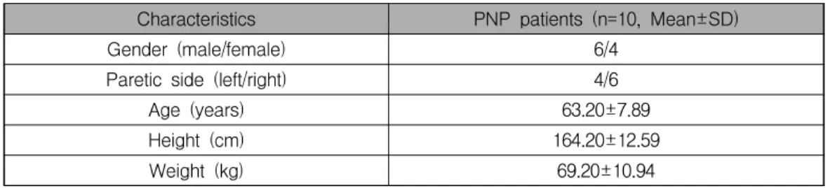
Table 1. General characteristics of subjects (N=10)
Characteristics PNP patients (n=10, Mean±SD)
Gender (male/female) 6/4
Paretic side (left/right) 4/6
Age (years) 63.20±7.89
Height (cm) 164.20±12.59
Weight (kg) 69.20±10.94
kinesio taping neither increased nor decreased muscle power immediately and 12 hours after application in athletes [20], suggesting that kinesio taping has no immediate effect on muscle strength. In our study, muscle strength and active ROM did improve after 8 weeks. This confirmed that the application of kinesio taping has no immediate effect on muscle strength.
However, the taping effect in this study could not exclude the placebo effect [21]. This is because there is no control group. Based on this study, further study is needed to compare with control group.
Pain levels (VAS and PPT) decreased. Pain may be a factor affecting the evaluation of muscle strength [22]. Pain and muscle strength are correlated. In our subjects, muscle strength and pain in the ankle joint may have been correlated. Therefore, the pain decreased due to muscle strength increasing.
The one-leg standing test improved for subjects after applying kinesiotaping. Control of one-leg standing requires muscle activity of the lower extremity. One-leg standing is a challenging posture, and control requires more complex motor activity than double-leg standing due to the narrower base of support [23].
In another study, the application of kinesio tape applied in the same manner resulted in immediate improvements in the Berg balance scale of stroke patients [11]. In addition, ankle kinesio taping improved the forward reach test and reduced the
mediolateral displacement of the center of pressure compared to a control group [11]. In previous studies, the results were attributed to stimulation of mechanoreceptors and correction of talipes equinovarus. The subjects of this study were thought to have improvements in ankle function because correction of the ankle joint was more influential than stimulation of the mechanoreceptors, since sensory loss was present.
In subjects with common PNP, kinesio taping increased MMT, active ROM, pain, and one-leg standing at 8 weeks after intervention. In this study, 8 weeks of taping was applied because the period required for recovery after peripheral nervous system injury is 8 weeks. In subsequent studies, it may be necessary to repeat the measurement of effects over time. In this study, motor and sensory nerve conduction or muscle activity of common PNP patients was not directly measured. These evaluations have limited usefulness, since neurologic pathways vary in individuals by age and race, and such tests are not easily accessible in clinical practice. The evaluation methods used in this study can be performed by anyone who is a physiotherapist by physical examination, and there is an expectation of their clinical utility in confirming the effect of kinesio taping on PNP patients. In future studies, it would be helpful to compare patients and controls in larger numbers, and it would be advantageous to add nerve conduction testing or electromyography.
Table 2. Change of MMT, ROM, PPT, VAS, One-leg standing
Variable Before test
(Mean±SD)
After test
(Mean±SD) P
MMT (grade)
Tibialis anterior 1.60±0.52 2.40±0.70 .001**
Peroneus longus 1.00±0.00 1.50±0.53 .015*
Active ROM (°)
Dorsiflexion 5.00±5.27 17.50±2.64 .001**
Eversion 2.50±2.64 7.50±2.64 .015*
PPT (㎏/㎠) 3.15±0.47 5.65±0.37 .001**
VAS (score) 5.00±1.05 2.50±0.53 .001**
One-leg standing (s) 2.00±1.83 11.00±3.46 .001**
Mean±SD, *p<.05, **p<.01
MMT: manual muscle test, ROM: range of motion, PPT: pressure pain threshold, VAS: visual analogue scale
5. CONCLUSION
In this study, we applied kinesio taping to produce ankle dorsiflexion and eversion in PNP patients with muscle weakness and pain. This study indicates that kinesio taping is effective in improving ankle strength, active ROM, pain and balance in PNP patients.
Therefore, kinesiotaping was effective in improving pain and weakness in PNP patients. Additional assessments should be performed to enhance the validity and reliability of the effects of kinesio taping on common PNP patients.
REFERENCES
[1] V. Rausch, M. Hackl, J. Oppermann, T. Leschinger, M. Scaal, L. P. Müller & K. Wegmann. (2019).
Peroneal nerve location at the fibular head: an anatomic study using 3D imaging. Archives of orthopaedic and trauma surgery, 139(7), 921-926.
DOI: 10.1007/s00402-019-03141-7.
[2] S. S. Divizyon. (2009). Variations in the high division of the sciatic nerve and relationship between the sciatic nerve and the piriformis.
Turkish neurosurgery, 19(2), 139-144.
[3] H. Berry & P. M. Richardson. (1976). Common peroneal nerve palsy: a clinical and electrophysiological review. Journal of Neurology, Neurosurgery & Psychiatry, 39(12), 1162-71.
DOI: 10.1136/jnnp.39.12.1162.
[4] A. L. Dellon, J. Ebmer & P. Swier. (2002). Anatomic variations related to decompression of the common peroneal nerve at the fibular head. Annals of plastic surgery, 48(1), 30-4. .
DOI: 10.1097/00000637-200201000-00004.
[5] J. H. Villafañe, P. Pillastrini & A. Borboni. (2013).
Manual therapy and neurodynamic mobilization in a patient with peroneal nerve paralysis: a case report. Journal of chiropractic medicine, 12(3), 176-181.
DOI: 10.1016/j.jcm.2013.10.007.
[6] P. Curley, K. Eyres, V. Brezinova, M. Allen, R.
Chan & M. Barnes. (1990). Common peroneal nerve dysfunction after high tibial osteotomy. The Journal of bone and joint surgery, British volume, 72(3), 405-408.
[7] D. Niall, R. W. Nutton & J. F. Keating. (2005).
Palsy of the common peroneal nerve after traumatic dislocation of the knee. The Journal of bone and joint surgery, British volume, 87(5), 664-667.
DOI: 10.1302/0301-620X.87B5.15607.
[8] J. F. Lehmann, S. M. Condon, B. J. De Lateur & R.
Price. (1986). Gait abnormalities in peroneal nerve paralysis and their corrections by orthoses: a biomechanical study. Archives of physical medicine and rehabilitation, 67(6), 380-386.
[9] J. L. Leung & A. Moseley. (2003). Impact of ankle-foot orthoses on gait and leg muscle activity in adults with hemiplegia: systematic literature review. Physiotherapy, 89(1), 39-55. DOI:
10.1016/s0031-9406(05)60668-2.
[10] P. N. Taylor, J. H. Burridge, A. L. Dunkerley, A.
Lamb, D. E. Wood, J. A. Norton & I. D. Swain.
(1999). Patients' perceptions of the Odstock Dropped Foot Stimulator (ODFS). Clinical Rehabilitation, 13(5), 439-446. DOI:
10.1191/026921599677086409.
[11] Z. Rojhani-Shirazi, S. Amirian & N. Meftahi. (2015).
Effects of ankle kinesio taping on postural control in stroke patients. Journal of Stroke and Cerebrovascular Diseases, 24(11), 2565-2571. DOI:
10.1016/j.jstrokecerebrovasdis.2015.07.008.
[12] L. Chinn, J. Dicharry, J. M. Hart, S. Saliba, R.
Wilder & J. Hertel. (2014). Gait kinematics after taping in participants with chronic ankle instability.
Journal of athletic training, 49(3), 322-330.
DOI: 10.4085/1062-6050-49.3.08.
[13] T. Halseth, J. W. McChesney, M. DeBeliso, R.
Vaughn & J. Lien. (2004). The effects of kinesio™
taping on proprioception at the ankle. Journal of sports science & medicine, 3(1), 1.
[14] E. Jaraczewska & C. Long. (2006). Kinesio®
taping in stroke: improving functional use of the upper extremity in hemiplegia. Topics in Stroke rehabilitation, 13(3), 31-42.
DOI: 10.1310/33KA-XYE3-QWJB-WGT6.
[15] Y. Suponitsky, O. Verbitsky, E. Peled & J.
Mizrahi. (2008). Effect of selective fatiguing of the shank muscles on single-leg-standing sway. Journal of Electromyography and Kinesiology, 18(4), 682-689. DOI: 10.1016/j.jelekin.2007.01.009.
[16] B. Ho, Z. Khan, P. J. Switaj, G. Ochenjele, D.
Fuchs, W. Dahl & A. R. Kadakia. (2014). Treatment of peroneal nerve injuries with simultaneous tendon
transfer and nerve exploration. Journal of orthopaedic surgery and research, 9(1), 67.
DOI: 10.1186/s13018-014-0067-6.
[17] B. D. Beynnon & P. A. Renström. (1991). The effect of bracing and taping in sports. In Annales Chirurgiae et Gynaecologiae, 80, (2), 230-238.
[18] D. Morris, D. Jones, H. Ryan & C. G. Ryan.
(2013). The clinical effects of Kinesio® Tex taping:
A systematic review. Physiotherapy theory and
practice, 29(4), 259-270. DOI:
10.3109/09593985.2012.731675.
[19] W. I. Kim, Y. K. Choi, J. H. Lee & Y. H. Park.
(2014). The effect of muscle facilitation using kinesio taping on walking and balance of stroke patients. Journal of physical therapy science, 26(11), 1831-1834. DOI: 10.1589/jpts.26.1831.
[20] T. C. Fu, A. M. Wong, Y. C. Pei, K. P. Wu, S. W.
Chou & Y. C. Lin. (2008). Effect of Kinesio taping on muscle strength in athletes—a pilot study.
Journal of science and medicine in sport, 11(2), 198-201.
DOI: 10.1016/j.jsams.2007.02.011.
[21] K. Sawkins, K. Refshauge, S. Kilbreath & J.
Raymond. (2007). The placebo effect of ankle taping on ankle instability. Medicine & Science in Sports & Exercise, 39(5), 781-787.
DOI: 10.1249/MSS.0b013e3180337371.
[22] G. M. Hare, P. J. Evans, S. E. Mackinnon, T. J.
Best, J. R. Bain, J. P. Szalai & D. A. Hunter.
(1992). Walking track analysis: a long-term assessment of peripheral nerve recovery. Plastic and reconstructive surgery, 89(2), 251-258.
DOI: 10.1097/00006534-199202000-00009.
[23] N. Shakoor, S. Furmanov, D. E. Nelson, Y. Li & J.
A. Block. (2008). Pain and its relationship with muscle strength and proprioception in knee OA:
results of an 8-week home exercise pilot study. J Musculoskelet Neuronal Interact, 8(1), 35-42.
박 시 은(Si-Eun Park) [정회원]
․Aug, 2015:Yongin University, Physical Therapy Department (PhD)
․Sep, 2018 ~ Present : Professor of Phsical Therapy Department at Woosong University
․ Research Interests : Orthopaedic Physiotherapy, Physiology
․ E-Mail : [email protected]
조균희(Kyun-Hee Cho) [정회원]
․Aug, 2018 :Yongin University, Physical Therapy Department (Ms)
․Jan, 2012 ~ present : AVENS Hospital
․ Research Interests : Stroke, Rehabilitation Physiotherapy
․ E-Mail : [email protected]
박 신 준(Shin-Jun Park) [정회원]
․Jun, 2018 : Yongin University, Physical Therapy Department (PhD)
․Sep, 2019 ~ Present : Professor of Physical Therapy Department at Gangdong University
․ Research Interests : Cardiopulmonary Physiotherapy, Orthopaedic Physiotherapy
․ E-Mail : [email protected]