Automatic Detection Method for Mura Defects on Display Films Using Morphological Image Processing and
Labeling
Sung-Je Cho
*, Seung-Ho Lee
*★Abstract
This paper proposes a new automatic detection method to inspect mura defects on display film surface using morphological image processing and labeling. This automatic detection method for mura defects on display films comprises 3 phases of preprocessing with morphological image processing, Gabor filtering, and labeling. Since distorted results could be obtained with the presence of non-uniform illumination, preprocessing step reduces illumination components using morphological image processing. In Gabor filtering, mura images are created with binary coded mura components using Gabor filters. Subsequently, labeling is a final phase of finding the mura defect area using the difference between large mura defects and values in the periphery. To evaluate the accuracy of the proposed detection method, detection rate was assessed by applying the method in 200 display film samples. As a result, the detection rate was high at about 95.5%. Moreover, the study was able to acquire reliable results using the Semu index for luminance mura in image quality inspection.
Keywords: mura, labeling, mophology, image processing, gabor filter
* Department of Electronic Engineering, Hanbat National University
★Corresponding [email protected] Manuscript received May. 12, 2014; revised Jun. 11, 2014
: accepted Jun. 12. 2014
I. Introduction
Along with the advances in the IT industry, the use of portable electronic devices such as smartphone or tablet PC and electric home appliances LCD monitor and TV has increased, and studies on different display types and sizes have been actively performed. In the display production process of display device , the presence of impurity or mura[1] defect on display surface distorts light emitted from backlight module, and brightness non-uniformity with blurry contour and smeared image occurs when defects persist until commercialization stage. Since these result in low
merchantability and consumer inconvenience, the quality inspection of display screen surface is crucial during manufacturing process. Therefore, inspection process is essential to detect mura defects on display surface. Mura defects can be created on the display panel during its production for various reasons including dust or impurities, scratches formed due to inspector’s mistake, and others. Defective products have been discriminated by inspecting mura defects with the naked eye.
However, the current inspection method is imprecise depending on persons, very costly and time consuming. To resolve these disadvantages, the use of image process is significantly more effective in mura detection in terms of time and cost compare to people detection methods. Therefore, this study proposes automatic detection method for mura defects on display film surface using morphological image processing and labeling.
Mura detection methods are divided into
detection in spatial domain and frequency domain.
Previous studies on detection in spatial domain are as follows: Lee et al. proposed a mura defect detection method on consecutively acquired panel images using multi-image accumulation and multi-resolution background subtraction [2], Lu et al. suggested image restructuring technique based on singular value decomposition (SVD) [3]. Li et al.
proposed a mura defect detection method by applying a threshold using the Hough transform [4].
These methods cannot detect mura defects when a mura pattern model is inaccurate.
Previous studies on detection in frequency domain are as follows: Bi et al. proposed detection technique which uses the Real Gabor filter and level set method [5]. Since this technique has to consider various characteristics of mura and use active contouring, its disadvantage is low speed. Chen et al. suggested a method to inspect mura using discrete cosine transform (DCT) and discrete wavelet transform (DWT) techniques [6]. However, this method also has a disadvantage of requiring a precise pattern model for mura detection.
This paper proposes a new automatic detection method that determines the presence of mura defects on display films by searching candidate regions of mura components and labeling them.
The proposed technique comprises three phases of morphological image processing through the removal of illumination components, Gabor filtering for the establishment of mura candidate regions on images, and labeling for final mura detection.
In Section 2, explains mura detection algorithms in each phase. Section 3 represents the results of tests and analyses performed using the proposed method to detect mura detects on display surface.
Ⅱ. RESEARCH ON AUTOMATIC DETECTION METHOD FOR MURA
DEFECTS ON DISPLAY FILM SURFACE USING MOPHOLOGTCAL IMAGE PROCESSING AND LABELING
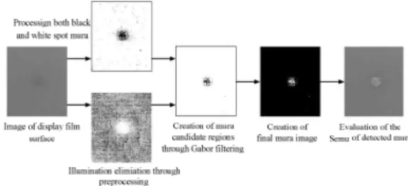
2-1. Flow diagram of detecting mura defects on display films with the proposed method Fig. 1 shows the flow diagram of detecting mura defects on display films using the method proposed in this study. In this process, illumination from images is first eliminated through preprocessing using morphological image processing, the candidate regions of mura is selected using the Gabor filter, and then mura defects are detected through labeling at the final inspection.
Fig. 1. Overview of the proposed method
When algorithms are applied to display surface without eliminating illumination components included in images, flawed results may be acquired by finding non-mura region. For this reason, preprocessing stage needs to be performed to remove illumination components. In the preprocessing stage, illumination is lowered by subtraction original image and background estimation image by estimating illumination components from candidate images created identical to original image. Mura parts are eliminated leaving only background components when opening and closing operations that use component factors are applied to an image among morphological image processing methods. When this image is subtracted from the original image, only mura parts remain from the original image and illumination is removed.
In the Gabor filtering stage, mura components are highlighted using the Gabor filter in the image eliminated with background components during preprocessing. In labeling stage, candidate regions are created by applying labeling to the image from the Gabor filtering stage to finally distinguish mura and background and final mura defects are detected using comparing algorithms for peripheral values.
2-2. Preprocessing with morphological image processing
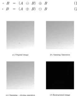
This section is about preprocessing stage where illumination components of the image are eliminated with morphological image processing. Small protrusions or noise pieces are removed with opening operation, and small holes or cracks are filled with closing operation. Therefore, only background components removed with mura components can be obtained through opening – closing operation as shown in the Fig. 2 images.
Opening and closing operations are shown in (1) and (2), respectively. Where, ⊖ is erosion operation and ⊕ is dilation operation [7].
∘ ⊖ ⊕ (1)
⋅ ⊕ ⊖ (2)
Fig. 2 Illumination component elminination process
Fig. 3 Histogram equalization of preprocessing image
As shown in Fig. 3, the images are restructured by expanding to the full range of grayscale using histogram equalization.
As shown in Fig. 4, when both white and black mura defects are present in the image, only either black or white spot mura is found after preprocessing on one side. For this reason, histogram equalization is performed once more by reversing the image.
Fig. 4 Histogram equalization on black and white spot mura
2-3. Mura selection after Gabor filtering and candidate regions labeling
Gabor filtering is done first in two images created after preprocessing stage, and then these images are restructured through AND operation.
Gabor filter can realize frequencies and compasses similarly to the visual system of humans and is favorable in distinguishing objects in the images.[8]
The Gabor filter is defined as (3). Where, g(x,y), the Gaussian function, is defined as (4). The Gabor filter is divided into real gabor filter (5) and imaginary gabor filter (6). Since real gabor filter is effective in separating image objects, real Gabor filtering is conducted in preprocessed images as shown in Fig. 5. After morphological preprocessing, black and white mura regions are expressed on the images through Gabor filtering done in an image divided into two parts. To present black and white mura regions on a single image, two images are restructured into a single image through AND operation.
exp
(3)
(4)
cos (5)
sin (6)
: frequency, : rotation angle,
: ,
: Fig. 5 Gabor filtering and image restructuring
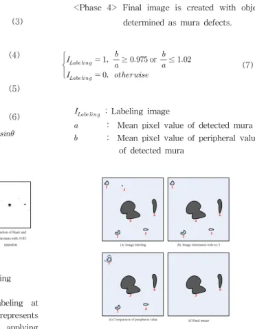
Mura is finally determined using labeling at Gabor filtering candidate regions. Fig. 6 represents images labeled with 8-connectivity by applying Gabor filtering after morphological preprocessing.
The four phases of labeling algorithm are as follows:
<Phase 1> Labeling is done in the candidate regions of Gabor filtering image with 8-connectivity.
<Phase 2> An entire labeling object with less than 3 pixels is not determined as mura, eliminated from the labeling image, and excluded from mura candidate.
<Phase 3> Mura is discrminated by applying peripheral value comparing equation (7) to each labeling object. The region judged as background is removed from the labeling image, whereas the region determined as mura is kept in the image to organize a final image.
<Phase 4> Final image is created with objects determined as mura defects.
≥ or
≤
(7)
: Labeling image
: Mean pixel value of detected mura
: Mean pixel value of peripheral value of detected mura
Fig. 6 Final mura detection with labeling
III. EXPERIMENTAL RESULT
This paper evaluated objective reliability using the Semu index[9]. The Semu index was first introduced by the SEMI in 2002 to objectively evaluate FPD mura. The Semu index is obtained using the ratio of CJND, the threshold value for human visual perception of mura, to Cx, the mean brightness of mura. The Semu is defined as the ratio of CJND value to Cx as shown in(8). The difference in brightness(Cx) between detected mura and background is proportional to the area(Sx) of detected mura, and increases as Cxand Sx increase.
(8)
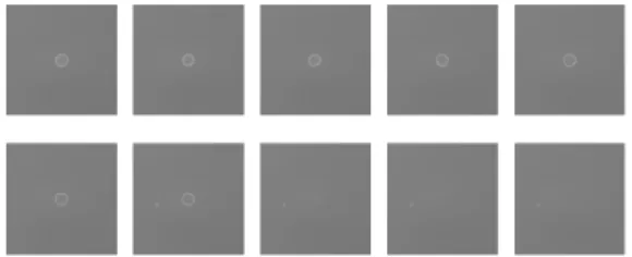
Fig. 7 represents the results of mura detection in images with reduced contrast in mura and background regions by one using the proposed method. Mura is detected from a contrast of 4.
Since mura is undetected in Fig. 7, the proposed method is identified to detect mura from a contrast of 4.
Fig. 7 Results of mura detection in Fig 3 image using the proposed method
Since mura is undetected in Fig. 7, the proposed method is identified to detect mura from a contrast of According to the evaluation results of the Semu from a study of Chen and Kuo[6] as shown in Table 2, contrast is 5. On the other hand, this study obtained a contrast of 4 using the proposed method, showing the improved result than that of Chen and Kuo. Meanwhile, the study predicted about 4.16 close to 4 for |Cx| for contrast 5 in Table 2. However, mura defects were not properly detected at stage 5 in the study of Chen andKuo because the Semu value was 3.07 in between 3 and 4 for |Cx| for contrast 5.
Table 1. Evaluation results of the Semu of the proposed method
Contrast
(%) (%) Semu Evaluation 10 8.1366 0.8958 9.0830 Success 9 7.3274 0.9080 8.0701 Success 8 6.5375 0.9056 7.2191 Success 7 5.7384 0.9034 6.3521 Success 6 4.9416 0.8990 5.4969 Success 5 4.1453 0.8978 4.6172 Success 4 3.3465 0.8972 3.7298 Success 3 2.8447 1.6414 1.7330 Failure 2 2.8540 1.6414 1.7387 Failure 1 2.8636 1.6414 1.7446 FailureTable 2. Evaluation results of the Semu in a study of Chen and Kuo
Contrast
(%) (%) Semu Evaluation10 8.32 0.88 9.3 Success
9 7.42 0.88 8.39 Success
8 6.61 0.88 7.48 Success
7 5.79 0.88 6.55 Success
6 4.78 0.88 5.39 Success
5 3.07 0.89 3.44 Success
4 0.52 1.22 0.43 Failure
3 0.48 1.28 0.31 Failure
2 0.37 1.28 0.29 Failure
1 0.18 1.5 0.12 Failure
Ⅳ. CONCLUSION
This study detected mura defects on display surface through morphological image processing and labeling. The proposed algorithm eliminates illumination components in preprocessing stage, and then highlights mura defects using the Gabor filter.
Consequently, this mura detection algorithm creates an final image using the labeling algorithm. This algorithm is verified to detect mura in images containing illumination since it applies algorithms after eliminating illumination components from an original image by estimating background using morphological image processing in preprocessing stage. The algorithm has outstanding detection rate by organizing an image with mura defects using the mura comparison algorithm in labeling stage where candidate objects are chosen as mura. This investigation was able to verify the detection ability of the proposed method by acquiring results regardless of a wide range of complex mura forms, colors, and others. The proposed detection method was tested in 200 display film samples and the detection rate was high at about 95.5%. Moreover, the study was able to acquire more desirable results than those of previous studies using the Semu index used to detect luminance mura for FPD image quality inspection.
Detection rate could be lowered when quite a number of labeling results are obtained in labeling process. For this reason, additional studies need to
be performed to improve detection rate by reducing candidate regions.
REFERENCES
[1]SEMI, "New Standard: Definition of Measurement Index (SEMU) for Luminance Mura in FPD", SEMI, 2002
[2]Y.C Lee, C.E Shie, Din-Chang Tseng, "LCD Mura Detection Based on Accumulated Differences and Multi-resolution Background Subtraction", 2009 [3]C. J. Lu, D. M. Tsai, "Automatic defect inspection for LCDs using singular value decomposition", International Journal of Advanced Manufacturing Technology, vol. 25, pp. 53-61, 2005 [4]W. C. Li, D. M. Tsai, "Defect Inspection in Low-Contrast LCD Images Using Hough Transform-Based Nonstationary Line Detection,"
IEEE Transactions on Industrial Informatics, vol. 7, no. 1, pp. 136-147, 2011
[5]X. Bi, C. Zhuang, H. Ding, "A New Mura Defect inspection way for TFT-LCD using level set method", IEEE Signal processing letters, vol. 16, no.
4, pp. 311-314, 2009
[6]L. C. Chen, C. C. Kuo, "Automatic TFT-LCD mura defect inspection using discrete cosine transform-based background filtering and 'just noticeable difference' quantification strategies", Measurement Science and Technology, vol. 19, pp.
1-10, 2008
[7]R. C. Gonzalez, R. E. Woods, "Digital Image Processing (3nd Edition) ", Prentice Hall, 2008 [8] J. R. Movellan, "Tutorial on Gabor Filters", 2008 [9]SEMI, "Definition of measurement index (SEMU) for luminance Mura in FPD image quality inspection", SEMI D31-1102, 2002
BIOGRAPHY
Sung-Je Cho (Member)
2012 : BS degree in Electronic Engineering, Hanbat National University
2014 : MS degree in Electronic Engineering, Hanbat National University
Seung-Ho Lee (Member)
1986 : BS degree in Electronic Engineering, Hanyang University 1989 : MS degree in Electronic Engineering, Hanyang University 1994 : Ph. D degree Electronic Engineering, Hanyang University 1994 ~ Present : Professor, Department of
Electronics&Control Engineering, Hanbat National University