• 검색 결과가 없습니다.

Idiopathic eosinophilic myositis in Korean native cattle (Bos taurus coreanae)

N/A
N/A
Protected

Academic year: 2021

Share "Idiopathic eosinophilic myositis in Korean native cattle (Bos taurus coreanae)"

Copied!
4
0
0

로드 중.... (전체 텍스트 보기)

전체 글

(1)

大韓獸醫學會誌(2008) 第48卷 第2號

Korean J Vet Res

(2008) 48(2) : 187~190

187

Idiopathic eosinophilic myositis in Korean native cattle ( Bos taurus coreanae )

Seong-Hee Rhee

1

, Il-Jeoung Yu

1

, Jong-Hoon Kim

1

, Jung-Kee Kwon

1

, Jinho Park

1

, Myung-Jo You

1

, Jeong-Won Lee

2

, Hee-Jin Park

1

, Irina Chekarova

1

, Gerry Amor Camer

3

,

Chae-Woong Lim

1

, Bumseok Kim

1,

*

1

Bio-safety Research Institute and College of Veterinary Medicine, Chonbuk National University, Jeonju 561-756, Korea

2

Jeongeup Branch, Institute of Live Stock and Veterinary Research, Jeongeup 580-814, Korea

3

College of Veterinary Medicine, University of Eastern Philippines, 6400, Catarman, Northern Samar, Philippines

(Accepted: April 3, 2008)

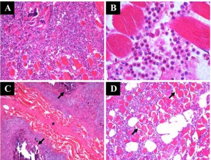
Abstract : Eosinophilic myositis lesions are characterized by severe eosinophil infiltration along muscles of affected animals. The exact cause of the lesion remains controversial and the carcass is condemned once this lesion is seen during meat inspection. A cow slaughtered in Chonbuk province, Korea was observed to have disseminated pale foci throughout the musculature; meat samples were obtained and macroscopically investigated. Cut ends of neck and thigh muscle tissues showed variably sized, multi- focal pale white-grayish nodular lesions. Histopathological examination consistently revealed inflammatory lesions with adjacent infiltration of eosinophilic granulocytes and focal necrotic calcification. However, no parasites, including Sarcocystis sp., could be discerned in the affected carcass. This case was diagnosed as idiopathic eosinophilic myositis in cattle.

Keywords : cattle, eosinophil, eosinophilic myositis, muscle

Introduction

Eosinophilic myositis (EM), a rare idiopathic inflammatory skeletal and cardiac muscle disease associated with eosinophilic infiltrates in cattle and sheep [17], is usually incidentally observed at slaughter. Despite the lack of specific information about its exact nature and etiological cause [15, 17], EM lesions are currently listed as valid grounds for meat condemnation; the unwholesome and unappealing look of the meat apparently justify its condemnation.

Some scientists have detected sarcocyst parasites in the lesions, prompting attribution of eosinophilic infiltrates to a dead or degenerating parasite [1, 5]. Others have reported similar lesions caused by trematodes and

Echinococcus

parasites [16]. Lesions of EM are inducible upon experimental infection of cattle with

Trichinella

larvae [11, 17], but it seems such infections are unlikely to be involved in natural cases of EM [17].

Traumatic muscle rupture has also been reported to

lead to eosinophilic infiltration [2].

Sarcocystis cruzi

specific IgG and IgE responses have been previously associated with EM lesions in cattle, as well [4, 8]. In the present study, a beef carcass with widespread localization of spot-like nodules in skeletal muscle was sampled and observed by light microscopy. This retrospective case report documents an unusual case of skeletal muscle necrosis with eosinophilic myopathy and calcification of obscured etiology.

Case

A carcass of slaughtered Korean native cattle (

Bos taurus coreanae

) in Chonbuk province was condemned due to disseminated white-grayish necrotic foci visibly evident along the thigh, abdominal, and neck musculatures. Samples of the thigh and neck muscles were retrospectively analyzed for histopathological diagnosis at the Department of Veterinary Pathology, College of Veterinary Medicine, Chonbuk National

*Corresponding author: Bumseok Kim

College of Veterinary Medicine, Chonbuk National University, Jeonju 561-756, Korea

[Tel: +82-63-270-4638, Fax: +82-63-270-3780, E-mail: [email protected]]

(2)

188 Seong-Hee Rhee

et al.

University, Jeonju, Korea. Neither detailed data nor clinical history was available as the lesion was noted only during meat inspection.

Grossly, the muscle appeared turgid with numerous demarcated, whitish, and disseminated nodules visible along the femoral (Fig. 1A) and neck (Fig. 1B) muscles sampled; lesions were not observed in cardiac or esophageal muscle. The size of lesions was variable, approximately 0.2 to 1 cm in diameter. Portions having nodular lesions and surrounding tissues were removed for histological study. Samples were fixed in neutral- buffered 10% formalin and processed by standard histological paraffin method, sectioned at 5

µ

m, stained with hematoxylin and eosin (H&E), and examined by light microscopy.

Histopathologically, the nodular lesions showed inflammation resembling that of granulomatous tissue, with large amounts of inflammatory cell infiltration mainly consisting of eosinophilic granulocytes and fibrous tissues (Fig. 2A). Infiltrating eosinophilic granulo- cytes were discerned to intersperse between muscle fibers (Fig. 2B). Foci of necrosis were remarkably noted. Dark, calcified materials were observed in the center of granulomatous lesions (Fig. 2C), and were

Fig. 2. Light micrograph of a typical section through a multiple granulomatous lesions in the bovine muscle affected with eosinophilic myositis (H&E stain). A. Muscle section showing the diffuse leukocytes infiltration and fibrous tissue formation (

×

100). B. Severe infiltration of eosinophilic granulocytes between muscle fibers (

×

400). C. Dark calcified material (arrow) seen in the center of eosinophilic granulomatous lesions. Fibroplasia (asterisk) was clearly evident between the muscle fibers (

×

20). D. Hypercontraction and degeneration of muscle fibers (arrow) along with infiltration of eosinophils (

×

100).

Fig. 1. Gross examination of nodular foci in muscle of

slaughtered cattle. A. Portion of thigh muscle shows nodular

whitish foci (arrow) extending through the incised muscle

(biceps femoris, cattle); B. Pale foci of different sizes from

cut portion of neck muscle (brachiocephalicus, cattle).

(3)

Idiopathic eosinophilic myositis in Korean native cattle (

Bos taurus coreanae

) 189 clearly surrounded by eosinophils and severe fibroplasia.

Hypercontraction and degeneration of segmental fibers with fat deposition were prominent in some lesions (Fig. 2D). Necrotic or hyalinized muscle fibers were also noted. Although it has been known that parasite infection is one of the major causes of eosinophilic infiltration in the musculature, numerous sections of adjacent muscle fibers revealed no evidence or remnant of parasites.

Discussion

Beef, an important meat for human consumption in Korea, is relatively expensive; meat condemnation due to myositis may thus cause unwarranted profit losses to ranchers and meat entrepreneurs. In Korea, cattle are slaughtered for human consumption between 2-3 years of age. The incidence of EM meat condemnation has not yet been established, though the presence of unusual pale necrotic foci in meat must be a cause for concern by ranchers, meat retailers and dealers, the meat inspectors and, most importantly, the consuming public. EM detection and diagnosis can only be achieved by careful meat inspection following slaughter, as there is a distinct lack of tests to diagnose the condition in living animals.

EM classically presents as a grayish or greenish color change, which may alter with time and could be attributed to bacterial invasion or toxin accumulation.

Under such circumstances, meat condemnation is justifiable. In the case reported here, the pale foci noted appear to be calcified whitish streaks, which may have resulted from myonecrosis, albeit of unknown etiology;

such lesions are often a sequel of dystrophic calcified changes. The finding of abundant eosinophils is intriguing. Such infiltrates connote excessive release of histamine or other similar vasoactive amine products ordinarily seen in the events of parasitic infiltration, vaccination reactions and Type I hypersensitivity reactions [7]. Recently Murata [14] demonstrated over- expression of IL-5, an inflammatory cytokines that induces eosinophil accumulation, and eosinophilic major basic protein in EM muscle, suggesting that such proteins adhere to the muscle fiber, resulting in muscle fiber damage. Furthermore, Krahn [12] reported that mutations in calpain-3, a muscle-specific protein that belongs to a subset of intracellular nonlysosomal proteases, occur in patients with idiopathic EM and can

cause EM. There is an on-going controversy as to the exact etiology of EM. One report confirmed that 58%

of the EM positive cattle showed sarcocysts positive [17]. However, larvae of

Trichinella

were not observed in the EM lesions indicating that trichinosis is not a main cause of EM in cattle. Despite mounting claims that EM can be attributed to the presence of degenerating sarcocyst parasites [6, 10, 13, 18], no evidence of parasitic presence, remnants or traces thereof was observed in this sample. Neither detailed data nor clinical history was available as the lesion was noted only during meat inspection. A classification system for idiopathic eosinophilic myositis was previously proposed by Hall [9], describing three distinct disorders including focal eosinophilic myositis, eosinophilic polymyositis and eosinophilic perimyositis. Based on histology of muscle, it seems that current EM case comes under eosinophilic polymyositis since most of eosinophils invaded perimysium and fibre invasion were unusual.

In the absence of parasitic infestation, a thorough investigation on factors that may contribute to allergic states must be undertaken. A variety of causes may induce differential eosinophilic responses, including adrenocortical insufficiency, anaphylaxis or recovery phases of acute inflammatory conditions, all of which should be considered [3]. In the current sample, immunological reactions, the intake of drugs, allergic states or a possibly obscured neoplasia of eosinophilic granulocytes were hypothesized as the probable cause of the lesion. EM is often problematic as the lesion can be noted only during post-mortem after slaughter.

It is suggested that in future encounter of unusual eosinophilic myositides, bone marrow sample must be obtained as this may help to rule out myeloid cell granulocytic eosinophilic neoplasia. Other infiltrating tumors in serous membranes and bones must likewise be explored.

Conclusion

The present report shows that idiopathic EM,

resulting in infiltration of eosinophilic granulocytes and

muscle damage, was diagnosed by histopathologic

results in Korean native cattle. However, the etiology

of the current case was not clear, as evidence of

parasitic infection was not observed, and the lesion was

noted during meat inspection, at which point the

clinical history of the animal was unavailable.

(4)

190 Seong-Hee Rhee

et al.

Acknowledgments

This work was supported by research funds of Bio- Safety Research Institute and Chonbuk National University. We thank Emily M. Deal (Stanford University, Stanford, USA) for editorial assistance with manuscript preparation.

References

1. Arness MK, Brown JD, Dubey JP, Neafie RC, Granstrom DE. An outbreak of acute eosinophilic myositis attributed to human Sarcocystis parasitism.

Am J Trop Med Hyg 1999, 61 , 548-553.

2. Bindseil E. Eosinophilic leucocyte infiltration in ruptured necrotic thigh muscle of downer cows. Vet Rec 1987, 120 , 183-184.

3. Coles EH. Veterinary Clinical Pathology. 4th ed. pp.

68-69, WB Saunders, Philadelphia, 1986.

4. Ely RW, Fox JC. Elevated IgG antibody to Sarcocystis cruzi associated with eosinophilic myositis in cattle. J Vet Diagn Invest 1989, 1 , 53-56.

5. Gajadhar AA, Marquardt WC. Ultrastructural and transmission evidence of Sarcocystis cruzi associated with eosinophilic myositis in cattle. Can J Vet Res 1992, 56 , 41-46.

6. Granstrom DE, Ridley RK, Baoan Y, Gershwin LJ.

Immunodominant proteins of Sarcocystis cruzi bradyzoites isolated from cattle affected or nonaffected with eosinophilic myositis. Am J Vet Res 1990, 51 , 1151-1155.

7. Granstrom DE, Ridley RK, Baoan Y, Gershwin LJ, Nesbitt PM, Wempe LA. Type-I hypersensitivity as a component of eosinophilic myositis (muscular sarcocystosis) in cattle. Am J Vet Res 1989, 50 , 571- 8. 574. Granstrom DE, Ridley RK, Yao B, Gershwin LJ, Briggs DJ. Immunofluorescent localization of Sarcocystis cruzi antigens, IgG and IgE, in lesions of eosinophilic myositis in cattle. J Vet Diagn Invest 1990, 2 , 147-149.

9. Hall FC, Krausz T, Walport MJ. Idiopathic eosinophilic myositis. QJM 1995, 88 , 581-586.

10. Jensen R, Alexander AF, Dahlgren RR, Jolley WR, Marquardt WC, Flack DE, Bennett BW, Cox MF, Harris CW, Hoffmann GA, Troutman RS, Hoff RL, Jones RL, Collins JK, Hamar DW, Cravans RL.

Eosinophilic myositis and muscular sarcocystosis in the carcasses of slaughtered cattle and lambs. Am J Vet Res 1986, 47 , 587-593.

11. Kennedy PC. Experimental bovine trichinosis: An attempt to produce eosinophilic myositis of cattle.

Cornell Vet 1995, 45 , 127-152.

12. Krahn M, Lopez de Munain A, Streichenberger N, Bernard R, Pécheux C, Testard H, Pena-Segura JL, Yoldi E, Cabello A, Romero NB, Poza JJ, Bouillot- Eimer S, Ferrer X, Goicoechea M, Garcia-Bragado F, Leturcq F, Urtizberea JA, Lévy N. CAPN3 mutations in patients with idiopathic eosinophilic myositis. Ann Neurol 2006, 59 , 905-911.

13. La Perle KM, Silveria F, Anderson DE, Blomme EA. Dalmeny disease in an alpaca (Lama pacos):

sarcocystosis, eosinophilic myositis and abortion. J Comp Pathol 1999, 121 , 287-293.

14. Murata K, Sugie K, Takamure M, Fujimoto T, Ueno S. Eosinophilic major basic protein and interleukin-5 in eosinophilic myositis. Eur J Neurol 2003, 10 , 35-38.

15. Perl S, Nitzulov R, Yakobson B, Lichawsky D, Gilboa D. Eosinophilic myositis in a beef steer - a case report. Isr J Vet Med 1999, 54 , 6.

16. Sierra MA, Gomez MA, Navarro JA, Bernabe A.

Bovine eosinophilic myositis caused by trematodes.

Anales de Veterinaria de Murcia 1986, 2 , 21-26.

17. Smith HJ, Snowdon KE, Finley GG. Eosinophilic myositis in Canadian cattle. Can J Vet Res, 1991, 55 , 94-95.

18. Wouda W, Snoep JJ, Dubey JP. Eosinophilic myositis

due to Sarcocystis hominis in a beef cow. J Comp

Pathol 2006, 135 , 249-253.

수치

Fig. 1.  Gross examination of nodular foci in muscle of slaughtered cattle. A. Portion of thigh muscle shows nodular whitish foci (arrow) extending through the incised muscle (biceps femoris, cattle); B

참조

관련 문서