292 292
16S rRNA 유전자를 이용한
Neisseria meningitidis,
Haemophilus influenzae, Streptococcus pneumoniae
및Streptococcus agalactiae
의 검출Detection of Neisseria meningitidis, Haemophilus influenzae, Streptococcus pneumoniae and Streptococcus agalactiae using 16S rRNA gene
Chae Hoon Lee, M.D., Kyung Dong Kim, M.D., Hyo Jin Chun, M.D.,* Dong Seok Jeon, M.D.,* and Jae Ryong Kim, M.D.*
Department of Clinical Pathology, School of Medicine, Yeungnam University;
Department of Clinical Pathology, School of Medicine, Keimyung University,* Taegu, Korea 이채훈∙김경동∙전효진*∙전동석*∙김재룡*
영남대학교 의과대학 임상병리과학교실, 계명대학교 의과대학 임상병리학교실*
292 292
접 수: 1999년 2월 8일 접수번호:KJCP1260
수정본접수: 2000년 4월 20일 교 신저 자: 이 채 훈
우 705-035 대구광역시 남구 대명동 317-1 영남대학병원 임상병리과 전화: 053-620-3632, Fax: 053-628-6682
Background : Delayed treatment of acute bacterial meningitis often causes death or serious neu- rological defects. Rapid and accurate diagnosis is very important for effective treatment. Although the Gram stain and latex agglutination test for major causative bacteria, Neisseria meningitidis,
Haemophilus influenzae, Streptococcus pneumoniae and Streptococcus agalactiae, are useful for
early detection of acute bacterial meningitis, their sensitivity is not satisfactory. In this study, we pur- pose to develop a PCR strategy for the simultaneous detection of N. meningitidis, H. influenzae, S.pneumoniae and S. agalactiae.
Methods : Primers were designed from the 16S rRNA genes of N. meningitidis, H. influenzae, S.
pneumoniae and S. agalactiae, which were constituted with 3 senses and 5 antisenses, and 7
primer pairs. The PCR assay was divided into two steps, the first PCR resulted in a general bacteri- al amplicon with universal primers, and the second were performed with species specific primer pairs in four combination reaction tubes. PCR sensitivity and specificity were tested to clinical iso- lates of 4 N. meningitidis, 3 H. influenzae, 14 S. pneumoniae, 6 S. agalactiae, 14 Staphylococcusepidermidis, and 25 reference strains of different species.
Results : The species specific primer pairs showed specific DNA amplification to all clinical iso- lates tested. All 25 reference strains of different species and S. epidermidis were distinguished from N. meningitidis, H. influenzae, S. pneumoniae and S. agalactiae except for Pseudomonas
aeruginosa, Neisseria and Candida species, The PCR detection limit for S. agalactiae was 200
CFU.Conclusions : The seminested PCR using bacterial 16S rRNA gene was sensitive and specific for the detection of Neisseria species, H. influenzae, S. pneumoniae and S. agalactiae. This results warranted the application of this method to cerebrospinal fluids to diagnose bacterial meningitis.
(Korean J Clin Pathol 2000; 20: 292-300)
Key words : Neisseria meningitidis, Haemophilus influenzae, Streptococcus pneumoniae, Strep-
tococcus agalactiae, 16s rRNA Gene, PCR, Bacterial meningitis
서 론
패혈증, 뇌수막염 등의 중증 감염증, 특히 급성 세균성 수막염 은 사망이나 심각한 신경학적 후유증을 유발할 수 있으므로 효과 적인 치료를 위해 원인균의 신속한 검출과 정확한 동정이 필요하 다. 현재 급성 세균성 수막염의 원인균을 규명하기 위해 현미경 관찰, 세균 배양, 라텍스 응집법 등이 널리 이용되고 있다. 현미 경 관찰은 쉽게 시행할 수는 있으나 특이도와 민감도가 떨어지고, 세균 배양 및 동정은 최소한 12시간에서 24시간이 경과되어야 균 검출이 가능하고 동정검사를 실시하는 경우에 추가적인 시간이 소요된다. 따라서 주요 원인균으로 알려진
Neisseria meningi- tidis, Haemophilus influenzae, Streptococcus pneumoniae
및S. agalactiae
등의 항원을 검출할 수 있는 뇌척수액 라텍스 응집 법이 신속한 결과를 제공함으로써 임상에서 널리 이용되고 있으 나, 세균성 수막염의 25-35%에서 세균수가 103-104 CFU/mL 이하로 검출되므로[1, 2], 103-104 CFU/mL 이상이 되어야 검 출이 가능한 라텍스 응집법의 검출감도에 문제점이 있다[3].최근에는 중합효소연쇄반응 등이 세균성 수막염을 유발하는 원 인균의 조기 검출에 이용되고 있으며[4-6] 뇌척수액 배양보다 민 감도가 높은 것으로 보고되고 있다[6]. 그러나 상기 보고된 검출 법은 특정 균종에 따라 시발체, 증폭조건 등이 다른 증폭환경을 이용함으로써 시간 및 방법의 번거러움이 있고 이러한 문제점을 해결하기 위해 16S rRNA 유전자를 이용하여 동일 증폭조건으 로 특정 세균을 검출하려는 시도가 있었다[7-9]. 16S rRNA 유 전자는 동일한 종의 세균은 염기서열이 거의 유사하지만 종간에 는 염기서열의 차이가 있어[10, 11], 균종의 분류[7, 10, 12], 특 정 균종의 검출[8, 9, 13, 14] 등에 이용되고 있다.
16S rRNA 유전자를 이용하여 급성 세균성 수막염의 주요 원 인균을 검출하기 위해 Greisen 등[15]은 universal 시발체로 중 합효소연쇄반응을 시행하고 이차적으로 균종별 특이 소식자를 이 용하는 방법을 소개하였으나 교잡과정이 1.5-18시간이 소요되어 조기 검출에 문제점이 있으며, Radstrom 등[16]은 universal 시 발체로 일차 증폭 후 균종별 특이 시발체로 이중 중합효소연쇄반 응을 시도하였으나
S. pneumoniae
의 25%에서 검출되지 않는 문제점을 보였다.이 연구에서는 상기 기술된 문제점을 보완하고 급성 수막염의 주요 원인균인
N. meningitidis, H. influenzae, S. pneumoniae
와S. agalactiae
의 조기 검출을 위해 세균의 16S rRNA 유전자 의 균종별 특이 시발체를 고안하여 이중 중합효소연쇄반응에 의 한 동시 측정방법을 시도하였다.재료 및 방법
1.대상
영남대학병원과 경상대학병원의 임상검체에서 분리된
N.
meningitidis
4주,H. influenzae
3주,S. pneumoniae
14주,S.
agalactiae
6주의 균주를 대상으로 균종별 특이 시발체의 증폭반 응 수행력을 검정하였고, 특이 시발체의 다른 균종과의 교차반응 은 ATCC 표준 균주 등 25주와 임상 검체에서 분리한Staphy- lococcus epidermidis
14주를 대상으로 실시하였다. 그리고 임상 검체는 검사전 혈액한천배지와 초콜렛 배지에 접종하여 자동 세 균 동정기인 ATB (BioMerieux, Marcy I’Etoile, France)와 수 기법을 이용하여 재동정하였다.2.
염기서열분석 및 시발체 고안
수막염의 주요 원인균인
N. meningitidis, H. influenzae, S.
pneumoniae
및S. agalactiae
의 염기서열을 분석하기 위해 표준 균주(N. meningitidis
ATCC 13090,H. influenzae
ATCC 35056,S. pneumoniae
ATCC 1905 및S. agalactiae
ATCC 13813)와 국내에서 분리된 균종의 염기서열의 차이점 유무를 확 인하기 위해 임상검체에서 분리된 각 1 균주에 대해 16S rRNA 유전자의 universal 시발체(PL06: 5’-GGTTAAGTCCCGA- ACGAGCGC-3’, DG74: 5’-AGGAGGTGATCAACCGCA- 3’)[15]를 이용하여 염기서열을 분석하였다. 염기서열분석은 universal 시발체로 증폭한 산물(452 bp)을 pGEM-T Vector Systems (Promega, Madison, USA)를 이용하여 cloning시켜Escherichia coli
에 접종하여E. coli
의 plasmid DNA를 분리하 였고, PL06와 DG74 시발체를 이용하여 dideoxynucleotide chain termination법[17]의 변법인 Silver Sequence DNA Sequencing System (Promega, Madison, USA)을 사용하여 시행하였다.시발체의 고안은 Dieffenbach 등[18], Kwok 등[19]과 Ed- ward 등[20]이 기술한 시발체 합성 기준을 참조하여 이 연구에 서 시행한 염기서열과 GenBank에 등록되어 있는 각 균종의 염 기서열(GenBank Accession Number,
N. meningitidis
: U029011, Z22776;H. influenzae
: Z22806, X87978, X87979, X87977, X87976, M35019, U02897;S. pneumoniae
: AJ001247, AJ001251, AJ001249, Z22807, AF003930, AB002522, U2920.;S. agalactiae
: Z22080, AF015927, AB002479, AB002480, X59032)에서 동일한 염기서열 부위를 기초로 제작하였고, 임상검체에서 흔히 분리되는 오염균인S.
epidermidis
의 교차반응을 방지하기 위해 GenBank에 등록되어 있는S. epidermidis
의 염기서열을 참조하였다.3.
주요 세균성 수막염 원인균의 동정을 위한 중합효소연쇄
반응
1) DNA 분리
배양된 균의 DNA 분리를 위하여 2-3개의 집락을 0.5 mL brain heart infusion 배양액에 풀어 0.5 mL 소원심분리관에 옮
。 . .
긴 후, 10,000×
g
에서 2분간 원심분리하여 침전물을 100 L 멸 균 증류수에 희석하였다. 희석된 균액 1 L와 GeneReleaser (BioVentures Incorporated, Murfreessboro, USA) 20 L를 혼합하여 microwave oven에서 500 W에서 7분간 방치하여 DNA를 분리하였고, 이중 10 L를 취하여 중합효소연쇄반응을 시행하였다.2) 중합효소연쇄반응
중합효소연쇄반응을 위한 thermocycler는 공기순환 방식인 FTC-2000 (Daehan Medical, Seoul, Korea)을 사용하였으며, universal 시발체를 이용하여 16S rRNA 유전자를 증폭하였다.
증폭조건은 표적 DNA 10 L와 Taq polymease 2 U를 가하 고, 최종농도가 Tris/HCl 50 mM (pH 8.3), MgCl2 3 mM, 우 혈청 알부민 500 g/mL, PL06 0.4 mM, DG74 0.4 mM, dNTP 200 M이 함유되도록 한 50 L 반응액을 94℃에서 20 초간 열변성 후 56℃에서 3초간 결합반응, 72℃에서 20초간 연장 반응, 94℃에서 3초간 열변성의 과정을 30회 반복한 후 72℃에서 2분간 연장반응을 실시하였다. universal 시발체로 일차 증폭을 한 후, 균 동정을 위해 다중시발체 중합효소연쇄반응을 이차적으 로 실시하였다. 이차 증폭은 일차 증폭된 산물 5 L에 고안한 각각의 시발체 20 pmol을 가하고 결합반응을 56℃와 61℃로 2회 실시하였으며, 그 이외의 반응조건은 1차 증폭과 동일하게 실시 하였다. 반응 산물의 확인은 0.5 g/mL ethidium bromide가 함유된 2% agarose gel에 증폭산물 10 L와 bromphenol blue 2 L를 혼합한 후 0.5×TBE (Tris-borate EDTA)용액에서 100 V에서 30분간 전기영동하여 UV transilluminator (파장 305 nm)로 관찰하고 폴라로이드로 촬영하여 균종에 따른 증폭 산물의 크기로서 분류하였다. 그리고 중합효소연쇄반응의 민감도 는
S. agalactiae
를 대상으로 1×10⁵ CFU에서 1 CFU까지 10 배수 희석하여 중합효소연쇄반응을 시행한 후 측정가능 희석액을 다시 10 배수 희석하여 측정하였다.결 과
1.시발체의 고안
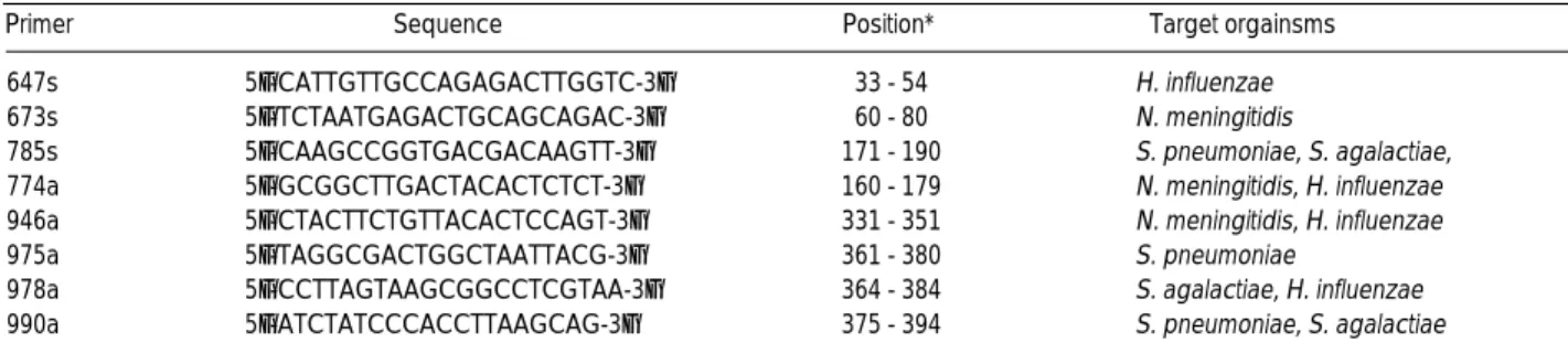
고안한 시발체는 647s, 673s 및 785s의 sense 3종과 774a, 946a, 975a, 978a 및 990a의 antisense 5종으로,
H. influenzae
는 647s와 946a의 시발체 쌍(이하 647s-946a라 함)과 647s와 978a 의 시발체 쌍(이하 647s-978a라 함),N. meningitidis
는 673s와 774a의 시발체 쌍(이하 673s-774a라 함)과 673s와 946a의 시발 체 쌍(이하 673s-946a라 함),S. pneumoniae
는 785s와 990a의 시발체 쌍(이하 785s-990a라 함)과 785s와 975a의 시발체 쌍(이 하 785s-975a라 함),S. agalactiae
는 785s와 990a의 시발체 쌍 (이하 785s-990a라 함)과 785s와 978a의 시발체 쌍(이하 785s-978a라 함)에서 각각 증폭되도록 하였다(Table 1, 2).
2.
다중시발체 중합효소연쇄반응
다중시발체 중합효소연쇄반응을 위한 시발체는 4가지 조합으 로 구성하여 각 조합별로 별도의 반응용기에서 수행하였으며 (Table 3), 조합 1은 673s, 785s, 774a 및 990a의 4종의 시발체 를 사용하여
N. meningitidis
는 673s-774a에서,S. pneumoniae
와S. agalactiae
는 785s-990a에서 각각 증폭되도록 하였다. 조합 2는 673s, 785s 및 975a의 3종의 시발체를 사용하여S. pneumo- niae
가 785s-975a에서 증폭되도록 하였다. 조합 3은 647s, 785s 및 978a의 3종의 시발체를 사용하여H. influenzae
는 647s-978a 에서,S. agalactiae
는 785s-978a에서 각각 증폭되도록 하였으며, 조합 4는 647s, 673s 및 946a의 3종의 시발체를 사용하여N.
meningitidis
는 673s-946s에서,H. influenzae
는 647s-946s에서 각각 증폭되도록 하였다.3.
시발체의 검증
고안한 각 시발체의 조합을 이용하여 각 균종에 따른 민감도를 검정하기 위해 임상검체에서 분리한
N. meningitidis, H.
influenzae, S. pneumoniae, S. agalactiae
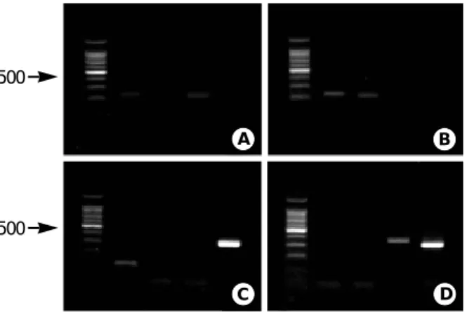
에 대해 결합온도를 56℃로 하여 중합효소연쇄반응을 실시하였으며, 각 균종은 고안한 특이 시발체에 모두 증폭산물이 관찰되었다(Table 4, Fig. 1).
그러나, 1주의
N. meningitidis
는 785s-990a에서, 1주의S.
pneumoniae
는 673s-774a 및 673s-946a에서, 2주의S. agalacti- ae
는 673s-774a에서 비특이 증폭산물이 관찰되어 27 대상 균주500
500
A
C
B
D
Fig. 1. PCR product patterns of S. agalactiae, S. pneumoniae, H.
influenzae and N. meningitidis. A) S. agalactiae showed 224 bp
in lane 1 and 214 bp in lane 3; B) S. agalactiae showed 224 bp in lane 1 214 bp in lane 2; C) N. meningitidis showed 120 bp in lane 4; D) H. influenzae showed 352 bp in lane 3 and 320 bp in lane 4. M means size marker (100 bp) and lane 1-4 mean reac- tion tube number of designed primer sets.Organism Sequence Position
S A 1 ggttaagtcc cgcaacgagc gcaaccccta t t g t t a g t t g ccatcattaa gttgggcact 1-60
S A 2 ggttaagtcc cgcaacgagc gcaaccccta t t g t t a g t t g ccatcattaa gttgggcact
S P 1 ggttaagtcc cgcaacgagc gcaaccccta t t g t t a g t t g ccatcattaa gttgggcact
S P 2 ggttaagtcc cgcaacgagc gcaaccccta t t g t t a g t t g ccatcattaa gttgggcact
H I 1 ggttaagtcc cgcaacgagc gcaaccctta t cc t t t g t t g ccagcgatac ggtcgggaac
H I 2 ggttaagtcc cgcaacgagc gcaaccctta t cc t t t g t t g ccagcgattc ggtcgggaac
NM1 ggttaagtcc cgcaacgagc gcaaccctta t c c t t t g t t g ccatcattca gttgggcact NM2 ggttaagtcc cgcaacgagc gcaaccctta t c c t t t g t t g ccatcattca gttgggcact
S A 1 ctagcgagac tgccggtaat aaaccggagg aaggtgggga tgacgtcaaa tcatcatgcc 61-120
S A 2 ctagcgagac tgccggtaat aaaccggagg aaggtgggga tgacgtcaaa tcatcatgcc
S P 1 ctagcgagac tgccggtaat aaaccggagg aaggtgggga tgacgtcaaa tcannntgcc
S P 2 ctagcgagac tgccggtaat aaaccggagg aaggtgggga tgacgtcaaa tcatnntgcc
H I 1 tcaaaggaga ctgccagtga taaactggag gaaggtgggg atgacgtcaa gtcatcatgg
H I 2 tcaaaggaga ctgccagtga taaactggag gaaggtgggg atgacgtcaa gtcatcatgg
NM1 ctaatgagac ctgccagtga aagccggagg aaggtgggga tgacgtcaag tcctcatggc
NM2 ctaatgagac tgccggtgac aagccggagg aaggtgggga tgacgtcaag tcctcatggc
S A 1 ccttatgacc tgggctacac acgtgctaca atggttggtn caacgagtcg caagccggtg 121-180
S A 2 ccttatgacc tgggctacac acgtgctaca atggttggta caacgagtcg caagccggtg
S P 1 ccttatgacc tgggctacac acgtgctaca atggctggta cancgagtcg caagccggtg
S P 2 ccttatgacc tgggctacac acgtgctaca atggctggta caacgagtcg caagccggtg
H I 1 cccttacgag tagggctaca cacgtgctac aatggcgtat acagagggaa gcgaagctgc
H I 2 cccttacgag tagggctaca cacgtgctac aatggcgtat acagagggtg gcgaaactgc
NM1 ccttatgacc agggcttcac acgtcataca atggtcggta cagagggtag ccaagccgcg NM2 ccttatgacc agggcttcac acgtcataca atggtcggta cagagggtag ccaagccgcg
S A 1 acggcaagct aatctcttaa agccaatctc agttcggatt gtaggctgca actcgcctac 181-240
S A 2 acggcaagct aatctcttaa agccaatctc agttcggatt gtagnctgca actcgcctac
S P 1 acggcaagct aatctcttaa agccaatctc agttcggatt gtaggctgca actcgcctac
S P 2 acggcaagct aatctcttaa agccaatctc agttcggatt gtaggcngca actcgcctac
H I 1 gaggtggagc gaatctcata aagtacntct aagtccggat tggagtctgc aactcgactc
H I 2 gaggtggagc gaatctcaga aagtacntct aagtncggat tggagnctgc aactcgactc
NM1 aggcggagcc aatctcacaa aaccgatcgt agtnnggatt gcactctgca actcgagtgc
NM2 aggcggagcc aatctcacaa aaccgatcgt agtnnggatt gcacnctgca actcgagtgc
S A 1 atgaagtcgg aatcnntagt aatcgcggat cancncgccg cggtgaatac gttcccgggc 241-300
S A 2 atgaagtcgg aatngctagt aatcgcggat cagcacnncg cggtgaatac gttcccgggc
S P 1 atgaagncgg aatcnctagt aatcgcgnat cagcacgncg cggtgaatac gttccngggc
S P 2 atgaagtcgg aatcnctagt aatcgcggat cagcacgccg cggtgnatac gttcccgggc
H I 1 catgangtcg gaatcgctag taatcgcgna tcagaatgtc gcggtgnata cgtnccnggg
H I 2 catgaagtcg gaatcgctag tnatcgcgaa tcagaatntc gcggtgaata cgttcccggg
NM1 atgnagtngg aatcgnnagt aancgcaggt cagcatactg cggtgaatac gttcncgggt
NM2 atgaagtcgg aatnnntagt aatcnnaggt cagcannctg cggtgaatac gttcccgggt
S A 1 cttgtacaca ccgccngtca cacnangaga gtttgtaaca ccngaagtcg gtnaggtaac 301-360
S A 2 cttgtanaca ccgcncgtca caccncgaga gtttgtaaca cccnaagtcg gtnnggtaac
S P 1 cttgtanaca ccgccngtca cannacgaga gtttgtaaca cccgnagtcg gtgaggtaac
S P 2 cttgtanaca ccgcccgtca caccacgaga gtttgtaaca cccgangtcg gtgaggtaac
H I 1 ccttgtacac accgcccgtc acaccatggg agtgggttgt accagaagtn gatagcttaa
H I 2 ccttgtacac accgnccgtc acaccatggg agtgggttgt accagaagna gatagcttaa
NM1 cttgtacaca ccgcccgtca caccatggga gtgggggata ccagaagtag gtaggntaac
NM2 cttgtacaca ccgcccgtca caccatggga gtgggggata ccagaagtag gtagggnaac
S A 1 cttttaggag cnagccgcct aaggtgggat agatnnttgg ggtgaanncg taacaaggta 361-420
S A 2 cttttaggag ccagccgcct naggtgggat agntgattgg ggtgaagtcg taacaaggta
S P 1 cgtaaggagc cagccgccta aggtgggata gatgnttggg tttctaagga taaggaactg
S P 2 cgnaagagnc agccgcctaa gntgggatag atgattgggg tcttgngngn gccttagctc
H I 1 ccttttggag ggcgtttacc acggtatgat tcatgactgg ggtgaagtcg taacaaggta
H I 2 ccttttggan ggcgtttacc acggtatgat tcatnnctgg ggtgaagtcg taacaaggta
NM1 cgcaaggagc ncgcttacca cggtatgctt catgactggg gtgaagtcgt nacanggtag NM2 cgcaaggagc ccgcttacca cggtatgctt catgannngg gtgaagtcgt aacaaggtag
S A 1 gccnnatcgg aagtgcggtt ggatcacctcct 421-452
S A 2 gccgtatcgg aagtgcggtt ggatcacctcct S P 1 cgcatntggt ctttgcggtt ggatcacctcct
S P 2 agntggnaga gcgtgcggtt ggatcacctcct
H I 1 accgtagggg aactgcggtt ggatcacctcct H I 2 accntagggg aactgcggtt ggatcacctcct NM1 ccgtngggga aactgcggtt ggatcacctcct NM2 ccgtanggga aactgcggtt ggatcacctcct
Table 1.Nucleotide sequences of 16S rRNA gene of S. agalactiae, S. pneumoniae, H. influenzae and N. meningitidis
: sense primer; : antisense primer; : overlapped region of antisense primer.
Abbreviations: SA1, S. agalactiae (ATCC 13813); SA2, S. agalactiae clinically isolated; SP1, S. pneumoniae (ATCC 1905); SP2, S. pneumoniae clinically isolat- ed; HI1, H. influenzae (ATCC 35056); HI2, H. influenzae clinically isolated; NM1, N. meningitidis (ATCC 13090); NM2, N. meningitidis clinically isolated.
에서 85.2%의 민감도를 보였다. 결합온도를 61℃ 증가시켜 반응 시켰을 때 모든 비특이 증폭산물의 소실되었으며
S. agalactiae
의 경우 비특이 산물인 673s-774a의 증폭산물은 소실되었으나, 785s-990a의 특이 증폭산물도 동시에 감소되어 92.6%의 민감도 를 보였다.시발체의 특이도 검증은 표준균주 및 임상검체에서 분리된
S.
epidermidis
로 실시하였으며N. meningitidis
의 특이 시발체인 673s-774a는Pseudomonas aeruginosa
,S. epidermidis
및Neisseria
species 등과 교차반응이 관찰되었으며, 이 중Neisse- ria gonorrhoeae, N. latamica
는 또 다른 특이 시발체인 673s- 946a에도 동시에 교차반응을 보여Neisseria
species는 구별할 수 없었다(Table 5).H. influenzae
의 특이 시발체 조합의 경우에는647s-946a는
V. fluvialis, Micrococcus luteus, Citrobacter fre- undii, Candida
species 등과 교차반응이 일어났고, 647s-978a는Candida
species에서도 증폭산물이 관찰되어Candida
species와 구별되지 않았다. 그러나,Candida
species 모두에서는 785s- 975a에서 추가 증폭산물이 관찰되었다.S. pneumoniae
와S.
agalactiae
에 대한 특이 시발체인 785s-990a에서는Acinetobac- ter calcoaceticus, Candida
species에서 증폭산물이 관찰되었으 나,S. agalactiae
의 또 다른 특이 시발체인 785s-978a에선 증폭 산물이 관찰되지 않았다.S. pneumoniae
의 다른 특이 시발체인 785s-975a에서Candida
speicies가 교차반응을 보여S. pneu- moniae
와Candida
species와의 구별에 어려운 점이 있었으나Candida
species에서는 추가적으로 647s-978a에서 증폭산물이Primer Sequence Position* Target orgainsms
647s 5’-CATTGTTGCCAGAGACTTGGTC-3’ 33 - 54 H. influenzae
673s 5’-TCTAATGAGACTGCAGCAGAC-3’ 60 - 80 N. meningitidis
785s 5’-CAAGCCGGTGACGACAAGTT-3’ 171 - 190 S. pneumoniae, S. agalactiae, 774a 5’-GCGGCTTGACTACACTCTCT-3’ 160 - 179 N. meningitidis, H. influenzae 946a 5’-CTACTTCTGTTACACTCCAGT-3’ 331 - 351 N. meningitidis, H. influenzae
975a 5’-TAGGCGACTGGCTAATTACG-3’ 361 - 380 S. pneumoniae
978a 5’-CCTTAGTAAGCGGCCTCGTAA-3’ 364 - 384 S. agalactiae, H. influenzae 990a 5’-ATCTATCCCACCTTAAGCAG-3’ 375 - 394 S. pneumoniae, S. agalactiae Table 2.Nucleotide sequences, position of primers and target organisms
*position from 5’end of PL06 primer on 1st PCR product.
Primer
Sense Antisense Primer pair Product Positive Organisms
673s-774a 120 N. meningitidis
785s-990a 224 S. pneumoniae, S. agalactiae 785s-975a 210 S. pneumoniae
673s-975a 321 -
785s-978a 214 S. agalactiae 647s-978a 352 H. influenzae 673s-946a 294 N. meningitidis 647s-946a 320 H. influenzae
*position from 5’end of PL06 primer on 1st PCR product.
1 673s, 785s 774a, 990a
2 673s, 785s 975a
3 647s, 785s 978a
4 647s, 673s 946a
Reaction Tube
Reaction Tube
1 2 3 4
120 224 210 321 214 352 294 320
N. meningitidis (4) 4 1* - - - - 4 -
S. pneumoniae (14) 1* 14 14 - - - 1* -
S. agalactiae (6) 2* 6 - - 6 - - -
H. influenzae (3) - - - - - 3 - 3
Table 4.PCR product of specific organisms for designed primer sets
*disappearance of PCR product at 61℃ annealing temperature.
Organism (N)
관찰되어 이 증폭산물의 유무로 구별이 가능하였다. 각 특이 시 발체의 조합을 대상으로 실시한 특이도는
N. meningitidis
의 시 발체 조합은 전체 26균종 중P. aerugninosa
와Neisseria
species 를 제외한 22균주에서 특이도(88%)를 보였고,Candida
species 을 제외하는 경우H. influenzae, S. pneumoniae, S. agalactiae
의 시발체 조합은 모든 대상균주에서 특이도를 보였다. 그리고,S. agalactiae
를 특이 시발체인 785s-978a로 결합온도를 56℃로 효소중합연쇄반응을 시행한 결과 검출한계는 세균수가 200 CFU 로 나타났으며, 61℃로 시행한 경우는 1000 CFU로 나타났다.고 찰
급성 세균성 수막염의 원인균을 검출하기 위해 사용되고 있는 검사 중 세균배양은 시간이 오래 걸리며, 항생제 투여 후 생길 수 있는 위음성 반응 등이 단점으로 지적되고 있다[21]. 이러한 단 점을 해결하기 위해 뇌척수액 내의 세균 항원을 측정하는 직접형 광면역법, 효소면역법과 라텍스 응집법 등의 면역학적 검사방법 을 이용하고 있으며[22, 23], 이중 라텍스 응집법이 검사상의 편
리함으로 널리 사용되고 있다. 그리고 최근 분자생물학의 발전으 로 세균성 수막염의 원인균을 검출하기 위해 중합효소연쇄반응, 교잡법 등 세균의 특정 염기서열의 검출 방법이 이용되고 있다[5, 6, 15, 16]. 특히 16S rRNA 유전자는 약 1,500 bp의 크기로 보 존성 영역(conserved region)과 가변성 영역(nonconserved region)으로 나눌 수 있으며[24], 가변성 영역은 종과 속을 구별 하는데 이용된다. 따라서 이러한 16S rRNA 유전자를 분석함으 로써 분자생물학적 계통분류[25], 세균의 검출 등에 이용할 수 있다[26, 27]. 특히 패혈증 및 무균성 체액[28], 양수[29] 등에 서 세균의 확인과 성장 조건이 까다롭고 혹은 병원성이 강한 균 을 배양을 실시하지 않고 세균의 존재를 증명하는데에 16S rRNA의 보존성 영역에 작용하는 시발체를 이용한 진단방법이 시도되기도 했다[30].
16S rRNA 유전자를 이용한 중합효소연쇄반응은 대부분의 세 균이 공통적으로 지니고 있는 염기서열을 일차적으로 증폭한 후 특정 균종에 따른 특이 시발체 혹은 소식자를 이용하여 검출할 수 있으므로 다른 유전영역을 사용하는 방법[4-6]보다 방법과 시 간 등이 유리한 편이다. 16S rRNA 유전자를 대상으로 급성 세 균성 수막염을 유발할 수 있는 주요 원인균의 동시검출을 위해
Reaction tube
1 2 3 4
120 224 210 321 214 352 294 320
S. aureus (ATCC 25923) - - - - - - - -
S. aureus (ATCC 29213) + - - - - - - -
S. epidermidis� + - - - - - - -
S. pneumoniae (ATCC 1905) - + + - - - - -
S. agalactiae (ATCC 1381) - + - - + - - -
S. pyogenes (ATCC 23970) + - - - - - - -
S. bovis (ATCC 49147) - - - - +* - - -
E. faecalis (ATCC 29212) - - - - - - - -
M. luteus (ATCC 49732) - - - +* - - - +
N. meningitidis (ATCC 13090) + - - - - - + -
N. gonorrhoeae (CDC 98) + +* - - - - + -
N. lactamica (ATCC 23970) + - - - + - + -
H. influenzae (ATCC 35056) - - - - - + - +
A. calcoaceticus (ATCC 19606) + +* - - - - - -
C. freundii (ATCC 8090) - - - - - - - +
E. aerogenes (ATCC 13048) - - - - - - +* +
E. coli (ATCC 25922) - - - - - - - -
K. oxytoca (ATCC 49131) - - - - - - - +
P. aeruginosa (ATCC 27853) + - - - - - + -
S. sonnei (ATCC 35931) + - - - - - - +
S. flexneri (ATCC 12022) + - - - - - - +
V. fluvialis (JNIH 26-68) + - - - - - - +*
C. albicans (ATCC 66027) + + + - - + - +
C. kefur (ATCC 66028) + + + - - + - +
C. lusitaniae (ATCC 66035) + + + - - + - +
C. tropicalis (ATCC 66029) - - + - - - - +
Table 5. PCR product of bacterial strains used for cross reaction of 16S rRNA primer
Organism
*disappearance of PCR products when annealing temperature is 61℃. �clinically isolated, 14 strains.
Radstrom 등[16]은 universal 시발체로 일차 증폭 후 균종별 특 이 시발체를 고안하여
H. influenzae, N. meningitidis, Strepto- coccus
species의 검출하고자 하였으나 사용한 universal 시발체 가 8주의S. pneumoniae
중 2주에서 증폭이 일어나지 않는 것으 로 보고하고 있어, 이 연구에서는 Greisen 등[15]이 고안한 PL06, DG74의 universal 시발체를 사용하였다. 그리고, Radstrom 등 [16]은 노르웨이와 수단에서 분리된N. meningitidis
의 16s rRNA 유전자 염기서열의 일부 염기에서 불일치를 보고하였고, Kotialinen 등[31]은 임상에서 분리된N. meningitidis
의 16S와 23S의 rRNA 염기서열의 동일성이 98.3-100%로 완전히 일치하 지 않는 것으로 보고하고 있어 이 연구에서는 임상검체에서 분리 된 대상균주 각 1주에 대한 염기서열를 분석하였으며, GenBank에 등록되어 있는 균종별 염기서열을 참조하여 동일한 염기 서열 부 위를 대상으로H. influenzae, N. meningitidis, S. agalactiae
및S. pneumoniae
의 균종별 특이 시발체를 고안하였다.그리고, 16S rRNA 유전자의 균종간 염기서열의 유사성으로 시발체의 염기서열 일부가 불일치인 경우에도 증폭이 일어나는 중합효소연쇄반응의 특성과 이 실험에서 미확인 균종의 위양성반 응을 최소화하기 위해 각 균종에 따른 특이 시발체를 다중으로 제조하였고, 반응용기는 동일 균종의 시발체가 동일용기에서 증 폭됨으로 생기는 상호간의 간섭현상을 방지하기 위해 각각 다른 반응용기에서 증폭반응이 일어나도록 고안하였다. PL06, DG74 의 시발체를 이용한 1차 중합효소연쇄반응에서 이 실험에서 사용 된 표준균주 및
H. influenzae, N. meningitidis, S. agalactiae, S. pneumoniae
등 모든 대상 균주에서 452 bp의 증폭산물이 관 찰되어, 무균성 체액에서 PL06, DG74으로 중합효소연쇄반응을 실시함으로써 세균의 존재 유무를 확인하는데 유용할 것으로 보 인다. 중합효소연쇄반응을 실시한 결과, 세균의 16S rRNA 유전 자의 유사성으로 위양성 증폭 산물이 관찰되었으며, 2가지의 양 성시발체 조합에서P. aeruginosa, Neisseria
species와Candida
species를 제외한 대상 균종에서 특이성을 보였다.Candida
species는H. influenzae
와S. pneumoniae
의 특이 시발체와의 교 차 증폭반응과 추가적인 증폭산물이 관찰되어,Candida
species 와 상기 균종의 동시 감염시H. influenzae
및S. pneumoniae
의 감염을 구별할 수 없었으며,N. meningitidis
의 특이 시발체는N.
gonorrhoeae
와N. lactamica
와 교차 반응을 보여Neisseria
species와 구별할 수 없는 점이 문제점으로 나타났다. 그리고,N.
lactamica
는 비 병원성 세균으로 뇌수막염을 거의 유발하지 않으 므로 뇌척수액을 대상으로 한 검체에는 큰 문제점이 없을 것으로 생각된다[32]. 임상검체에서 흔히 분리되는S. epidermidis
와의 교차반응 검증에서 14균주에서 고안한 시발체 조합으로 위양성 반응은 나타나지 않아 임상 적용시S. epidermidis
의 오염에 의 한 위양성 반응은 배제할 수 있을 것으로 보인다.증폭시 나타나는 비특이 산물을 감소시키기 위해 결합온도를 61℃로 상승시킨 경우, 비특이 산물의 감소가 관찰되었으나 1주 의
S. agalactiae
는 비특이 시발체인 673s-774a의 증폭산물은 소실되었으나 특이 시발체인 785s-990a의 특이 증폭산물이 관찰되 지 않아 판독에 어려운 점이 있었다. 그리고 Greisen 등[15]은 특이 소식자를 이용하는 경우
E. coli
16S rRNA의 유전자 copy 10개, 즉 3 CFU를 검출할 수 있는 것으로 보고하고 있으나, 이 연구에서S. agalactiae
를 대상으로 측정한 결과 56℃의 200 CFU에서 61℃의 1000 CFU로 검출감도가 떨어졌으며, 이는 Radstrom 등[16]의 보고와는 유사한 결과를 보여주었다. 위양성 반응을 감소시키기 위해 Radstrom 등[16]은 증폭횟수를 감소시 킴으로써 위양성 반응을 억제하였으나, 개방적 환경에서 실시한 이 실험에서는 오염에 의한 위양성 반응은 나타나지 않았다. 그 러나, 시발체의 상호 조합으로 균종을 분류함으로써 검체 채취시 와 검사상의 상재균 오염으로 인한 검사 결과의 판독이 문제가 될 수 있으므로 검사 시작과정에서 음성 대조군이 필요할 것으로 생각된다.Greisen 등[15]은 수막염을 유발할 수 있는 세균 등 병원성 세 균에 대해 일차적으로 16S rRNA를 대상으로 PL06와 DG74 시 발체로 증폭시킨 후 균종별 특이 소식자로 병원성 세균의 검출 방법을 고안하였으나, 이 연구는 균종에 따른 다중 특이 시발체 를 고안함으로써 교잡법보다 쉽게 임상에 응용할 수 있을 것으로 생각되며, 뇌척수액에서 16S rRNA 유전자를 이용하여
H.
influenzae, N. meningitidis, Streptococcus
species의 검출을 시 도한 Radstrom 등[16]의 보고에서는S. pneumoniae
와S.
agalactiae
의 구별이 불가능하였고, 추후 보고에서S. pneumoni- ae, S. agalactiae
와Listeria monocytogenes
에 특이 시발체를 추가적으로 고안하였다[3]. 이 실험에서는 고안한 특이 시발체는S. pneumoniae
와S. agalactiae
의 구별이 가능하였으며,E. coli
및L. monocytogenes
에 대한 특이 시발체의 고안과 일부 균종 에서 나타나는 교차반응에 대한 문제점도 개선해야 할 것으로 생 각된다. 그리고 뇌척수액은 DNA분리 등의 전처리 없이 중합효 소연쇄반응을 시행할 수 있으므로[16], 임상에 적용할 경우 소요 되는 시간은 약 5시간 정도로 세균 배양보다는 빠른 시간에 결과 를 판정할 수 있을 것으로 생각된다.요 약
배경 : 사망률이 높고 심각한 신경학적 후유증을 유발할 수 있 는 급성 세균성 수막염의 효율적인 치료를 위해 원인균의 빠르고 정확한 검출이 필요하다. Gram 염색과 라텍스 응집법은 조기 검 출에 이용되고 있으나, 민감도가 떨어져 최근 분자생물학적 방법 으로 급성 세균성 수막염의 원인균을 검출하고자 하는 시도가 이 루어지고 있다. 이 연구에서는 급성 세균성 수막염의 주요 원인 균인
H. influenzae, N. meningitidis, S. pneumoniae
및S.
agalactiae
의 조기 검출을 위해 16S rRNA 유전자에 대해 균종 별 다중 시발체를 고안하여 이중 중합효소연쇄반응을 이용한 동 시 검출방법을 시도하였다.。 . .
。 . .
。 . .
。 . .
。 . .
방법 :
H. influenzae, N. meningitidis, S. pneumoniae
및S.
agalactiae
의 16S rRNA 유전자에 작용하는 sense 3종과 anti- sense 5종의 시발체를 고안하여 7종의 시발체 조합으로 분류하였 다. 중합효소연쇄반응은 universal 시발체로 균종의 16S rRNA 유전자를 공통적으로 일차 증폭한 후, 균종별 동정이 가능하도록 시발체를 적절히 구성한 4가지 조합의 다중시발체 중합효소연쇄 반응을 시행하였다. 민감도와 특이도는 임상검체에서 분리된H.
influenzae
3주,N. meningitidis
4주,S. pneumoniae
14주,S.
agalactiae
6주,S. epidermidis
14주와 표준균주 25주를 대상으 로 실시하였다.결과 : 임상검체에서 분리된
H. influenzae, N. meningitidis, S. pneumoniae
와S. agalactiae
27균주를 대상으로 다중시발체 중합효소연쇄반응은 결합온도를 56℃로 시행한 경우 85.2%, 61℃의 경우 92.6%의 민감도를 보였다. 균종별 특이 시발체의 타 균종과의 교차반응은
N. meningitidis
의 시발체 조합은 전체 26 균종 중P. aerugninosa
와Neisseria
species를 제외한 22균주에 서 특이도를 보였고,Candida
species는H. influenzae
와S.
pneumoniae
의 특이 시발체에 교차반응이 있었으나, 다른 시발체 에서도 추가로 증폭산물이 관찰되어 구별할 수 있었다.Candida
species을 제외한 표준균주에서H. influenzae, S. pneumoniae, S. agalactiae
의 시발체 조합은 대상 균주 모두에서 특이도를 보 였다.S. agalactiae
을 대상으로 실시한 검출감도는 결합온도를 56℃로 시행시 200 CFU로 나타났다.결론 : 이 연구에서 16S rRNA 유전자를 대상으로 고안한 다 중시발체 중합효소연쇄반응이 급성 세균성 수막염을 유발할 수 있는
H. influenzae, N. meningitidis, S. pneumoniae, S. aga- lactiae
에 높은 민감도와 특이도를 가지고 있었다. 이후 실험 대상 에 포함되어 있지 않은 균종 및 상재균에 대한 검증과 임상 적용 에 대한 연구가 따라야 할 것으로 생각된다.참고문헌
1. La Scolea L Jr and Dryja D. Quantitation of bacteria in cerebrospinal fluid and blood of children with meningitis and its diagnostic significance.
J Clin Microbiol 1984; 19: 187-90.
2. Bingen E, Lambert-Zechovsky N, Mariani-Kurkdyian P, Doit C, Aujard Y, Fournerie F, et al. Bacterial counts in cerebrospinal fluid of children with meningitis. Eur J Clin Microbiol Infect Dis 1990; 9: 278-81.
3. Olcen P, Lantz PG, Backman A, Radstrom P. Rapid diagnosis of bac- terial meningitis by a seminested PCR strategy. Scand J Infect Dis, 1995;
27: 537-9.
4. Van Ketel J, de Wever B, van Alphen L. Detection of Haemophilus influenzae in cerebrospinal fluids by polymerase chain reaction DNA amplification. J Med Microbiol 1990: 33: 271-6.
5. Kristiansen BE, Ask E, Jenkins A, Fermer C, Radstrom P, Skold O.
Rapid diagnosis of meningococcal meningitis by polymerase chain reac- tion. Lancet 1991; 337: 1568-9.
6. Radstrom P, Fermer C, Kristiansen BE, Jenkins A, Skold O, Swed- berg G. Transformational exchanges in the dihydropteroate synthase gene of Neisseria meiningitidis: a novel mechanism for acquisition of sulfon- amide resistance. J Bacteriol 1992; 174: 6386-93.
7. Bentley RW and Leigh JA. Development of PCR-based hybridization protocol for identification of streptococcal species. J Clin Microbiol 1995;
33: 1296-301.
8. Tjhie JH, van Kuppeveld FJM, Roosendaal R, Melchers WJG, Gordijn R, MacLaren DM, et al. Direct PCR enables detection of Mycoplasma pneumoniae in patients with respiratory tract infections. J Clin Microbiol 1994; 32: 11-6.
9. Battles JK, Williamson JC, Pike KM, Gorelick PL, Ward JM, Gonda MA. Diagnostic assay for Helicobacter hepaticus based on nucleotide sequence of its 16S rRNA gene. J Clin Microbiol 1995; 33: 1344-7.
10. Lane DJ, Pace B, Olsen GJ, Stahl DA, Sogin ML, Pace NR. Rapid determination of 16S ribosomal RNA sequences for phylogenetic analysis.
Proc Natl Acad Sci USA 1985; 82: 6955-9.
11. Wilson KH. New vistas for bacteriologist. Analysis based on compar- isons of 16S rRNA sequences provide a rapid and reliable approach to identify human pathogens. ASM News 1992; 58: 318-21.
12. Widjojoatmodjo MN, Fluit ADC, Verhoef J. Rapid identification of bacteria by PCR-single-strand comformation polymorphism. J Clin Microbiol 1994; 32: 3002-7.
13. Romero C, Gamazo C, Pardo M, Lopez-Goni I. Specific detection of Brucella DNA by PCR. J Clin Microbiol 1995; 33: 615-7.
14. Cardarelli-Leite P, Blom K, Patton CM, Nicholson MA, Steigervalt AG, Hunter SB, et al. Rapid identification of Campylobacter species by restriction fragment length polymorphism analysis of a PCR-amplified fragment of the gene coding for 16S rRNA. J Clin Microbiol 1996; 34:
62-7.
15. Greisen K, Hoeffelholz M, Purohit A, Leong D. PCR primers and probes for the 16S rRNA gene of most species of pathogenic bacteria, including bacteria found in cerebrospinal fluid. J Clin Microbiol 1994; 32:
335-51.
16. Radstrom P, Backman A, Qian NY, Kragsbjerg P, Pahlsom C, Olcen P. Detection of bacterial DNA in cerebrospinal fluid by an assay for simultaneous detection of Neisseria meningitidis, Haemophilus influenzae, and Strepcocci using a seminested PCR strategy. J Clin Microbiol 1994; 32: 2738-44.
17. Sanger F, Nicklen S, Coulson AR. DNA sequencing with chain-termi- nating inhibitors. Proc Natl Acad Sci USA 1977; 74: 5463-7.
18. Dieffenbach CW, Lowe TMJ, et al. General concepts for PCR primer design. In: Dieffenbach CW and Dveksler GS, eds. PCR primer: a laboratoy manual. New York: Cold Spring Harbor Laboratory Press, 1995: 133-42.
. . 。 . .
. . . .
。 。
′
′
′
′
. . ′ . .
。
. . . .
。
19. Kwok S, Chang SY, et al. Design and use of mismatched and degenerat- ed primer. In: Dieffenbach CW and Dveksler GS, eds. PCR primer: a labo- ratoy manual. New York: Cold Spring Harbor Laboratory Press, 1995:
143-55.
20. Edward MC and Bibbs RA. Multiplex PCR. In: Dieffenbach CW and Dveksler GS, eds. PCR primer: a laboratoy manual., New York: Cold Spring Harbor Laboratory Press, 1995: 157-71.
21. Smith GP and Kjeldsberg CR. Cerebrospinal, synovial and serous body fluids, In: Henry JB, ed. Clincal diagnosis and management by laboratory methods. 19th ed. Philadelphia: WB Saunders Company, 1996: 465.
22. Salih MAM, Ahmed HS, Hofvander Y, Danielsson D, Olcen P.
Rapid diagnosis of bacterial meningitis by an emzyme immunoassay of cerebrospinal fluid. Epidemiol Infect 1989; 103: 301-10.
23. Nato F, Mazie JC, Fournier JM, Slizewicz B, Sagon N, Guibour- denche M, et al. Production of polyclonal and monoclonal antibodies against group A, B, C capsular polysaccharides of Neisseria meningitidis and preparation of latex reagents. J Clin Microbiol 1991; 29: 1447-52.
24. Gray MW, Sankoff D, Cedergren RJ. On the evolutionary descent of organisms and organelles: a global phylogeny based on a highly conserved structural core in small subunit ribosomal RNA. Nucleic Acids Res 1984;
12: 5837-52.
25. Gutell RR, Larsen N, Woese CR. Lessons from an evolving rRNA: 16S and 23S rRNA structures from a comparative perspective. Microbiol Rev
1994; 58: 10-26.
26. Chen K, Neimakr H, Rumore P, Steinman C. Broad range DNA probes for detecting and amplifying eubacterial nucleic acids. FEMS Microbiol Lett 1989; 48: 19-24.
27. Wilson KH, Blitchington RB, Greene RC. Amplification of bacterial 16S ribosomal DNA with polymerase chain reaction. J Clin Microbiol 1990; 28: 1942-6.
28. McCabe KM, Khan G, Zhangnndn YH, Mason EO, McCabe ERB.
Ampilification of bacterial DNA using highly conserved sequences: auto- mated analysis and potential for molecular triage of sepsis. Pediatrics 1995; 95: 165-9.
29. Jalava J, Mantymaa ML, Ekblad U, Toivanen P, Skurnik M, Lassila O, et al. Bacterial 16S rDNA polymerase chain reaction in the detection of intra-amniotic infection. Br J Obstet Gynaecol 1996; 193; 664-9.
30. Weisburg WG, Barns SM, Pelletier DA, Lane DJ. 16S ribosomal DNA amplification for phylogenetic study. J Bacteriol 1991; 173: 697- 703.
31. Kotilainen P, Jalava J, Meurman O, Lehtonen OP, Rintala E, Sep- palra OP, et al. Diagnosis of meningococcal meningitis by broad-range bacterial PCR with cerebrospinal fluid. J Clin Microbiol, 1998; 36:2205-9.
32. Baron EJ, Peterson LR, et al. Aerobic gram-negative cocci (Neisseria and Moraxella catarrhalis). In: Baron EJ, Peterson LR, et al. eds. Bailey and Scott’s diagnostic microbiology. 9th ed. St. Louis: Mosby, 1994: 353-61.
′