ISSN 2288-1069 (Online)
http://dx.doi.org/10.12925/jkocs.2021.38.1.186
Effect of stem cell culture on thermal stability
Ji-Sun Moon
✝Professor Dept. of Medical Bueauty Care Jungwon University
(Received February 4, 2021; Revised February 15, 2021; Accepted February 17, 2021)
줄기세포배양액이 열 안정성에 미치는 영향
문지선✝
중원대학교 의료뷰티케어학과, 교수
(2021년 2월 4일 접수: 2021년 2월 15일 수정: 2021년 2월 17일 채택)
Abstract : In this study, when stem cell culture solution is used as a cosmetic ingredient, one of the most prominent problems is that the ingredients generally have low thermal stability. Therefore, in this study, in order to find out how the stem cell culture medium is heated or preserved at high temperature, the effect of various effects of stem cells on the various effects of the stem cells was investigated. Investigated. As a result of the experiment, the wound healing assay confirmed that the cell migration increased after 6 hours, and after 24 hours, it was confirmed that the cell mobility was increased and cell division was promoted, thereby being concentrated. As a result of investigating the amount of transdermal water loss by preparing a cosmetic product containing stem cell culture solution, it was confirmed that the culture solution addition group showed an improvement rate of 31% compared to the non-added group, thereby helping in skin wound recovery. As a result of this, it is considered that this point should be considered when the stem cell culture medium is used as an active ingredient in cosmetics in the future.
Keywords : Stem cell conditioned medium, Wound recovery, Active ingredient, Transepidermal water loss, Cosmetics
1. Introduction
Stem cells are defined as cells with a self-renewal that produce cells of the same capacity as themselves, and a pluripotency as
✝
Corresponding author (E-mail:mjs@jwu.ac.kr)
primitive cells that have the ability to differentiate into specific cells when placed in appropriate circumstances [1]. Stem cells are cells capable of self-replicating and capable of differentiating into various cells, and can be used in the fields of regenerative medicine, tissue engineering, and cell therapy, and recently, their usefulness and scope of use have expanded to cosmetic surgery and cosmetic
surgery. Is becoming [2, 3]. Since it was discovered in 1998 by plastic surgeons at the University of Pittsburgh that a large amount of adult stem cells exist in adipose tissue, trials of clinical treatment using them have been actively conducted in Korea [4]. A new aspect of medical care in the 21st century is stem cell therapy in which stem cells are differentiated into desired tissues and then injected into a patient, which goes to the damaged area and regenerates itself. As a cosmetic ingredient for manufacturing and human-derived cosmetics, the United States has no restrictions on the use of raw materials registered with the American Cosmetics Association. Canada designates human-derived cell culture materials as human-derived raw materials. Quality control, cell sources, and cosmetic name labeling data are presented to health authorities before making them available for sale [5].
Stem cells can be applied to cosmetics: First, stem cells are directly applied to cosmetics, second, stem cell culture fluid is applied to cosmetics, third, specific ingredients are identified from stem cell culture fluid, and applied to cosmetics, or fourth, There is a method of directly screening the stem cell activator and then identifying the ingredients and applying it to cosmetics. Stem cell cosmetics do not directly use live stem cells as a raw material for cosmetics, but mean that a culture medium or extract obtained by culturing stem cells is used as a cosmetic raw material [6, 7]. It is reported that the stem cell culture solution prepared with ingredients permitted as a cosmetic composition can be used as a useful composition for the development of various products for the purpose of promoting skin regeneration, showing whitening, wrinkle improvement, and skin soothing effects, and enhancing moisturizing function. Is done [8, 9]. One of the most prominent problems when stem cell culture is used as a cosmetic ingredient is that the ingredients generally have low thermal
stability. According to the Korea Food and Drug Administration's guidelines for stability testing of cosmetics, high temperature storage is a method of evaluating the stability of quality over time, such as the storage method of cosmetics in the stability test of cosmetics.
Therefore, in this experiment, stem cell culture cosmetics are effective through comparative experiments on the effect of the stem cell culture solution on skin cells when heated or stored at high temperature, the effect of preservation period and thermal stability on the active ingredient, and the effect on cosmetic efficacy. It was confirmed whether there is a possibility of use as an ingredient.
2. Materials and Methods
2.1. Experiment material
2.1.1. Cell line and cell culture
HDF human dermal fibroblast cells, the cell line used in this experiment, were purchased and used from the Korean Cell Line Bank (Korea), and in culture, they were used in High glucose Dulbecco's modified Eagle's medium (DMEM, Hyclone, USA), and the medium 10% fetal bovine serum (FBS;
Sigma-Aldrich, USA), 1% penicillin (100 IU/mL, GE Healthcare Life Sciences, USA), and 1% penicillin/streptomycin (100 IU/ 50 mg/mL) were added to, Human Adipose Stem Cells (ASCs) are α-MEM (α-modification of Eagle's minimum essential medium, Hyclone, USA) in 10% fetal bovine serum (FBS, Sigma) and 1% penicillin/streptomysin (100 IU/50 mg/mL) was added and cultured in a 5%
CO2 wet incubator maintained at 37°C.
2.1.2. Stem cell culture medium heat treatment manufacturing method
When the stem cells reached 70-80%
confluency after passage 3-4 times, the α -MEM medium was removed and replaced with DMEM/F12 low glucose serum-free
Ingredients B C D
A
D.W up to 100 mL up to 100 mL up to 100 mL
Glycerine 15 15 15
Butylene glycol 15 15 15
Aloe vera water 15 15 15
Acetyl cysteine 0.5 0.5 0.5
EDTA-2Na 0.01 0.01 0.01
Tocopheyl acetate 0.5 0.5 0.5
B Rose hip oil 0.3 0.3 0.3
Jojoba oil 0.3 0.3 0.3
C
Xanthan gum (1%) 5 5 5
SCM (10%) - 10 10
KOH (1N) 2 2 2
D Vitamin C - - 0.05
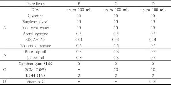
SCM: stem cell conditioned media (liquid), B: without SCM 10%, C: with SCM 10%, D: with SCM 10% + vitamin C 50 mg
Table 1. The experimental formulation of the cosmetic product containing stem cell conditioned media
medium. On the third day of the culture conditions, the culture solution was collected, filtered with a 0.2 mm syringe filter (13 mm, 100 units/ Whatman Cat No. 6779 1302), and used (stem cell conditioned media (liquid);
SCM). The collected culture solution was heated at 100 °C for 5 min, filtered with a syringe filter (0.2 mm), and used after obtaining a freeze-dried powder form. (boiled
& freeze dried stem cell conditioned media (powder); SCMBF).
2.2. Experiment method
2.2.1. Cell migration assay measurement In order to observe cell proliferation and migration under cell culture conditions, damaged cells are repaired to observe how substances affect the wound healing process.
Dispense into a 6 well plate at a concentration of 2 x 10⁵ cells/well, incubate for 24 hours to allow cells to adhere to the bottom, and draw an injury line with a yellow pipette tip. And after washing the separated cells in PBS, the samples were treated by concentration. The
rate of decrease in width and movement of cells were observed with a microscope for each concentration of the sample at 0, 3, 6, 9, and 24 hours.
2.2.2. Stem cell culture liquid cosmetics manufacturing method and prescription The serum formulation used in the experiment was prepared as follows according to the prescription in Table 1. After the (A) phase is heated to 30°C, the (B) phase is added to the (A) phase to completely dissolve.
Then, (C) phase was added to phases (A) + (B), stirred at 3,000 rpm in a homomixer for 15 minutes, and then a serum formulation was obtained and used in the experiment.
2.2.3. Clinical evaluation method
2.2.3.1. Research subject
This study targeted 10 women in their twenties who satisfied the following conditions and agreed to participate in the study.
According to the guidelines for evaluating the effectiveness of functional cosmetics suggested
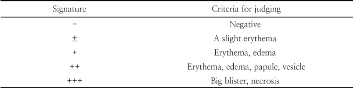
Signature Criteria for judging
- Negative
± A slight erythema
+ Erythema, edema
++ Erythema, edema, papule, vesicle
+++ Big blister, necrosis
Table 2. A criterion of patch test
by the Korea Food and Drug Administration when selecting a study subject, the following items are excluded.
1) Women of childbearing potential or pregnant or lactating
2) People with a history or concern of sensitivity, irritability, or photosensitization 3) Persons who use skin cosmetic products containing steroids for more than 1 month 4) 12 months have passed since participating in the same test
6) Persons with skin abnormalities such as spots, acne, erythema, and expansion of capillaries at the irradiated area
7) When similar cosmetics or drugs were used within 12 months before the start of the study
8) Others who are considered unsuitable for the experiment by the judgment of the main tester
2.2.3.2. Clinical research method
The experiment period was carried out for a total of 5 days. During the experiment, the inner part of the arm was kept out of water, and the use of the body product that was normally used was stopped, and a transparent flat was made to minimize variation in the result, and the correct position was drawn on the inside of the arm.
The experimental methods of this study are experimental groups A, B, C, D (A: no application group, B: stem cell culture solution without cosmetics, C: stem cell culture solution 10% added group, D: stem cell culture solution 10% and vitamin C added
group) and applied after peeling, and instructed to apply at 10 am and 5 pm of the day. It was measured and compared at 3 pm once a day.
2.2.3.3. Skin irritation
0.5 mL of serum and vitamin C were used on the inner side of the subject's upper arm, and an untreated patch was used as a control.
As a method, a tape with finn chambers (Epitest®, Tuusula, Finland) attached to each subject was used, and the tape was attached and removed 48 hours later, and visual analysis was performed 1, 2 hours and 48 hours after removal. Table 2, which is the criteria of the International Research Council on Contact Dermatitis, was applied as the criteria.
2.2.4. Skin measurement tools
2.2.4.1. Measurement method
For accurate skin measurement, the experimenters use the same seaweed peel prepared after setting a certain period of time, and the indoor environmental conditions at the time of measurement after damage to the skin measurement site is a constant temperature that maintains an indoor temperature of 22 to 24°C and an indoor humidity of 40 to 60%.
It was measured after damage in the indoor conditions of humidity and 30 minutes or 1 hour after application. When measuring, divide the inner part of the lower arm into 4 places and measure it by dividing it into the right (2 cm from the side), the left (2 cm from the
TEWL (%) =
side), the lower right (2 cm from the top), and the lower left (2 cm from the top). In order to increase the accuracy, a constant area was measured repeatedly 4 times.
2.2.4.2. Measurement of skin's transdermal water loss evaporation
To measure the level of moisture loss from the skin, transepidermal water loss (TEWL) was measured using a Vapometer (Delfin, Finland) before and after skin damage, respectively. Before skin damage, TEWL comes out low, after skin damage, TEWL comes out low, and after damage, skin damage occurs, which means that the skin barrier function is deteriorated. At the time of measurement, in order to increase the accuracy, the average value was recorded by repeating a constant area 4 times at all times, and the same person measured TEWL of the skin from start to finish. TEWL was measured once a day after sample treatment for 5 days at the measurement site under the arm.
Sample treatment after
damage TEWL ☓ 100 (5)
Before damage TEWL
2.3. Statistics processing
All experiments in this study were independently conducted three or more times under the same conditions to obtain experimental results, and all experimental results were expressed as mean±standard deviation. Statistical analysis was performed using SPSS Window Version 17.0 (SPSS Inc., Illinois, USA).
3. Results and Discussion
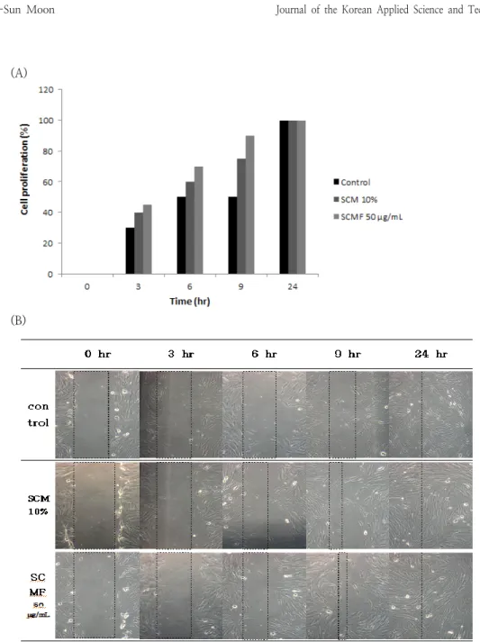
3.1. Cell migration assay measurement To analyze the effect of stem cell culture media on cell migration, a wound healing
assay was performed on HDF cells. All the cells in the line were removed by scratching the HDF cells uniformly grown as a single layer with a yellow tip. Thereafter, the cells were cultured for 0, 3, 6, 9, and 24 hours, and the degree of cell migration was compared and observed at concentrations of control, SCM 10%, and SCMF 50 mg/mL. As a result, it was observed that with 10% SCM and 50 mg/mL of SCMF, the number of cells increased significantly after 9 hours, resulting in the fastest healing of the wound [10]. It was observed that the effect of the stem cell culture medium in the whole sample treatment in wound healing increases the mobility of cells and promotes cell division to help wound healing. These results seem to be affected by cytokines in the stem cell culture medium, and cell migration to the wound area rapidly occurs.
3.2. Effects of the Thermal Stability of Stem Cell Culture Media on Cell Migration
In order to analyze the effect of stem cell culture medium on cell migration, a wound healing assay was performed on HDF cells [11]. All the cells in the line were removed by scratching the HDF cells uniformly grown as a single layer with a yellow tip. Thereafter, the degree of migration of the cells to the area in the line from which the cells were removed by culturing for 0, 3, 6, 9, and 24 hours was compared and observed at concentrations of SCMHF 3-7 and SCMBF 50 mg/mL. As a result, it was confirmed that cell mobility was compared after 3 hours of SCMHF 3-7 and 3 hours of SCMBF. In SCMBF, it was confirmed that the mobility of cells increased after 3 hours and 9 hours than SCMHF 3-7.
Likewise, after 9 hours in SCMBF, it was observed that the cells were concentrated by increasing the mobility and promoting cell division compared to other samples. As shown in Figure 19, it was observed that the overall sample treatment of SCMHF 3-7 and SCMBF
(A)
(B)
Fig. 1. Effects of SCM and SCMF on (A)proliferation and (B)migration of HDF cells.
SCM: stem cell conditioned media (liquid), SCMF: freeze dried stem cell conditioned media (powder)
increases cell mobility and promotes cell division to help wound healing.
It was observed that the mobility of cells increased even after 24 hours even during heat treatment and high temperature preservation, so that the intervals of 0, 3, 6, and 9 hours
were compared, but after 24 hours, there was no effect on the thermal stability of the stem cell culture solution. Cell migration to the wound area seems to occur rapidly under the influence of cytokines in stem cell culture media.
(A)
(B)
Fig. 2. Thermal stability testing of SCMF with (A)proliferation and (B)migration on HDF cells.
SCMBF: boiled & freeze dried stem cell conditioned media (powder), SCMHF: heated &
freeze dried stem cell conditioned media (powder)
3.2.1. Skin irritation by patch test
In order to obtain a valid result through the patch test, care must be taken in the implementation method and interpretation of the result [12]. In particular, if a positive reaction is found, it is said that it is
impossible to distinguish between irritant and allergic reactions only by the shape of the reaction. [13]. In this case, it can be distinguished through the change over time of the reaction, medical history, usage test, and re-test [13, 14]. In case of positive reactions
Erythema Allergy Edema Swelling
Subject 1 (-) (-) (-) (-)
Subject 2 (-) (-) (-) (-)
Subject 3 (-) (-) (-) (-)
Subject 4 (-) (-) (-) (-)
Subject 5 (-) (-) (-) (-)
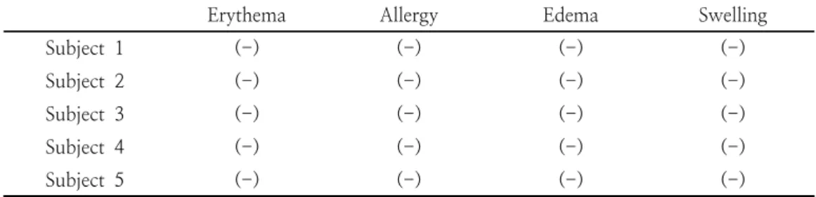
Table 3. Observation of skin disorder after 48 hours closed patch test at the same time on the 2nd and 4th days
after the patch test, continuous reaction, 2 Cases that were positive on the first day but negative on the fourth day were classified as disappearing reactions, and negative on the second day but positive on the fourth day were classified as delayed reactions [15]. As a result of Table 3, which conducted a closed patch test on the serum to which the stem cell culture medium used in the clinical trial was added in this experiment, there were no volunteers showing side effects during the test, and the patch was removed after 48 hours.
After a period of time, the disappearance of the transient erythema due to removal was waited and observed with the naked eye, and then the evaluation was made. In this experiment, no volunteers tested positive on the 2nd and 4th days, so the serum containing the stem cell culture medium used was evaluated as having no skin irritation.
3.3. Effect on transdermal water loss The skin barrier is the most important first line of defense that prevents loss of body fluids and protects the body from harmful environments, and refers to the skin's barrier function to toxic substances, microorganisms, mechanical irritation or ultraviolet rays [16].
Vapometer measures TEWL through relative humidity [17]. Vapometer is known as a portable device while showing results consistent with Tewameter, a device that has been widely used in the past [18]. There are many studies in which TEWL has been conducted on
various skin diseases or skin damage-induced individuals, and in the preceding paper [19], in order to conduct a study on the factors that affect skin barrier recovery, the induction of skin barrier damage was performed by tape stripping. Cellophane tape was used to repeatedly induce damage by attaching and peeling off the tape of the skin with a certain force.In this experiment, the effect of stem cell culture fluid on wound healing was investigated after increasing the amount of transdermal water loss by wounding the skin.
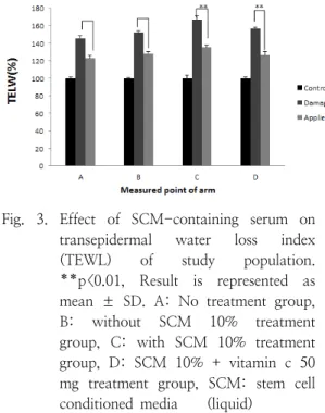
The skin barrier was damaged by seaweed peeling. As a result of the experiment in Fig.
3, as a result of comparing TEWL before and after skin damage, TEWL after skin damage was significantly higher than TEWL before skin damage. Group A was a group that did not treat anything, and the improvement rate was 22%, group B was a serum group without SCM 10% added, and the improvement rate was 24%, group C was a serum group added with 10% SCM, 31%, and group D. With the addition of 10% SCM and 50 mg of vitamin c, the improvement rate was 30%. This result shows that vitamin c is very difficult to penetrate the skin of vitamin c, which is one of the problems of cosmetics, so if applied after skin damage, it helps absorption. It can be assumed that the improvement rate was high because of the cycle, and it was confirmed that the improvement rate was 31%
in the C group to which 10% SCM was added, helping to recover skin wounds.
Fig. 3. Effect of SCM-containing serum on transepidermal water loss index (TEWL) of study population.
**p<0.01, Result is represented as mean ± SD. A: No treatment group, B: without SCM 10% treatment group, C: with SCM 10% treatment group, D: SCM 10% + vitamin c 50 mg treatment group, SCM: stem cell conditioned media (liquid)
4. Conclusion
One of the most prominent problems when stem cell culture is used as a cosmetic ingredient is that the ingredients generally have low thermal stability. Therefore, in this experiment, the physiological activity effect and thermal stability were examined for how the stem cell culture medium was heated or preserved at a high temperature on various effects of stem cells. The effect of stem cell culture solution on cell migration was analyzed, the effect on cell migration and proliferation after heat treatment, and cosmetic formulations were prepared to investigate the amount of transdermal water loss. As a result of the experiment, it was confirmed that cell mobility was increased in HDF cells, and the heat treatment of stem cell culture solution had no effect on cell migration and proliferation. Cosmetics showed a 31%
improvement in wound recovery rate, and stem cell culture solution recovered skin
wounds. It was confirmed to be helpful to After the heat treatment of the stem cell culture solution, the effect of the stem cell culture solution does not appear than before the heat treatment, so long-term preservation has been shown to have an effect as an active ingredient in cosmetics.
References
1. J. Thomson, A., Itskovitz-Eldor, J., Shapiro, S. S., Waknitz, M. A., Swiergiel, J. J., Marshall, V. S., & Jones, J. M,
“Embryonic stem cell lines derived from human blastocysts“, S
cience,
Vol.282, No.5391, pp.1145-1147, (1998).2. Zuk, P. A., Zhu, M. I. N., Mizuno, H., Huang, J., Futrell, J. W., Katz, A. J., &
Hedrick, M. H, “Multilineage cells from human adipose tissue: implications for cell-based therapies”,
Tissue engineering,
Vol.7, No.2, pp.211-228, (2001).3. Tsai, M. S., Lee, J. L., Chang, Y. J., &
Hwang, S. M, “Isolation of human multipotent mesenchymal stem cells from second‐trimester amniotic fluid using a novel two‐stage culture protocol”,
Human reproduction,
Vol.19, No.6, pp.1450-1456, (2004).4. Shin, M. S. “Present and future of aesthetic plastic plastic surgery in Korea“,
J. Korean Med. Assoc,
Vol.54, No.6, pp.581-588, (2011).5. Sordi V, Malosio M.L, Marchesi F, Mercalli A, Melzi R, Giordano T, Belmonte N, Ferrari G,
et el.
“Bone marrow mesenchymal stem cells express a restricted set of functionally active chemokine receptors capable of promoting migration to pancreatic islets”,Blood,
Vol.106, No.41, pp.9-427, (2005).6. Kim W.S, Park B.S, Sung J.H, Yang J.M, Park S.B, Kwak S.J, Park J.S. “Wound healing effect of adipose-derived stem cells: A critical role of secretory factors
on human dermal fibroblasts”,
J.
Dermatol. Sci.,
Vol.48, No.1, pp.15—24, (2007).7. Varma, Maikel J. Oedayrajsingh, et al.
“Phenotypical and functional characterization of freshly isolated adipose tissue-derived stem cells",
Stem cells and development,
Vol.16. No.1, pp.91-104, (2007).8. Xu, Yu-Xin, et al. “Mesenchymal stem cell therapy for diabetes through paracrine mechanisms”,
Medical hypotheses,
Vol.71, No.3, pp.390-393, (2008).9. Ji, S. Q., Cao, J., Zhang, Q. Y., Li, Y.
Y., Yan, Y. Q., & Yu, F. X. “Adipose tissue-derived stem cells promote pancreatic cancer cell proliferation and invasion. Brazilian”,
Journal of Medical and Biological Research
, Vol.46 No.9, pp.758-764, (2013).10. Strem, B. M., Hicok, K. C., Zhu, M., Wulur, I., Alfonso, Z., Schreiber, R. E., &
Hedrick, M. H. “Multipotential differentiation of adipose tissue-derived stem cells”,
The Keio journal of medicine,
Vol.54, No.3, pp.132-141, (2005).11. Kim, J. M., Lee, S. T., Chu, K., Jung, K.
H., Song, E. C., Kim, S. J.,& Roh, J. K.
“Systemic transplantation of human adipose stem cells attenuated cerebral inflammation and degeneration in a hemorrhagic stroke model”,
Brain research,
Vol.1183, pp.43-50, (2007).12. Constantin, G., Marconi, S., Rossi, B., Angiari, S., Calderan, L., Anghileri, E., &
Bonetti, B. “Adipose‐derived mesenchymal stem cells ameliorate chronic experimental autoimmune encephalomyelitis”,
Stem cells,
Vol.27, No.10, pp.2624-2635, (2009).13. Camberlain, G., Fox, J., Ashton, B., &
Middleton, J. “Mesenchymal stem cells:
their phenotype, differentiation capacity, immunological features, and potential for homing”,
Stem Cells,
Vol.25, No.11, pp.2739-2749, (2007).14. Carpenter, M. K., Rosler, E., & Rao, M.
S. “Characterization and differentiation of human embryonic stem cells”,
Cloning &
Stem Cells
, Vol.5, No.1, pp.79-88, (2003).15. Buonocore, D., Nobile, V., Michelotti, A.,
& Marzatico, F. “Clinical Efficacy of a cosmetic treatment by Crescina® human follicle stem cell on healthy males with androgenetic alopecia”,
Dermatology and therapy,
Vol..3, No.1, pp.53-62, (2013).16. Food and Drug Administration,
Director of the Food and Drug Administration.
Regulations on the designation of ingredients for cosmetics.
No.2010, pp.99, (2010).17. Jouni N, Esko A, Pekka A, Lahtinen MR, Ilkka H, Tapani L. “A closed unventilated chamber for the measurement of transepisermal water loss”,
Skin Res.
Technol.,
Vol..9, No.2, pp.85-89, (2003).18. Kristien DP, Evi H, Ralf A, Wiesemann F, Rogiers V. “Validation of the VapoMeter, a closed unventilated chamber system to assess tranespidermal water loss vs. the open chamber Tewameters”,
Skin Res.
Technol.,
Vol..11, No.1, pp.61-69, (2005).19. Cha J. H, Kim Y. B, “The study on the factors which improve skin barrier recovery”,