Expression and Purification of Three Lipases (LipAD1, LipAD2, and LipAD3) and a Lipase Chaperone (LipBD) from Acinetobacter schindleri DYL129
Sun-Hee Kim
†, Yong-Suk Lee
†, Hae-Rin Jeong, Hyo-Min Pyeon, Ju-Soon You and Yong-Lark Choi*
Department of Biotechnology, College of Natural Resources and Life Science, Dong-A University, Busan 49315, Korea Received February 23, 2019 /Revised April 24, 2019 /Accepted April 25, 2019
Previously, three kinds of lipases, lipAD1, lipAD2, and lipAD3, and lipase chaperone, lipBD, of Acinetobacter schindleri DYL129 isolated from soil sample were reported. In this report, three lipase and lipase chap- erone were cloned into the pET32a(+) or pGEX-6P-1 vectors for protein expression in Escherichia coli, and named as pETLAD1, pETLAD2, pETLAD3 and pETLBD or pGEXLAD1, pGEXLAD 2, pGEXLAD3 and pGEXLBD, respectively. Protein expression rate was higher in pET system than in pGEX system.
Although LipAD1 and LipAD2 were produced as inclusion bodies, their expression levels were high.
So LipAD1 and LipAD2 could be solubilized in 8 M urea buffer and purified. LipAD3 and LipBD were overexpressed in soluble form and purified. Those proteins were purified by His-tag affinity chroma- tography connected in AKTA prime system. The activities of the purified lipases were demonstrated with 1% tributyrin agar plate. After purification, molecular mass was determined with sodium dodecyl sulfate-polyacrylamide gel electrophoresis. LipAD1 showed high activity toward ρ-nitrophenyl acetate and ρ-nitrophenyl butyrate, LipAD2 showed high activity toward ρ-nitrophenyl acetate and ρ-nitro- phenyl myristate, and LipAD3 showed high activity toward ρ-nitrophenyl acetate, ρ-nitrophenyl buty- rate, and ρ-nitrophenyl miristate, respectively. Three lipases, LipAD1, LipAD2, and LipAD3, showed optimal reaction at 50℃ using ρ-nitrophenyl butyrate, as substrate.
Key words : Acinetobacter schindleri, lipase, lipase chaperone, protein expression, purification
†Authors contributed equally.
*Corresponding author
*Tel : +82-51-200-7585, Fax : +82-51-200-6536
*E-mail : [email protected]
This is an Open-Access article distributed under the terms of the Creative Commons Attribution Non-Commercial License (http://creativecommons.org/licenses/by-nc/3.0) which permits unrestricted non-commercial use, distribution, and reproduction in any medium, provided the original work is properly cited.
Journal of Life Science 2019 Vol. 29. No. 4. 492~498 DOI : https://doi.org/10.5352/JLS.2019.29.4.492
Introduction
Many microbial lipases are available as commercial prod- ucts, and the majority of these enzymes are used in de- tergents, cosmetics, organic synthesis, and as food flavoring agents [1]. Lipases are valued biocatalysts owing to advan- tages such as their ability to display chemoselectivity, re- gioselectivity, and stereoselectivity [6, 12]. Furthermore, these enzymes are readily available in large quantities, as many of these may be produced in high yields from micro- organism such as fungi and bacteria [27]. The crystal struc- tures of several lipases have been solved, facilitating the de- sign of rational engineering strategies [1, 9]. Lipases do not usually require cofactors and do not catalyze side reactions;
moreover, these enzymes act under extremely mild con- ditions and are stable in organic solvents [1].
Acinetobacter is a strictly aerobic, gram-negative coccoba- cillus that is ubiquitous in geographical distribution [29]. The genus is best known for its capacity for bioremediation of alkanes and aromatic hydrocarbons as well as for the pro- duction of high molecular weight heteropolysaccharides, which may serve as powerful emulsifiers with high commer- cial potential [8, 21]. These properties make lipases from Acinetobacter the most widely used biocatalysts in organic chemistry. So far, several Acinetobacter lipases have been suc- cessfully cloned and expressed in heterologous hosts [2, 5, 21, 24].
The expression of any foreign protein in prokaryotic sys- tems is a common approach to achieve high-level expression in fundamental studies as well as for commercial purposes [23]. The fast growth rate and ease of cultivation of Eschericoli coli make this organism suitable for industrial application.
However, gene expression is mainly performed with two
main goals, i.e., to achieve high cell density and high-level
gene expression [17]. The expression vector and host are im-
portant factors that determine the maximal expression of the
cloned genes. However, molecular cloning of a foreign gene
may not ensure its successful expression [15, 23]. The most
difficult problems with bacterial expression are proteolytic
degradation and production of proteins that accumulate in
- Note -
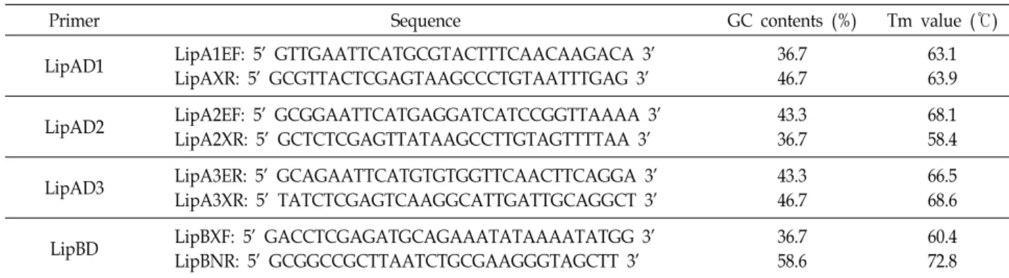
Table 1. PCR primers used for the expression of three lipases and the lipase chaperone
Primer Sequence GC contents (%) Tm value (℃)
LipAD1 LipA1EF: 5' GTTGAATTCATGCGTACTTTCAACAAGACA 3'
LipAXR: 5' GCGTTACTCGAGTAAGCCCTGTAATTTGAG 3'
36.7 46.7
63.1 63.9
LipAD2 LipA2EF: 5' GCGGAATTCATGAGGATCATCCGGTTAAAA 3'
LipA2XR: 5' GCTCTCGAGTTATAAGCCTTGTAGTTTTAA 3'
43.3 36.7
68.1 58.4
LipAD3 LipA3ER: 5' GCAGAATTCATGTGTGGTTCAACTTCAGGA 3'
LipA3XR: 5' TATCTCGAGTCAAGGCATTGATTGCAGGCT 3'
43.3 46.7
66.5 68.6
LipBD LipBXF: 5' GACCTCGAGATGCAGAAATATAAAATATGG 3'
LipBNR: 5' GCGGCCGCTTAATCTGCGAAGGGTAGCTT 3'
36.7 58.6
60.4 72.8
their misfolded forms as inclusion bodies [23].
In this study, histidine-tagged lipases, LipAD1, LipAD2, and LipAD3 from A. schindleri DYL129 were overexpressed in E. coli and purified with His-tag affinity chromatography.
Materials and Methods
Expression vector and media
E. coli BL21 (DE3) was used as the host for the expression of lipases and lipase chaperone genes under the control of T7 promoter. For E. coli expression studies, pET-32a(+) (Novagen, Madison, WI, USA) was used. The pET-32a(+) vector carried six histidine and the trx gene was fused to facilitate purification after the process of protein expression.
E. coli BL21 (trxB) transformants were cultured at 37℃ in 2‘ YTA supplemented with 50 μg/ml of kanamycin. The me- dium (g/l) used for expression comprised 16 g of yeast ex- tract, 10 g of tryptone, and 5 g of sodium chloride (NaCl), while its pH was adjusted to 7.0. After autoclaving, ampi- cillin was added at 100 μg/ml concentration.
Amplification and subcloning of lipase and lipase chaperone genes
The open reading frame (ORF) of the lipase and lipase chaperone genes were amplified from the genomic DNA of A. schindleri DYL129 with Taq DNA polymerase using the following polymerase chain reaction (PCR) conditions: an in- itial denaturation step at 95℃ for 5 min, 30 cycles at 95℃
for 1 min, annealing at 60-65℃ for 40 s, and extension at 72℃ for 100 s [13]. The final extension was performed for 10 min and preservation was carried out at 4℃. The primers used were designed as per the previous report, as indicated in Table 1 [10]. The amplified product was purified with a PCR purification kit (SolGent, Korea) and digested with EcoRI/XhoI. After digestion, the lipase and lipase chaperone
genes were subcloned in pET-32a(+) or pGEX-6P-1 vectors digested with the same restriction enzymes. The eight re- combinant plasmids were selected and named as pETLAD1- 3 and pETLB or pGEXLAD1-3 and pGEXLB, respectively.
Protein expression and solubilization
E. coli BL21 (trxB) cells containing the recombinant plas- mids were grown at 37℃ in 500 ml of 2‘ YTA up to an OD
600nmvalue of 0.5-0.6. The culture was treated with 0.1 mM isopropyl β-D-1-thiolgalactopyranoside (IPTG) and the incubation was continued at 37℃ for 4 hr. Cells were har- vested by centrifugation at 6,000 rpm for 10 min and washed twice with buffer A (20 mM Tris-HCl [pH 7.4], 0.1 M NaCl, 5% glycerol and 1% Triton X-100). The cells collected by cen- trifugation were resuspended in buffer A and sonicated with a sonicator (SONICS & MATERIALS INC. DANBURY, CT, USA) for 30 s. The cell-free extract was centrifuged at 13,000 rpm for 20 min to remove cell debris. For aggregated pro- teins, the cell debris was washed once with buffer A and solubilized in buffer B (8 M urea, 0.1 M NaCl, 1 mM dithio- threitol [DTT], 20 mM Tris-HCl [pH 7.4]) for 1 hr at room temperature. After solubilization, the samples were centri- fuged for 10 min and the supernatants were collected. The protein concentration of enzyme preparations was de- termined by Bradford method with bobine serum albumin as a standard.
Protein purification
LipAD3 and LipBD were purified on a His-high trap col-
umn (Amersham-Pharmacia Biotech, Sweden). The cell-free
extracts were loaded onto His-high trap column run by
AKTA Prime. The column was washed with 30 ml of bind-
ing buffer (20 mM Tris-HCl [pH 7.4], 0.1 M NaCl, 5 mM
imidazole) to remove the unbound proteins. Elution was
performed with 30 ml of elution buffer (20 mM Tris-HCl
[pH 7.4], 0.1 M NaCl, 500 mM imidazole). Every 1 ml frac- tion was collected and analyzed by 12% sodium dodecyl sul- fate-polyacrylamide gel electrophoresis (SDS-PAGE). The purified sample was dialyzed and concentrated with 20 mM Tris-HCl (pH 7.4) buffer using Centricon (Amicon ultra, Millipore, cut-off size 10,000 Da).
Inclusion body purification and refolding
For solubilization of proteins, LipAD1 and LipAD2 were loaded onto a His-high trap column. The column was wash- ed with 30 ml of binding buffer (8 M urea, 20 mM Tris-HCl, 0.1 M NaCl [pH7.4]) and eluted with 30 ml of elution buffer (8 M urea, 20 mM Tris-HCl, 0.1 M NaCl, 500 mM imidazole [pH 7.4]). After purification, the collected sample was di- luted with buffer B and refolded using buffer C (2 M urea, 20 mM Tris-HCl, 0.1 M NaCl, 1 mM DTT [pH 7.4]) at 4℃
for 2 days. The refolded sample was dialyzed with 20 mM Tris-HCl (pH 7.4) buffer at 4℃ for 2 days and concentrated.
Electrophoresis
We performed SDS-PAGE as described by Laemmli [11]
using a 5% stacking gel and 12% resolving gel. A broad range of protein standards (ELPIS-BIOTECH, Daejeon, Korea) were used as molecular weight markers. The insoluble pellet was resuspended in 300 μl of Tris-HCl buffer (pH 7.4), and 4× sample loading buffer was added. The mixture (8 μl) was boiled for 10 min and loaded onto a 12% polyacrylamide gel (Fig. 3A).
Characterization of enzyme activity
After protein separation, the characteristics of lipases (2 μg), with lipase chaperone (2 μg) for refolding, were de- termined. The release of ρ-nitrophenyl (ρ-NP) from ρ-NP de- rivative substrates was measured as described. A total of 0.025 M ρ-NP butyrate was dissolved in 99% ethanol and mixed with 20 mM Tris-HCl buffer (pH 7.4). After in- cubation at 50℃ for 30 min, the lipase activity was measured by monitoring the absorbance at 420 nm that indicated the amount of released ρ-NP. One unit of activity was defined as the amount of enzyme that released 1 μmol of ρ-NP per minute under the assay conditions.
Results and Discussion
Subcloning of the three lipases and a lipase chaperone The nucleotide sequences of the four fragments (LipAD1-
3 and LipBD) were obtained with primers (Table 1) from the genomic DNA of A. schindleri DYL129 and cloned into the expression vector pET-32a(+) after digestion with EcoRI and XhoI. The primers were designed specific to the 5‘ and 3‘ region of the ORFs 1-4. The recombinant plasmids with LipAD1-3 and LipBD were named pETLAD1-3 and pETLB, respectively, and transformed into E. coli BL21 (trxB). The overexpression of the lipase genes was confirmed by analyz- ing the total proteins from the non-induced and IPTG-in- duced cells on the SDS-PAGE gel (data not shown).
Protein expression of three lipases
A. schindleri DYL129 lipase and lipase chaperone genes were expressed as fusion proteins in E. coli. A good combina- tion of expression system and host is necessary to obtain high-level expression. E. coli is an organism widely used for the overproduction of recombinant proteins. However, in spite of the extensive knowledge about the genetics and mo- lecular biology of E. coli, not every gene may be effectively expressed in this organism. In general, high-level gene ex- pression may be achieved (up to 50% of the total cell protein) through gene manipulation and appropriate choice of the vector-host combination and IPTG concentration. The pET [7, 16, 19, 31] and pGEX [4, 13, 14, 18, 26] vector systems have been extensively used for the protein overexpression in E. coli. In this study, LipAD1 was expressed with pET and pGEX under the regulation of T7 and tac promoter, respectively. These systems allow regulation of the expression of the gene of interest under the control of different pro- moters to achieve high-level expression in response to chem- ical induction. We compared these two vector systems and chose the one that showed higher levels of lipase expression.
The plasmids pGEXLAD1 and pETLAD1 were expressed for
4 hr at 37℃ in E. coli BL21 (trxB) after induction with 0.1
mM IPTG at an OD
600nmvalue of 0.5-0.6. We confirmed the
expression level using a 12% SDS-PAGE gel after loading
equal amounts of total proteins (30 μg) into the gel. LipAD1
expression was greatly increased with both the expression
systems, but the expression level observed with pET system
(32 μg/μl) was around 1.5-fold higher than that reported
with pGEX system (22 μg/ μl) under same conditions. Based
on this result, we tested the effect of IPTG concentration on
protein expression (data not shown). Each LipAD1 fusion
protein was induced with different concentrations of IPTG
(0.01 and 0.03 mM). In the presence of 0.01 mM IPTG, the
expression was higher with pETLAD1 than with pGEXLAD1,
M 1 2 3 4 5 6 7 8
M 1 2 3 4 5 6 7 8
A
B
Fig. 1. Overexpression of LipAD1 and LipAD2. A) M, size mark- er (100, 70, 50, 40, 30, 20, 15 kDa); 1-4 insoluble proteins (1: NIP pET, 2: IP pET, 3: NIP His-LipAD1, 4: IP His-LipAD1); 5-8 soluble proteins (5: NI pET, 6: I pET, 7: NI His-LipAD1, 8: I LipAD2), B) M, size marker; 1-4 soluble proteins (1: NI pET, 2: I pET, 3: NI His-LipAD2, 4: I His-LipAD2); 5-8 insoluble proteins (5: NIP pET, 6:
IP pET, 7: NIP His-LipAD2, 8: IP His-LipAD2). Arrow indicates expressed protein (about 56 kDa) NIP: unin- duced protein pellet, IP: induced protein pellet, NI: unin- duced protein, I: induced protein.
M 1 2 3 4 5 6 7 8 M 1 2 3 4 5 6 7 8
A
B
Fig. 2. Expression of LipAD3 and LipBD. A) The soluble pro- teins were loaded onto a 12% polyacrylamide gel. M:
size marker; 1: NI pET; 2: I pET; 3: NI His-LipAD3; 4, 5: I His-LipAD3; 6: NI LipBD; 7, 8: IP His- LipBD. B) Insoluble proteins. M: size marker; 1: NIP pET; 2: IP pET;
3: NIP His-LipAD3; 4, 5: IP His-LipAD3; 6: NIP His- LipBD; 7, 8: IP His- LipBD. Arrow indicates expressed proteins (about 36 and 57 kDa, respectively). NIP: unin- duced protein pellet, IP: induced protein pellet, NI:
uninduced protein, I: induced protein.
while opposite effects were observed with 0.03 mM IPTG.
Hence, we chose the pET vector system, and pETLAD1 was overexpressed for further purification process.
Purification of three lipases and the lipase chaperone The lysates from the induced and non-induced bacteria and control bacterium (vector only) were analyzed by SDS- PAGE (Fig. 1). Fig. 1 shows two bands about 56 kDa in size (Fig. 1A: lane 4, LipAD1; Fig. 1B: lane 8, LipAD2) corre- sponding to the His-lipase fusion protein. LipAD1 and LipAD2 were expressed only as inclusion bodies. The over- expressed LipAD3 and LipBD proteins were observed in Fig.
2A (lane 4 and 5, LipAD3; lane2, 7, and 8, LipBD as soluble protein). LipAD3 and LipBD were not only expressed in the soluble form but also as inclusion bodies. Fig. 2B, lanes 4, 5, 7, and 8 show inclusion bodies of LipAD3 and LipBD.
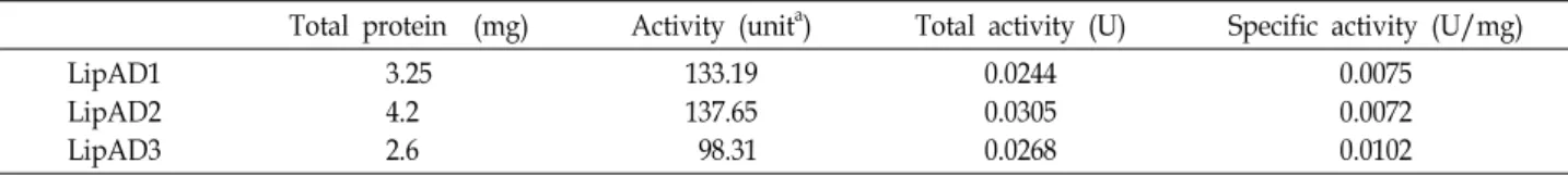
The size of LipAD3 and LipBD was about 36 and 57 kDa, respectively. The inclusion bodies are solublized using high concentration of urea [25]. No protein band was detected at same positions in the induced and non-induced control (vector only). These observations confirm the expression of the lipase gene. After expression, the fusion proteins were purified by affinity chromatography (Table 2) and analyzed by SDS-PAGE (Fig. 3A). The activity of the purified enzyme was measured with a plate assay, wherein the enzyme was added (20 μl) and the reaction was performed at 37℃ for 12 hr. Fig. 3B shows a yellow clear zone, indicative of the released fatty acids.
Substrate specificity of lipases
The substrate specificity of LipAD1-3 was examined using
various fatty acid esters of ρ-NP and the activity was meas-
ured after 30 min at 37℃. The enzymes showed activity to-
Table 2. Purification of lipases from Acinetobacter schindleri DYL129
Total protein (mg) Activity (unita) Total activity (U) Specific activity (U/mg) LipAD1
LipAD2 LipAD3
3.25 4.2 2.6
133.19 137.65 98.31
0.0244 0.0305 0.0268
0.0075 0.0072 0.0102
aOne unit of enzyme activity catalyzes the production of 1μmol of
ρ
-NP per min.A B
Fig. 3. The three lipases were purified by affinity chromatog- raphy. A) SDS-PAGE analysis of the samples of purified lipases and lipase chaperone. Samples were resolved on 12% polyacrylamide gel and stained with Coomassie blue R-250. Lane 1: molecular size markers (100, 70, 50, 40, 30, 20, 15 kDa); lane 2: N His- LipBD; lane 3: P His- LipBD, 57 kDa; lane 4: native His-LipAD1; lane 5: P His-LipAD1, 56 kDa; lane 6: N His-LipAD2; lane 7: P His-LipAD2, 56 kDa; lane 8: N His-LipAD3; lane 9: P His-LipAD3, 34 kDa (N: native protein, P: purified protein). B) Activity test for the purified and dialyzed lipases. The enzyme activities of fractions 6-9 were tested with 1% tributyrin agar plate containing 0.01% phenol red. The yellow color indicates the released fatty acids from tributyrin.
Fig. 4. Substrate specificity of His-LipAD1, His-LipAD2, and His-LipAD3. Acyl chain length specificity of purified li- pases was determined from their activities toward vari- ous esters of
ρ
-NP (0.5 mM). Percentages shown are rel- ative to maximum activity.Fig. 5. Effect of temperature on the activity of lipases. The en- zyme was incubated in 20 mM Tris-HCl buffer (pH 7.4) for 30 min at various temperatures. The activity was de- termined using
ρ
-NP butyrate as substrate. The value obtained for His-LipAD2 at 50℃ was considered as 100%.ward a broad range of acyl chain lengths (Fig. 4). LipAD1, LipAD2, and LipAD3 showed maximum activity toward C2, C4, and C14. In particular, LipAD2 showed a strong activity
toward C14 and was thought to attack C2 and C14.
Acinetobacter sp. RAG-1 was shown to exhibit maximum ac- tivity toward medium-length fatty acid esters (C6-C8).
Lipases and esterases share common substrate specificities [28]. However, unlike esterases, lipases often demonstrate interfacial activation, i.e., a marked increase in activity upon the formation of a lipid-water interface [22]. Therefore, lipase substrates are typically long-chain (≥ C10) fatty acid esters available in the micellar form [30]. The properties of LipAD1-3 reported herein are slightly different, and the three enzymes had different substrate activities. The differ- ent substrate specificities of each enzyme are thought to help break down the various types of hydrocarbon chains present in the environment [5].
Temperature effects on lipases
The optimum reaction temperature for LipAD1-3 activity
toward ρ-NP butyrate was 50℃ (Fig. 5). At this temperature,
LipAD1-3 showed about three fold increase in activity com-
pared with that observed at 37℃. The optimal reaction tem-
perature reported in the present study was similar to that
reported for many bacterial lipases under similar ex- perimental conditions. Activity at high temperature is a use- ful characteristic for lipases used in detergent formulations and biotransformation. Many bacterial lipolytic enzymes showed the highest activity at around 50℃. For example, the lipase from Ralstonia sp. showed optimal activity at 50-55
℃ [32], the alkaline lipase from Serratia sp. W3 showed its maximal activity at 55℃ [3], and the halophilic lipase from Marinobacter litoralis SW-45 showed optimal activity at 50℃
[20].
Acknowledgment
This research was supported by the Basic Science Re- search Program through the National Research Foundation of Korea (NRF) funded by the Ministry of Education (2017R1D1A3B03034514) and the Ministry of Science and ICT (2017R1C1B5014876).
References
1. Aravindan, R., Anbumathi, P. and Viruthagiri, T. 2007.
Lipase applications in food industry. Ind. J. Biotechnol. 6, 141-158.
2. Cho, A. R., Yoo, S. K. and Kim, E. J. 2000. Cloning, sequenc- ing and expression in Escherichia coli of a thermophilic lipase from Bacillus thermoleovorans ID-1. FEMS Microbiol. Lett. 186, 235-238.
3. Eddehech, A., Zied, Z., Aloui, F., Smichi, N., Noiriel, A., Abousalham, A. and Gargouri, Y. 2018. Production, purifi- cation and biochemical characterization of a thermoactive, alkaline lipase from a newly isolated Serratia sp. W3 tunisian strain. Int. J. Biol. Macromol. 123, 792-800.
4. Gavya, S. L., Arora, N. and Ghosh, S. S. 2018. Retention of functional characteristics of glutathione-S-transferase and lactate dehydrogenase-A in fusion protein. Prep. Biochem.
Biotechnol. 48, 128-135.
5. Gupta, K. K., Jagtap, S., Priya, R. and Ramadas, K. 2018.
Purification, characterization of alkaline cold active lipase from Acinetobacter radioresistens PR8 and development of a new zymography method for lipase detection. Pretein Pept.
Lett. 25, 897-907.
6. Jaeger, K. E. and Eggert, T. 2002. Lipase for biotechnology.
Curr. Opin. Biotechnol. 13, 390-397.
7. Jawed, M., Pi, J., Xu, L., Zhang, H., Hakeem, A. and Yan, Y. 2016. Enhanced H2 production and redirected metabolic flux via overexpression of fhlA and pncB in Klebsiella HQ-3 strain. Appl. Biochem. Biotechnol. 178, 1113-1128.
8. Kaplan, N. and Rosenberg, E. 1982. Exopolysaccharide dis- tribution and bioemulsifier production by Acinetobacter cal- coaceticus BD4 and BD413. Appl. Environ. Microbiol. 44, 1335- 1341.
9. Kim, H. K., Park, S. Y., Lee, J. K. and Oh, T. K. 1998. Gene cloning and characterization of thermostable lipase from Bacillus stearothermophilus L1. Biosci. Biotechnol. Biochem. 62, 66-71.
10. Kim, S. H., Park, I. Y., Lee, S. C., Lee, Y. S., Ahn, S. C., Kim, C. M. and Choi, Y. L. 2008. Discovery of three novel lipase (LipA1, LipA2, LipA3) and lipase specific chaperone (LipB) gene present in Acinetbacter sp. DYL129. Appl. Microbiol.
Biotechnol. 77, 1041-1051.
11. Laemmli, U. K. 1970. Most commonly used discontinuous buffer system for SDS electrophoresis. Nature 227, 680-686.
12. Lee, Y. S. 2016. Isolation and characterization of a novel cold-adapted esterase, MtEst45, from Microbulbifer thermoto- lerans DAU221. Front. Microbiol. 7, 218.
13. Lee, Y. S., Park, I. H., Yoo, J. S., Chung, S. Y., Lee, Y. C., Cho, Y. S., Ahn, S. C., Kim, C. M. and Choi, Y. L. 2007.
Cloning, purification, and characterization of chitinase form Bacillus sp. DAU101. Bioresour. Technol. 98, 2734-2741.
14. Lee, Y. S., Yoo, J. S., Chung, S. Y., Lee, Y. C., Cho, Y. S.
and Choi, Y. L. 2006. Cloning, purification, and character- ization of chitosanase from Bacillus sp. DAU101. Appl.
Microbiol. Biotechnol. 73, 113-121.
15. Leow, T. C., Rahman, Z. A., Basri, M. and Salleh, A. B. 2004.
High level expression of thermostable lipase from Geobacillus sp. strain T1. Biosci. Biotechnol. Biochem. 68, 96-103.
16. Li, H. and Xia, Y. 2018. Recombinant production of the in- secticidal scorpion toxin BjαIT in Escherichia coli. Protein Expr. Purif. 142, 62-67.
17. Lin, W. J., Huang, S. and Chou, C. P. High-level extracellular production of penicillin acylase by genetic engineering of Escherichia coli. J. Chem. Tech. Biotechnol. 76, 1030-1037.
18. Lou, D., Tan, J., Zhu, L., Ji, S., Tang, S., Yao, K., Han, J. and Wang, B. 2018. Engineering Clostridium absonum 7α-hydrox- ysteroid dehydrogenase for enhancing thermostability based on flexible site and ∆∆G prediction. Protein Pept.
Lett. 25, 230-235.
19. Luo, H., He, C. and Han, L. 2018. Heterologous expression of ZjOMT from Zoysia joponica in Escherichia coli confers alu- minum resistance through melatonin production. PLoS One 13, e0196952.
20. Musa, H., Hafiz Kasim, F., Nagoor Gunny, A. A., Gopinath, S. C. B. and Azmier Ahmad, M. 2019. Enhanced halophilic lipase secretion by Marinobacter litoralis SW-45 and its poten- tial fatty acid esters release. J. Basic Microbiol. 59, 87-100.
21. Navon-Venezia, S., Zosim, Z., Gottlieb, A., Legmann, R., Carmeli, S., Ron, E. Z. and Rosenberg, E. 1995. A new bio- emulsifier from Acinetobacter radioresistens. Appl. Environ.
Microbiol. 61, 3240-3244.
22. Reis, P., Holmberg, K., Watzke, H., Leser, M. E. and Miller, R. 2009. Lipases at interfaces: a review. Adv. Colloid Interface Sci. 147-148, 237-250.
23. Rosano, G. R. and Ceccarelli, E. A. 2014. Recombinant pro- tein expression in Escherichia coli: advances and challenges.
Front. Microbiol. 5, 172.
24. Schmidt-Dannert, C., Rua, M. L., Atomi, H. and Schmid, R.
D. 1996. Thermoalkalophilic lipase of Bacillus thermocatenu-
초록: Acinetobacter schindleri DYL129 유래의 3개 lipases와 chaperone의 발현과 정제
김선희
†․이용석
†․정해린․편효민․유주순․최용락*
(동아대학교 생명자원과학대학 생명공학과)
기존 연구를 통하여 토양에서 분리한 Acinetobacter schindleri DYL129로부터 3개의 lipase 유전자(lipAD1, lipAD2 와 lipAD3)들과 1개의 chaperone (lipBD) 유전자를 보고하였다. 본 연구에서는 각 유전자들의 발현을 위해서 pET32a(+)와 pGEX-6P-1 벡터에 클로닝하여 각각을 pETLAD1-3와 pETLBD 또는 pGEXLAD1-3와 pGEXLB로 명 명하였으며, 단백질의 발현량은 pET 시스템을 사용할 때 1.5 배 정도 향상됨을 확인하였다. LipAD1과 LipAD2는 inclusion body 형태로 발현이 되었으며, LipAD3과 LipBD는 soluble type으로도 발현되었다. Inclusion body 형 태의 LipAD1과 LipAD2는 고농도의 우레아를 처리하여 refolding 시켰다. LipAD1은 C4와 C2를, LipAD2는 C2와 C14를 그리고 lipAD3은 C2, C4와 C14를 기질로 잘 이용하는 것을 확인하였다. 그리고 모든 효소들은 50℃에서 최적 활성을 나타내었다.
lantus. I. Molecular cloning, nucleotide sequence, purifica- tion and some properties. Biochim. Biophys. Acta 1301, 105- 114.
25. Singh, A., Upadhyay, V., Upadhyay, A. K., Singh, S. M. and Panda, A. K. 2015. Protein recovery from inclusion bodies of Escherichia coli using mild solubilization process. Microb.
Cell Fact. 14, 41.
26. Singh, P. K., Tang, M., Kumar, S. and Shrivastava, A. K.
2018. Decoding the role of hypothetical protein All32255 of anabaena PCC7120 in heavy metal stress management in Escherichia coli. Arch. Microbiol. 200, 463-471.
27. Singh, R., Kumar, M., Mittal, A. and Mehta, P. K. 2016.
Microbial enzymes: industrial progress in 21st century. 3 Biotech. 6, 174
28. Snellman, E. A., Sullivan, E. R. and Cowell, R. R. 2002.
Purification and properties of the extracellular lipase, LipA,
of Acinetobacter sp. RAG-1. Eur. J. Biochem. 269, 5771-5779.
29. Tripathi, P. C., Gajbhiye, S. R. and Agrawal, G. N. 2014.
Clinical and antimicrobial profile of Acinetobacter spp.: an emerging nosocomial superbug. And. Biomed. Res. 3, 13.
30. van Tilbeurgh, H., Egloff, M. P., Martinez, C., Rugani, N., Verger, R. and Cambillau, C. 1993. Interfacial activation of the lipase procolipase complex by mixed micelles revealed by X-ray crystallography. Nature 362, 814-820.
31. Wang, H., Zhong, X., Li, J., Zhu, M., Wnag, L., Ji, X., Fan, J. and Wang, L. 2018. Cloning and expression of H. influen- zae 49247 IgA protease in E. coli. Mol. Biotechnol. 60, 134-140.
32. Yoo, Y. H., Simkhada, J. R., Cho, S. S., Park, D. H., Kim, S. W., Seong, C. N. and Yoo, J. C. 2011. A novel alkaline lipase from Ralstonia with potential application in biodiesel production. Bioresour. Technol. 102, 6104-6111.