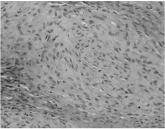
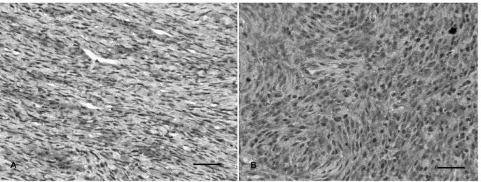
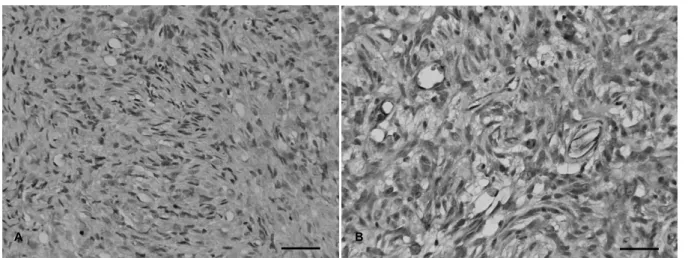
Canine nervous-tissue tumors with features of peripheral nerve sheath tumor: histopathological and immunohistochemical findings
전체 글
수치
관련 문서
The frequency of impacted tooth are the following orders : upper.. canine, upper incisor, lower premolar, upper premolar, lower canine. Traction period
-The presence of giant cells indicates that the phagocytic system is incapable of ridding the body of foreign matter, and that tissue destruction often occurs and results in
The present study investigated that CAFs in xenografted tumors had higher amounts of fatty acids, particularly OA, compared to normal fibroblasts, and promoted the
The most distinct cytological feature of DSRCT compared to the other small round cell tumors, is its stromal fi- brillary fragment, which is not a commonly found in body flu-
Moreover, the number of branches of the axillary nerve was associated with the distal insertion site of the deltoid muscle and proximal crossing point of the axillary
Results : The expression of p21 was increased in boderline serous tumor and serous cystadenocarcinoma in contrast to benign serous tumors. The expression of
• Generation of different specialized kinds of cells from zygote (fertilized egg) or other precursor cells.. – Generate blood cells, muscle
