Characterization of HA/PCL composite scaffolds fabricated by layer manufacturing technology
Seung Eon Kim1, Yong Taek Hyun1, Hui Suk Yun1, Taek Rim Yoon2, Su Jin Heo3, Jung Woog Shin3
1Center for Future Technology, Korea Institute of Materials Science, Changwon, Gyeongnam, Korea
2Center for Joint Deseases, Chonnam National University Hwasun Hospital, Hwasun, Jeonnam, Korea
3Department of Biomedical Engineering, Inje University, Gimhae, Gyeongnam, Korea Key Words : Layer manufacturing, HA/PCL, Scaffolds, Tissue engineering
Abstract
Layer manufacturing technology has been recently spotlighted as a promising candidate to fabricate porous scaffolds for tissue engineering, because it can provide three dimensional interconnectivity and different pore structures and on-demand scaffold design. This study aims to fabricate HA/PCL composite scaffolds for bone tissue engineering by a layer manufacturing technology, paste extruding deposition, and to characterize in vitro and in vivo biocompatibilities of the scaffolds. This study discusses the mechnical properties, proliferation and differentiation of osteogenic cells, and tissue in-growth and bone regeneration behavior using animal models.
1. Introduction
Scaffolds for tissue engineering should be highly porous, tissue supportable and biodegradable.
Furthermore, scaffolds for bone regeneration should be osteoinductive as well as osteoconductive.
Hydroxyapatite (HA) and poly ε-caprolactone (PCL) composites have been paid much attention as one of the prospective bone scaffold materials, due to their good combination of biodegradability and osteoconductivity (1).
By the way, pore structure of the scaffolds is very important to lead cell or tissue ingrowth. The 3D scaffolds fabricated by layer manufacturing process have been recently spotlighted, because they can provide sufficient interconnectivity of pores and mechanical stiffness. This study aims to fabricate HA/PCL composite 3D scaffolds by a modified layer manufacturing process and to characterize in vitro and in vivo biocompatibilities of the scaffolds as a candidate for bone tissue engineering applications. This study discusses the effect of HA particle size in nano- and micro-scale on the mechnical properties, proliferation and differentiation of osteogenic cells of the scaffolds(2). For the nano-HA/PCL composite scaffolds, tissue in-growth and bone regeneration behavior using animal models were also characterized.
2. Materials and Methods
Nano-hydroxyapatite (n-HA) was synthesized by a precipitation method using calcium nitrate and
ammonium phosphate. Micro-HA (m-HA) was purchased from Sigma Aldrich. Poly ε- caprolactone (PCL) (Sigma Aldrich, Mw 65 kDa) was dissolved in chloroform at 40 oC. Nano- and micro- hydroxyapatite (HA) was mixed with the PCL solution. The mixture paste was extruded and stacked layer by layer into a rectangular lattice shaped scaffolds using a PED machine.
Compression tests were conducted at room temperature using a Microload System (R&B Inc., Korea). Crosshead speed was 0.5 mm/min and 2 kgf load cell was employed. The compressive modulus was determined at initial strain range less than 30%.
Osteoblast-like MG63 cells (ATCC, USA) were cultured on the PCL scaffolds. Cell seeding density was 1x10⁶ cells/ml and culture medium was Dulbecco’s Modified Eagle Medium with 10% FBS and 1% P/S. To determine cell viability, MTT assay was carried out after 4 h, 1, 4, 7 day culture, using a ELISA reader.
To evaluate osteogenetic differentiation behavior, hBMSC was cultured on the scaffolds in culture medium with dexamethasone, glycerophosphate and L-ascorbic acid, and ALP assay was carried out. To evaluate in vivo tissue in- growth and bone regeneration, subcutaneous implantation in rats and bony implantation in New Zealand white rabbits, respectively.
3. Results and Discussion
The 3D scaffolds composed of n-HA/PCL and
1 대한기계학회 2008년도 추계학술대회 논문집
1409
2 m-HA/PCL composites were successfully fabricated by a layer manufacturing process as shown in Fig. 1. The compressive modulus of n- HA/PCL scaffolds was much higher than that of m- HA/PCL. This is presumed that the nanoparticles of HA contribute dispersion hardening. Initial cell attachment and growth of osteoblast-like MG63 cells on n-HA/PCL were better than those on m- HA/PCL, as shown in Fig. 2. The superior cell proliferation behavior on n-HA/PCL is related to hydrophilicity and osteoblastic cell affinity of HA.
Fig. 1. n-HA/PCL and m-HA/PCL scaffolds.
Fig. 2. MTT assay for MG63 on scaffolds.
The n-HA/PCL scaffolds showed better ALP activity of hBMSC than the m-HA/PCL ones as shown in Fig. 3. This implies that nanoparticles of HA play a role of chemical stimulants for osteogenic differentiation.
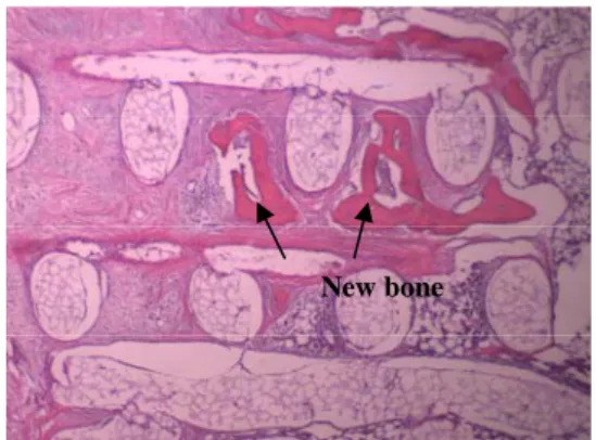
Subcutaneous implantation of n-HA/PCL scaffolds in rats for 4 weeks showed good tissue in- growth behavior as shown in Fig. 4. Bony implantation of n-HA/PCL scaffolds in rabbits for 8 weeks showed excellent bone regeneration as well as bone in-growth, as shown in Fig 5. It can be therefore argued that three dimensionally designed pore structure is obviously effective in tissue in- growth and the nanoparticles of HA enhance osteogenic cell differentiation as well as proliferation.
Fig. 3. ALP activity of hBMSC in scaffolds.
Fig. 4. Subcutaneous implantation of n-HA/PCL scaffolds in rats for 4 weeks.
Fig. 5. Bony implantation of n-HA/PCL scaffolds in rabbits for 8 weeks.
4. Conclusion
HA/PCL composite 3D scaffolds exhibited good osteogenic cell proliferation differentiation and reasonable mechanical stiffness. They also showed excellent tissue in-growth and bone regeneration behavior in animal models.
5. References
(1) L. Shor, S. Guceri, X. Wen, M. Gandhi, W. Sun, 2007, Biomaterials, Vol. 28, p.5291.
(2) S. J. Heo, S. E. Kim, J. Wei, Y. T. Hyun, H. S.
Yun, D. H. Kim, J. W. Shin, 2008, J. Biomed.
Mater. Res., Part A, DOI:10.1002/jbm.a.31726.
0.
0 0.02 0.04 0.06 0.08 0.1 0.12 0.14
DAY 3 DAY 7 DAY 10 DAY 14
umol/ng/DN
* n-HA/PCL ALP
m-HA/PCL
A
n- HA/PCL
m- HA/PCL
0 0.1 0.2 0.3 0.4 0.5 0.6 0.7
4h Day 1 Day 4 Day 7
Day
O.D.
n-HA/PCL
m-HA/PCL MTT
New bone
*
1410