INTRODUCTION
When considering a lateral approach to the sinus, the major differences between the various surgical methods are the type of graft material used and the choice of immediate or delayed implant placement
1). In case of severe atrophy of the maxillary alveolar process, sinus floor elevation and implant insertion are usually per- formed in two stages
2). When an autogenous bone graft is used, it takes approximately 6 months following aug- mentation for the transplanted bone to be integrated and substituted by osteoconduction (creeping substitution).
Alternatively, autogenous bone transplants can be replaced by bone substitutes, such as Bio-Oss (Geistlich, Wolhusen, Switzerland), to avoid donor site morbidity
3).
Bio-Oss, a frequently used alternative bone substitute, has been evaluated in several animal
4,5)and clinical stud- ies
6,7). This bone substitute is derived from porous bovine bone material processed to yield natural bone material lacking the organic component; it is reported to have good tissue acceptance and to provide a scaffold for new bone deposition with natural osteotrophic properties
8).
Histologic data regarding the outcomes of treatments involving sinus grafting in humans are scarce
9). Even when specimens were obtained at different clinical cen- ters, they were retrieved from patients for whom the sur- gical procedure was successful.
In implant dentistry, numerous techniques have been studied for promoting and accelerating the osseous heal- ing of dental implants and bone grafts by increasing the
* Corresponding author Su-Gwan Kim
Dept. of OMFS, College of Dentistry, Chosun University 421, Seosuk-dong, Dong-Gu, Gwangju, 501-825, Korea Tel: 82-62-220-3815 Fax: 82-62-228-7316
E-mail: [email protected]
Effects of fibrin glue on bone formation in combination with deproteinized bone xenografts in humans
Moon-Su Kim
1, Su-Gwan Kim
1, Sung-Chul Lim
2, Hak-Kyun Kim
1, Seong-Young Moon
11
Department of Oral & Maxillofacial Surgery, College of Dentistry, Chosun University
2
Department of Pathology, College of Medicine, Chosun University, Gwangju, Korea
Thirty-six sinus grafts were performed in 34 patients with an alveolar crest bone height in the posterior maxilla of 3 to 5 mm before grafting. The sinuses were grafted using Bio-Oss alone or mixed with fibrin glue. Group 1 was the control group and included 25 patients who received a xenograft mixed in saline. Group 2 comprised 9 patients who received a xenograft and fibrin glue. The study was further subdivided at the time of 9 months. This histologic study evaluated by hematoxylin-eosin (H&E) and histomorphometric analysis whether fibrin glue in combination with Bio-Oss enhances bone regeneration in sinus floor elevation in humans. The new bone formation was better in Group 2 than in Group 1, but the difference was not significant. The absorption of the graft material was faster in Group 2 than in Group 1, in the short term, but better in Group 1 over the long term, although the difference was not significant.
Lamellar bone was formed earlier in Group 1 compared to Group 2, but the difference was not significant. Overall, the surgery site sta- bilized earlier with new bone formation in Group 2 than in Group 1, but the difference was not significant. Combining a fibrin sealant and Bio-Oss could lead to improved scaffolds for bone tissue engineering based on the synergistic effects of the biomaterials. Therefore, Bio-Oss or Bio-Oss plus Tisseel may be used depending on the situation.
Key words
Fibrin glue; Bone formation; Deproteinized bone xenografts
Abstract
bone regenerative potential
10). These techniques include the application of platelet-rich plasma (PRP)
11), bone mor- phogenetic protein
12), growth factors
13), and fibrin glue
14,15). Fibrin glue in various formulations has been used widely in various surgical procedures by means of estab- lishing hemostasis and for tissue approximation. Fibrin sealants have been used in such diverse locations as the heart and pericardial cavity, lung, bowel, ovary, nasal sinuses, and skin. Formulations include both autologous preparations, which require peri-operative preparation, and prepackaged commercial kits
16).
Fibrin glue is a physiological matrix whose principal component, fibrin, plays fundamental roles in the process of blood clotting and wound healing
17). It is a potentially suitable biological vehicle for cell transplanta- tion since it has proven biocompatibility, biodegradabili- ty, and binding capacity to cells
18). Fibrin-stabilizing fac- tor XIII, contained in fibrin glue, favors the migration of undifferentiated mesenchymal stem cells (MSCs) on the highly cross-linked structure of the glue, and it enhances the proliferation of these cells
19).
The use of fibrin glue to improve bone regeneration is well documented
20-28), and platelet-rich fibrin (PRF) is an autologous fibrin matrix used to enhance bone genera- tion
20). Tayapongsak et al.
14)found that it facilitates the placement of graft material in the recipient cavity by pre- venting the displacement of the bone graft particles dur- ing placement. It also helps the remodeling process begin earlier by accelerating the migration of fibroblasts and the revascularization process.
In bone reconstruction, the combination of bioceramics and fibrin glues may have synergistic effects. The mechanical strength of the composite is superior to that of the ceramic alone
21). Furthermore, the bioceramic/fib- rin glue composite is stabilized initially through its adap- tation and adhesion to the walls of the bone defect. The fibrin might also enhance its biological properties by act- ing positively on angiogenesis, cell attachment, and pro- liferation
22).
Few study has examined composites consisting of Bio- Oss and fibrin glue. Therefore, this histologic study eval- uated whether fibrin glue in combination with Bio-Oss enhances bone regeneration in sinus floor elevation in humans.
MATERIALS AND METHODS
with an alveolar crest bone height in the posterior maxil- la of 3 to 5 mm before grafting. The sinuses were grafted using Bio-Oss alone or mixed with fibrin glue. Informed consent was obtained from all patients.
Group 1 was the control group and included 25 patients who received a xenograft mixed in saline. Group 2 comprised 9 patients who received a xenograft and fib- rin glue. The study was further subdivided at the time of 9 months.
Patient selection
Patients were enrolled in this study if they had no sys- temic or local contraindications: no history of uncon- trolled diabetes, no radiation therapy to the head or neck in doses over 5,000 rads, no chemotherapy within the 12 months preceding surgery, no active sinus infection, no uncontrolled periodontal disease, and no psychological problems that would prevent long-term treatment.
Smokers were advised to reduce or refrain from smok- ing.
Surgical technique
Immediately before surgery, the patients rinsed with a 0.2% chlorhexidine digluconate solution for 2 minutes.
Local anesthesia was obtained with lidocaine containing epinephrine 1:100,000.
A crestal incision, slightly displaced toward the palate, was made, and a vertical releasing incision was placed in the canine area to facilitate flap elevation. A mucope- riosteal flap was elevated, exposing the lateral wall of the sinus. A bony window, averaging 15 × 10 mm, was out- lined using a no. 6 round carbide bur without perforat- ing the sinus membrane. After mobilizing the window, the sinus membrane was elevated starting from the infe- rior border of the osteotomy site. The lateral window was pushed inward and elevated superiorly, creating a new horizontal ceiling, as the membrane was carefully dissected from the medial and inferior walls of the sinus.
The graft material (Bio-Oss) was hydrated with saline
solution and gently packed into the sinus until it filled
the entire cavity. In Group 2, the Bio-Oss was mixed with
fibrin glue. Immediate implant placement was indicated
when sufficient native bone was available to achieve pri-
mary stability after placement. The procedure was
delayed 6 to 18 months after grafting for cases in which it
implant in the subsinus ridge. Screw-type, machined- surface implants were used in the patients.
All the implants were submerged. The abutments were connected during two distinct postoperative periods, at 6 and 18 months post-implant placement. Bone cores from the graft sites were taken for histologic examination.
Three to 4 weeks after soft tissue healing, the final abut- ments were connected, implant stability was tested man- ually, and prosthetic treatment was carried out.
Histologic examination
Bone cores were harvested from the lateral wall using a 2-mm-diameter trephine bur under sterile saline irriga- tion. The biopsies were retrieved from areas located between the implants, about 10 mm from the alveolar ridge, at a mean depth of 7 mm.
Microscopic examination and histomorphometric analysis
After fixing the harvested bone in 10% formalin using the conventional method and treating the bone in nitric acid (De-Cal Rapid, Pational Diagnosis, Atlanta, GA) for approximately 4 hours to decalcify, blocks were made to obtain representative sections. After washing and pro- cessing the tissues using an autoprocessing machine (Hypercenter XP, Shandon, UK), the blocks were embed- ded in paraffin. The embedded blocks were cut into 4-μ m-thick slices, stained with hematoxylin-eosin (H&E), and observed under an optical microscope. The degree of new bone formation (bone forming activity) and compo- sition ratio of the tissue sample were determined by measuring and comparing the area of new bone forma- tion using computer-assisted histomorphometry
3).
Images of each tissue sample were taken with a digital camera (Polaroid, Cambridge, MA) and analyzed using Image Pro Plus (Media Cybernetics, Silver Spring, MD).
∙ Bone forming activity = area of new bone formation / whole surface area of the specimen × 100 (%)
∙ Composition ratio = areas of woven bone : lamellar bone : residual implant material (%)
Statistical analysis
To confirm the significance after measuring the area of new bone formed, Student’s t-test was performed. P
<0.05 was considered statistically significant.
RESULTS
Group 1, <9 months
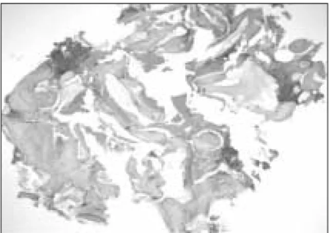
New bone was formed around the implanted Bio-Oss chips. Under higher magnification, organized trabecular woven bone around the implanted Bio-Oss chips was noted (Figs. 1, 2). The bone forming activity was 46.3 ± 30.0% (Table I). The composition ratio of the tissue sam- ple was 47.7 : 14.6 : 37.7 (areas of woven bone : lamellar bone : residual implant material, Table 1).
Group 1, ≥ ≥9 months
New bone was formed around the implanted Bio-Oss chips. The new bone forming activity increased slightly (56.5 ± 31.5%) compared to Group 1 at <9 months (Table I), although the difference was not significant (p=0.21).
Under higher magnification, organized trabecular woven bone was seen around the implanted Bio-Oss chips.
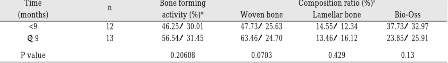
Table 1. Bone forming activity and composition ratio after implant surgery in Group 1
Time n Bone forming Composition ratio (%)
#(months) activity (%)* Woven bone Lamellar bone Bio-Oss
<9 12 46.25±30.01 47.73±25.63 14.55±12.34 37.73±32.97
≥9 13 56.54±31.45 63.46±24.70 13.46±16.12 23.85±25.91
P value 0.20608 0.0703 0.429 0.13
*Bone forming activity = area of new bone formation/area of total sample × 100 (%)
#
Composition ratio = area of woven bone : lamellar bone : residual implant materials (%)
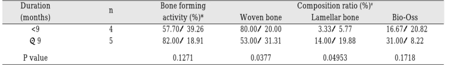
Table 2. Bone forming activity and composition ratio after implant surgery in Group
Duration n Bone forming Composition ratio (%)
#(months) activity (%)* Woven bone Lamellar bone Bio-Oss
<9 4 57.70±39.26 80.00±20.00 3.33±5.77 16.67±20.82
≥9 5 82.00±18.91 53.00±31.31 14.00±19.88 31.00±8.22
P value 0.1271 0.0377 0.04953 0.1718
*Bone forming activity = area of new bone formation/area of total sample × 100 (%)
#
Composition ratio = area of woven bone : lamellar bone : residual implant materials (%)
Fig. 1. New bone was seen around the implanted Bio- Oss chips (asterisks) (hematoxylin and eosin stain, orig- inal magnification ×40).
Fig. 2. Higher magnification of the histopathologic findings. Organized trabecular woven bone (open as- terisks) was seen around the implanted Bio-Oss chips (closed asterisks) (hematoxylin and eosin stain, original magnification ×100).
Fig. 3. New bone was seen around the implanted Bio- Oss chips (asterisks). The new bone forming activity in- creased slightly (hematoxylin and eosin stain, original magnification ×40).
Fig. 4. Higher magnification of the histopathologic
findings. Organized trabecular woven bone (open as-
terisks) was seen around the implanted Bio-Oss chips
(closed asterisks). Focal lamellar bone formation (ar-
rows) is noted (hematoxylin and eosin stain, original
magnification ×100).
Focal lamellar bone formation was also noted (Figs. 3, 4).
The composition ratio of the tissue sample was 63.5 : 13.5 : 23.9 (Table 1). In this group, the proportion of woven bone formation increased compared to Group 1 at
<9 months, although the difference was not significant (p=0.07).
Group 2, <9 months
New bone was seen around the implanted Bio-Oss chips. Under higher magnification, organized woven bone formation was seen around the implanted Bio-Oss chips (Figs. 5, 6). The new bone forming activity
increased slightly (57.7 ± 39.3%) compared to Group 1 at
<9 months (Table 2), but the difference was not signifi- cant (p=0.277; Table 3).
The composition ratio of the tissue sample was 80.0 : 3.3 : 16.7 (Table 2). In this group, the proportion of woven bone increased significantly (p=0.034) compared to Group 1 at <9 months (Table 4).
Group 2, ≥ ≥9 months
Thick woven bone was seen around a few implanted Bio-Oss chips. Although the new bone forming activity (82.0 ± 18.9) increased (Table 2), it was not significantly Fig. 5. New bone was seen around the implanted Bio-
Oss chips (asterisks) (hematoxylin and eosin stain, orig- inal magnification ×40).
Fig. 6. Higher magnification of the histopathologic findings. Organized woven bone (open asterisks) was seen around the implanted Bio-Oss chips (closed aster- isks) (hematoxylin and eosin stain, original magnifica- tion ×100).
Fig. 7. Thick woven bone around a few implanted Bio- Oss chips (asterisks) is seen. The new bone forming ac- tivity increased markedly (hematoxylin and eosin stain, original magnification ×40).
Fig. 8. Higher magnification of the histopathologic
findings. Thick woven bone is seen with a few implant-
ed Bio-Oss chips. (hematoxylin and eosin stain, original
magnification ×100).
different from Group 1 at ≥9 months (p=0.056, Table 3) or from Group 2 at <9 months (P=0.13). Under higher magnification, thick woven bone with a few implanted Bio-Oss chips was seen (Figs. 7, 8).
The composition ratio of the tissue sample was 53.0 : 14.0 : 31.0 (Table 2). The proportion of woven bone decreased significantly (p=0.038) compared to Group 2 at
<9 months, while the proportion of lamellar bone increased significantly (P=0.049).
DISCUSSION
Autografts are the ideal material for reconstructing hard tissue defects. They undergo osteogenesis, osteoin- duction, and osteoconduction, do not pose a risk of immune rejection, and require short recovery times.
ed, and absorption and the secondary defects at the donor site are major drawbacks. To avoid harvesting an autograft and thereby eliminating the additional surgical procedure and associated risks, bone grafting materials and substitutes are used as alternative grafting materials for ridge augmentation
23). The ideal grafting materials should be biocompatible, noncarcinogenic, and nonaller- genic; show early vascularization; be replaced by new host bone tissue; strength, resist infection, and be sterile;
and undergo surface resorption by the host
24).The use of bovine hydroxyapatite (Bio-Oss) has been suggested for maxillary sinus grafting procedures before or in conjunction with implant placement
25). Bio-Oss is deproteinized, sterilized bovine bone with 75 to 80%
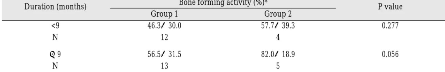
porosity and a crystal size of approximately 10 nm in the form of cortical and cancellous granules and blocks. It Table 3. Comparison of the bone forming activity after implant surgery between Groups 1 and 2
Duration (months) Bone forming activity (%)* P value
Group 1 Group 2
<9 46.3±30.0 57.7±39.3 0.277
N 12 4
≥9 56.5±31.5 82.0±18.9 0.056
N 13 5
* Bone forming activity = area of new bone formation/area of total sample × 100 (%)
Table 4. Comparison of the composition ratio after implant surgery between Groups 1 and 2
Duration (months) Composition ratio (%)*
P value
Group 1 Group 2
<9
woven bone 47.7±25.6 80.0±20.0 0.034
lamellar bone 14.6±12.3 3.3±5.8 0.080
Bio-Oss 37.7±33.0 16.7±20.8 0.161
≥9
woven bone 63.5±24.7 53.0±31.3
¶0.198
lamellar bone 13.5±16.1 14.0±19.9
§0.472
Bio-Oss 23.9±25.9 31.0±8.2 0.291
* composition ratio=percentage area of woven bone formation vs. percentage area of lamellar bone formation vs. percentage area of residual implant materials (%)
¶
The proportion of woven bone was statistically significant decreased compared to Group 2, <9 months, p< .05.
§