• 검색 결과가 없습니다.

A Case of Isolated Pulmonary Mucormycosis in an Immunocompetent Host

N/A
N/A
Protected

Academic year: 2021

Share "A Case of Isolated Pulmonary Mucormycosis in an Immunocompetent Host"

Copied!
5
0
0

로드 중.... (전체 텍스트 보기)

전체 글

(1)

Tuberc Respir Dis 2013;74:269-273

CopyrightⒸ2013. The Korean Academy of Tuberculosis and Respiratory Diseases. All rights reserved.

A Case of Isolated Pulmonary Mucormycosis in an Immunocompetent Host

Jung Su Lee, M.D.1, Ho Cheol Kim, M.D.1, Sang Woo Park, M.D.1, Hoon Sub So, M.D.1, Chang Yun Woo, M.D.1, Jong Han Choi, M.D.1, Sang Hyung Kim, M.D.1, Se Jin Kim, M.D.2, Yeon-Mok Oh, M.D.1

1Department of Medicine, Asan Medical Center, University of Ulsan College of Medicine, 2Division of Pulmonary Medicine, Department of Internal Medicine, The Armed Forced Capital Hospital, Seoul, Korea

Mucormycosis is a rare fungal disease that holds a fatal opportunistic fungal infection in diabetes mellitus, hematological malignancy, and immunocompromised host. Isolated pulmonary mucormycosis is extremely rare.

Optimal therapy is a combined medical-surgical approach and a management of the patient's underlying disease.

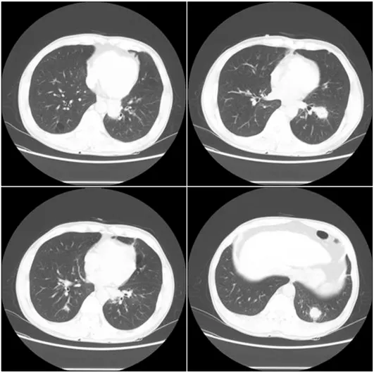
Herein, we report a case-study of isolated pulmonary mucormycosis which was being presented as multiple lung nodules in a patient with no underlying risk factors. Considering that the patient had poor pulmonary functions, we treated him with only antifungal agent rather than a combined medical-surgical approach. After treatment with antifungal agent for six months, the nodules of pulmonary mucormycosis were improved with the prominent reductions of size on the computed tomography.

Key Words: Mucormycosis; Lung; Immunocompetence; Posaconazole

Address for correspondence: Yeon-Mok Oh, M.D.

Department of Medicine, Asan Medical Center, University of Ulsan College of Medicine, 88 Olympic-ro 43-gil, Songpa- gu, Seoul 138-736, Korea

Phone: 82-2-3010-3136, Fax: 82-2-3010-6968 E-mail: ymoh55@amc.seoul.kr

Received: Sep. 10, 2012 Revised: Sep. 13, 2012 Accepted: Oct. 11, 2012

CCIt is identical to the Creative Commons Attribution Non-Commercial

Introduction

Mucormycosis is a life-threatening fungal infection which occurs in patients with diabetes mellitus, hemato- logical malignancy. Although the infection commonly begins by invading the respiratory tract, isolated pulmo- nary mucormycosis is extremely rarely reported

1,2

. Typically an airborne infection, mucormycosis is able to extent to the whole body by invading alveolar blood vessel after germinating in paranasal sinuses

3

. Isolated pulmonary mucormycosis is rare, and usually occurs in the patients with diabetes mellitus, hematological malig- nancy or cancer undergoing induction chemotherapy

4-7

.

In this case reports, we report isolated pulmonary mucormycosis that arose as multiple nodule in health patient without apparent immune deficiency which was suspected as lung cancer.

Case Report

A 52-year-old male presented to our hospital with a chief complaint of sputum. Productive cough with yel- lowish sputum for two months, four pulmonary nodule in both lungs which was detected in chest computed tomography (CT).

1. Past medical history

Using inhalers (tiopropium, salmeterol/fluticasone) because of chronic obstructive pulmonary disease (COPD) which was diagnosed 15 years ago. Seven years ago, he received occasionally a intravenous steroids be- cause of COPD flare-ups which occurs one or two time.

Pulmonary tuberculosis had been treated for three-years

medical care in 13 years ago. Hepatitis C was diagnosed

in 10 years ago.

(2)

Figure 2. Computed to- mography of chest show- ed three long nodules in lower left lobe of lung and one lung nodule in lower right lobe.

Figure 1. Initial chest radiography revealed two lung nod- ules in lower left lung field.

2. Physical examination

His vital signs were within the normal range. There was no lymphadenopathy in the cervical, axillary, or in- guinal regions. His had barrel chest. On auscultation of the chest, a coarse breathing sound with wheezing in left upper lung fields was heard.

3. Laboratory analysis

Laboratory data were within the normal range. Blood

glucose was 116 mg/dL. HbA1c was 6.1%. Hepatitis C

virus antibody test was positive. Human immunodefi-

ciency virus antigen and antibody were negative, Para-

gonimus antibody and Aspergillus antigen were nega-

tive.

(3)

Figure 3. Pathological findings of lung tissue are shown as thick, irregular and non-septated fungal hyphaes with right-an- gle branching in hematoxylin and eosin stain (A, ×400) and Gomori's methenamine silver stain (B, ×400).

Figure 4. After treatment with antifungal agent for 3 months, computed tomog- raphy of chest showed the prominent reduction of size in the nodules of pulmo- 4. Radiological findings

A chest radiography showed an inactive tuberculosis scar in right upper lobe and two nodules in left lower lobe (Figure 1). A CT scan of the chest showed three

nodules in left lower lobe, a nodule in right lower lobe, and emphysema (Figure 2).

5. Treatment and progress

We confirmed no endobronchial lesion by broncho-

(4)

Figure 5. After the end of the treatment for 3 months, chest radiography revealed no definitive relapse of mucor- mycosis.

scopy. The bronchoscopic washing specimen was neg- ative for the acid-fast bacilli smear and culture test, and also for the microbial and fungal culture test. A trans- cutaneous needle biopsy of the nodule at left lower lobe showed thick fungal hyphe with right-angle branching, highly suggestive for mucormycosis (Figure 3). Because the patient was immunocompetent, we screened other af- fected lesion. However brain magnetic resonance imaging and nasal endoscopy showed no apparent lesion.

The patient was started on amphotericin B. Surgical resection of the affected lobe was not possible treatment option, because pulmonary function test showed that forced expiratory volume in one second of the patient was 45%. Amphotericin B was used during 1 week and discontinued after discharge. Posaconazole oral suspen- sion was started for home treatment. Total treatment du- ration is 3 months. After treatment, the chest CT in the follow-up showed that the size of the nodules was re- duced (Figure 4). After the end of the treatment for 3 months, chest radiography revealed no definitive re- lapse of mucormycosis (Figure 5).

Discussion

Pulmonary mucormycosis is the one of a fatal oppor-

tunistic fungal infection in immunocompromised host.

The infection was accompanied by a high mortality rate (50–70%)

2,8

. Although fever and cough are mainly re- ported as the main symptom of pulmonary mucormy- cosis, those are not specific for the infection. Hemopty- sis is also observed, because mucormycosis has a high affinity for invading blood vessels. Although those symptoms and signs manifest chronically over one month, massive hemoptysis is fatal for a patient.

Therefore, proper diagnosis and treatment for the in- fection are needed

9

. Sputum culture which is non-in- vasive test for fungal infection is not possible for a pa- tient, if there was no productive cough. A resected specimen is often not suitable for diagnosis, because ne- crosis is main portion of the lesion. Without suspecting, the fungal culture test of specimen is not often done in the laboratory. Above factors are the obstacles for diagnosis of the mucormycosis infection

3,9

. Although a chest radiography has no specific sign for mucormy- cosis, unilateral infiltrate in upper lobe are mainly reported. Because the air-crescent sign in chest radiog- raphy is the sign of massive fatal hemoptysis which is cause of the death, careful assessments are needed

9

. The patient in this case report was immunocompetent, and has multiple nodules in both lower lobes, which are not common sign of the pulmonary mucormycosis.

Therefore, the nodules could be mistaken for lung cancer.

The main principles of the therapy for mucormycosis are management of the patient's underlying disease and a combined medical-surgical approach. The motality rate of the mucormycosis is reported as 50%, and that of pulmonary mucormycosis is reported up to 80%.

Surgical resection of invaded lobe and anti-fungal agent are required for the pulmonary mucormycosis accom- panied with massive hemoptysis

3,8

. However, the pa- tient in this case report has the affected lesion in both side of lung and the low pulmonary function, surgical resection was not possible. Medical approach, combin- ing amphotericin B and posaconazole oral suspension, was successfully reduced the size of the affected lesion.

Because it is rare that isolated pulmonary mucormy-

(5)

cosis occur as the nodules in chest radiography without immunodeficiency, the proper diagnosis and treatment was not possible without suspecting the fungal infection in the transcutaneous biopsy. The important negative factor for the good prognosis in fungal infection is the delayed diagnosis and treatment

9

. We confirmed that medical approach using posaconazole oral suspension without surgical resection and long-term hospitalization is suitable treatment for the immunocompetent patient with pulmonary mucormycosis.

References

1. Butala A, Shah B, Cho YT, Schmidt MF. Isolated pul- monary mucormycosis in an apparently normal host:

a case report. J Natl Med Assoc 1995;87:572-4.

2. Ibrahim AS, Spellberg B, Walsh TJ, Kontoyiannis DP.

Pathogenesis of mucormycosis. Clin Infect Dis 2012;54 Suppl 1:S16-22.

3. Petrikkos G, Skiada A, Lortholary O, Roilides E, Walsh TJ, Kontoyiannis DP. Epidemiology and clinical mani- festations of mucormycosis. Clin Infect Dis 2012;54

Suppl 1:S23-34.

4. Kim TJ, Park CY, Jin YG, Park KE, Her KM, Kim YH, et al. A case of pulmonary mucormycosis associated uncontrolled diabetes mellitus. Korean J Med 1997;

53(Suppl 3):S792-6.

5. Lee SJ, Yoon SJ, Kim EJ, Cha SI, Park JY, Jung TH, et al. A case of pulmonary mucormycosis complicated by pneumomediastinum and subcutaneous emphyse- ma after chemotherapy in a patient with small cell lung cancer. Korean J Med 2007;72:551-7.

6. Pagano L, Offidani M, Fianchi L, Nosari A, Candoni A, Piccardi M, et al. Mucormycosis in hematologic patients. Haematologica 2004;89:207-14.

7. Cho YJ, Kang MH, Kim HS, Jeong YY, Jang IS, Kim HC, et al. A case of angio-invasive pulmonary mu- cormycosis with uncontrolled diabetes mellitus. Tuberc Respir Dis 2008;64:451-5.

8. Spellberg B, Kontoyiannis DP, Fredricks D, Morris MI, Perfect JR, Chin-Hong PV, et al. Risk factors for mortal- ity in patients with mucormycosis. Med Mycol 2012;50:

611-8.

9. Hamilos G, Samonis G, Kontoyiannis DP. Pulmonary mucormycosis. Semin Respir Crit Care Med 2011;32:

693-702.

수치

Figure 1. Initial chest radiography revealed two lung nod- nod-ules  in  lower  left  lung  field.
Figure 3. Pathological findings of lung tissue are shown as thick, irregular and non-septated fungal hyphaes with right-an- right-an-gle  branching  in  hematoxylin  and  eosin  stain  (A,  ×400)  and  Gomori's  methenamine  silver  stain  (B,  ×400).
Figure  5.  After  the  end  of  the  treatment  for  3  months,  chest radiography revealed no definitive relapse of  mucor-mycosis.

참조

관련 문서

This book contains hundreds of complete, working examples illustrating many common Java programming tasks using the core, enterprise, and foun- dation classes APIs.. Java Examples

GRIPHON Prostacyclin (PGI2) Receptor Agonist In Pulmonary Arterial Hypertension IPAH idiopathic pulmonary arterial hypertension MCTD mixed connective tissue

Anti-fungal effect of isolated bactrial strains was tested in vitro by incubating in potato dextrose agar with isolates of four fungal plant pathogens

Generally, in case that we predict the vibration by blasting, we compute it on the basis of the charge weight per delay but in case of shocking

In this paper, it was examined whether there is a possibility that the medical institution might be mistaken as a specialized hospital designated by the Ministry of Health

Just as we achieved good control with a joint-based controller that was based on a linearizing and decoupling model of the arm, we can do the same for the Cartesian case.

This paper has significant as a study of case marker reflecting the colloquialism in Yanbian region in China which neglected by the studies on relying on

The purpose of this study was to explore research trends in child assessment system, to examine problems that were arose in process of child assessment, and