Conditions for Preparing Glycyrrhiza uralensis Extract for Inhibiting Biofilm Formation of Streptococcus mutans 1
Youngseok Ham
2⋅Tae-Jong Kim
2,†ABSTRACT
1)Licorice, which has an extensive history of use as an herbal medicine, has been suggested to have oral health benefits.
However, to date, no systematic study has been conducted on the preparation method of licorice extracts for oral health.
In this study, licorice extracts prepared using water and ethanol were investigated for its ability to inhibit the biofilm formation of Streptococcus mutans. The licorice extract prepared with around 60% ethanol effectively inhibited the biofilm formation of S. mutans. Licorice extracted with 50% ethanol almost completely inhibited the biofilm formation at 1.5 g/L of licorice extract. This inhibitory activity was confirmed in a microplate assay and a flow cell system.
Glycyrrhetic acid was extracted from licorice effectively with 60% ethanol concentration. The strong inhibitory activity of glycyrrhetic acid and the synergistic inhibition with glycyrrhizin on biofilm formation were suggested as major reasons for a concentration-specific extraction. These results suggest that licorice extract prepared using around 60% ethanol effectively inhibits the biofilm formation of S. mutans.
Keywords: licorice, ethanol extract, biofilm, Streptococcus mutans, glycyrrhetic acid
1. INTRODUCTION
Licorice, the root and rhizome of Glycyrrhiza sp., has been traditional used as a medicinal herb and a natural sweetener. It prevents gastrointestinal ulcers (Momeni et al., 2014; Jalilzadeh-Amin et al., 2015);
neutralizes toxic properties of compounds (Isbrucker and Burdock, 2006; Gong et al., 2015); and has antiviral (Wang et al., 2013; Fukuchi et al., 2016), antibiotic (He et al., 2006; Gupta et al., 2008; Lau and Plotkin, 2013; Chakotiya et al., 2016; Rohinishree and Negi, 2016), antifungal (Messier and Grenier, 2011; Seleem et al., 2016; Sharma et al., 2016), and anticancer
activities (Shen et al., 2015; Fukuchi et al., 2016). Over the past 20 years, the number of studies on the use of licorice for oral health has steadily increased.
Compounds in licorice improve bad breath by reducing the bacterial production of volatile sulfur compounds (Shin-ichi et al., 2012). Licorice has also been shown to ameliorate mouth dryness more effectively than water (Yu et al., 2016). In particular, licorice is thought to enhance oral health by inhibiting the growth of Streptococcus mutans, a primary causative agent of tooth decay (Segal et al., 1985; Hwang et al., 2004; Jain et al., 2013; Ajagannanavar et al., 2014). Consequently, the licorice lollipop was developed to control dental
1Date Received August 21, 2018, Date Accepted February 21, 2019
2Department of Forest Products and Biotechnology, Kookmin University, Seoul 02707, Republic of Korea
†Corresponding author: Tae-Jong Kim (e-mail: [email protected], ORCID: 0000-0002-7483-0432) Original Article
caries (Hum et al., 2011; Almaz et al., 2017).
Oral bacteria produce biofilms, called plaque, in the oral cavity. The oral cavity is a continuous flow system of food, drink, and saliva, and therefore, oral bacteria would be easily washed away without the ability to form a biofilm. Nonetheless, the oral cavity is a favorable environment for bacterial growth, and its many surfaces are coated with a plethora of bacteria, with >500–700 bacterial strains residing in the oral cavity (Rosan and Lamont, 2000; Dewhirst et al., 2010;
Huang et al., 2011). Some of these bacteria are implicated in oral diseases. Moreover, specific oral bacteria strains have been associated with several systemic diseases.
S. mutans, a gram-positive bacterium, is a regular member of the mature dental biofilm community. S. mutans has four serotypes c, e, f, and k based on its polysaccharides.
Almost 70-80% of S. mutans found in the oral cavity is serotype c. However, under certain conditions, S.
mutans can become prevalent and cause dental caries (Loesche, 1986; Ahn et al., 2008).
S. mutans produces insoluble glucan using sugars present in food. The insoluble glucan aids in plaque formation on the tooth surface where S. mutans exists (Krzysciak et al., 2014; Ren et al., 2016). S. mutans in the plaque cannot be easily removed by physical and chemical treatments (Welin-Neilands and Svensater, 2007; Bowen and Koo, 2011). The plaque also facilitates the intraoral adhesion of other bacteria, and these bacteria have been reported to cause not only oral diseases but also other complications, including endocarditis (Berbari et al., 1997), pneumonia (Scannapieco, 1999), low birth weight (Buduneli et al., 2005), and cardiovascular disease (Beck et al., 1996).
These previous studies have suggested that chemical inhibition of biofilm formation in the oral cavity may play an important biological role in human health. One way to inhibit biofilm formation is by killing bacterial cells (Kondo et al., 2007). Recently, plant extracts that inhibited biofilm formation without the bactericidal
effect of S. mutans have also been reported (Ham and Kim, 2018).
Although many studies have reported the beneficial effects of licorice extracts on oral health, no systematic study has been conducted on the extraction methods for preparing licorice extracts for inhibiting the biofilm formation of S. mutans. In this study, the activity of licorice extract to inhibit the biofilm formation of S.
mutans was investigated according to ethanol con- centration of extraction solvent. In addition, the extraction of the chemicals inhibiting the biofilm formation according to the ethanol concentration was observed to explain the specific extraction conditions.
2. MATERIALS and METHODS
2.1. Media for cell culture and biofilm formation assays
Streptococcus mutans serotype c was obtained from LG Household & Health Care Ltd (Daejeon, Korea).
Brain heart infusion (BHI) broth (BD Biosciences Korea, Seoul, Korea) was used for growing S. mutans. BHI agar was prepared by adding 1.5% agar to BHI broth. BHI-S medium, the biofilm formation medium, was prepared by supplementing BHI broth with 10-g/L sucrose.
Glycyrrhizin (product number: G0150), glycyrrhetic acid (product number: G0149), and glucuronic acid (product number: G0302) were purchased from Tokyo Chemical Industry Co., Ltd. (Tokyo, Japan). Liquiritin was purchased from Kono Chemical Co., Ltd. (Xian, China). Glycyrrhizin was dissolved in 50% ethanol, liquiritin and glycyrrhetic acid were dissolved in dimethyl sulfoxide, and glucuronic acid was dissolved in distilled water before use, respectively.
2.2. Preparation of licorice extracts
Licorice was purchased from Jiundang Oriental
Medicine Co. (Seoul, Korea). The dried licorice extracts were prepared using licorice pulverized to a diameter of ≤1 mm. Subsequently, 10 g of the powdered licorice were mixed with 50-mL of water and ethanol and incubated at 50°C for 3 h. The content of ethanol in the extract solution was indicated in each experiment.
The residual solid materials were removed by filtration (Whatman filter paper Grade 1). The filtrates were concentrated using a rotary evaporator (RV10, IKA
®Korea, Ltd., Seoul, Korea). Without making powder, the original volume of the pre-concentrate extract was restored by adding distilled water. Insoluble materials were removed by centrifugation at 16,810 × g for 20 min and filtration through a syringe filter (25-mm diameter, cellulose acetate, 0.20-μm pore size; GVS North America, Inc., Sanford, ME, USA). The transparent extracts were stored at −80°C.
2.3. Biofilm formation assay and quantitative analysis
S. mutans, which was stored at −80°C, was streaked onto a BHI agar plate and cultured at 37°C for 2 days.
A single colony was inoculated into 5-mL BHI broth and incubated at 37°C for 24 h without shaking. The cell density of cultured S. mutans was measured at an absorbance of 600 nm (Abs
600) using an Optizen 2120 UV Plus spectrophotometer (Mecasys Co., Ltd., Daejeon, Korea). A 96-well polyvinyl chloride microplate containing 100-μL BHI-S medium was inoculated with the cultured cells at an optical density of 0.05 at Abs
600. After incubation at 37°C for 24 h, the cell density was measured at Abs
595using an Opsys MR microplate reader (Dynex Technologies, Chantilly, VA, USA).
After removing planktonic cells with distilled water, 100 μL of 1% crystal violet solution was dispensed into each well and allowed to stand for 15 min. After washing gently three times with distilled water, 100 μL of 95% ethanol was added to each well. After
incubating for 15 min to allow the elution of the stained crystal violet, Abs
595was measured using an Opsys MR microplate reader. The one-way analysis of variance was used to distinguish differences from controls at the 95% confidence level by Tukey test using SPSS software (Ver. 23.0, SPSS Inc., Chicago, Illinois, USA).
2.4. Biofilm formation in a flow cell system
To observe biofilm formation in a flow state similar to the oral cavity, biofilm formation of S. mutans was monitored in a continuous flow cell system. The flow cell system was purchased from the Center for Biomedical Microbiology (Technical University of Denmark, Lyngby, Denmark). The flow cell channel was covered with a polyvinyl chloride coverslip on which a biofilm was formed. Licorice extracted with 50% ethanol was added to the BHI-S medium at the indicated concentration.
As a preculture, S. mutans was inoculated into 5-mL
BHI broth and incubated at 37°C for 24 h without
shaking. The precultured cells were diluted to an optical
density of 0.05 at Abs
600, and 350 μL of diluted cells
were inoculated into each channel. The coverslip was
placed facing downward for 1 h, giving the cells a
chance to attach to the surface of the coverslip. The
flow cell system was turned on with the coverslip
pointing upward and cultured for 24 h at a constant
flow rate of 13 mL/h. The biofilm medium was
continuously supplied to the flow cell system using
a peristaltic pump (Masterflex L/S Digital Drive EW-
07523-90, Masterflex L/S 12-channel, 8-roller cartridge
pump head EW-07519-25, Masterflex L/S small
cartridges EW-07519-85, Cole-Parmer Instrument
Company, LLC., Vernon Hills, IL USA). The biofilm
formed under the coverslip was observed using an Axio
Scope.A1 microscope (Carl Zeiss Co., Ltd., Seoul,
Korea) equipped with ZEN microscope software (Carl
Zeiss Co., Ltd).
2.5. Analysis of glycyrrhizin and glycyrrhetic acid in licorice extract using high performance liquid chromatograph
For analysis of glycyrrhizin and glycyrrhetic acid in licorice extracts, the method of a previous study (Wang and Yang, 2007) has been used. The equipment of high performance liquid chromatograph was Acme 9000 (YL Instruments Co., Ltd, Anyang, Korea). The analytical column used was YMC-Triart C18 (Product code: TA12S05-2546WT, YMC Korea Co., Ltd, Seongnam, Korea). The flow rate of mobile phase was 1.2 mL/min. The wavelength of light for UV detector was 254 nm. The column temperature was 40°C. The injection volume of sample was 10 L. The licorice extract was separated using a gradient mobile phase according to a previous study (Wang and Yang, 2007).
The data were analyzed with Autochro-3000 software (version 2.0.0).
3. RESULTS and DISCUSSION 3.1. Ethanol content of extract solvents
for inhibited biofilm formation of Streptococcus mutans
Licorice has recently been shown to be effective against dental caries and oro-dental diseases (Touyz, 2009; Messier et al., 2012). In previous studies, various solvents were used for licorice extraction (He et al., 2006; Al-Turki et al., 2008; Nitalikar et al., 2010; Ahn et al., 2012; Rodino et al., 2015; Karahan et al., 2016).
In the present study, water and ethanol were tested to prepare the licorice extract for evaluating its inhibitory activity on the biofilm formation of S. mutans.
Biofilm formation and cell growth inhibition were measured as a function of the ethanol content in the extracting solvent (Fig. 1). When 5% by volume of the extract was treated with the culture, a robust
inhibitory effect on biofilm formation was observed using licorice extracts made with 40%–80% ethanol (Fig. 1A). The cell growth of S. mutans was correlated with biofilm formation (Fig. 1B). To determine the range of effective ethanol concentrations more accurately, we tested with extracts having 1.25% of the cell culture volume tested (Fig. 1C and 1D). Clearly, the most effective inhibitory activity on the biofilm formation of S. mutans was shown the licorice extract made with 60% ethanol.
To confirm the effect of ethanol content in the extracting solvent in Fig. 1, the amount of the solvent required for inhibiting the biofilm formation of S.
mutans was examined (Fig. 2). The licorice extracts prepared using water or 95% ethanol as the extraction solvent showed no inhibitory effect on biofilm Fig. 1. Effect of ethanol content of extract solvent on the biofilm formation (A and C) and the cell growth (B and D) of S. mutans. Extracts of 5% (A and B) and 1.25% (B and C) of the tested culture volume were treated. The control value is the result of the untreated extract, which is indicated on the right side of the figure by a black arrow. The standard deviation was calculated from five independent experimental results. Statistical analysis was performed using a tukey test. Values that differ from the control with the 95%
confidence level are marked with a star on the top
of symbols.
formation, irrespective of the amount of solvent (Fig.
2A and 2E, respectively). In case of 50% ethanol, the inhibitory activity on biofilm formation started appearing using the licorice extract prepared with 100-mL solvent and most biofilm formation was inhibited by licorice extracts prepared with a solvent volume of less than 50-mL (Fig. 2C). The effect of the licorice extract on the cell growth of S. mutans
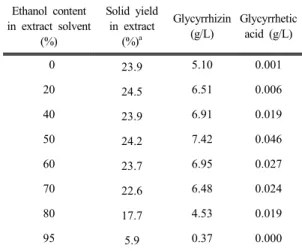
was comparable to that on biofilm formation (Fig. 2D and 2F), except for the water extract (Fig. 2B). The results in Fig. 2 confirmed that licorice extracted with around 50% aqueous ethanol effectively inhibited the biofilm formation of S. mutans in Fig. 1. The yield in extraction was measured according to the ethanol content (Table 1). There was no significant change in the solid yield up to 70% ethanol, but it decreased rapidly at more than 80% ethanol. This result support that there was a large portion of water soluble materials in licorice (Kondo et al., 2007). However, the specific inhibitory effect on the biofilm formation shown in the licorice extract using 60% ethanol as the extraction solvent cannot be explained by the yield of the extract.
The inhibitory effect on biofilm formation was not observed using the water extract, which is the traditional method for making licorice extract, or the 95% ethanol extract. The biofilm formation of S. mutans was inhibited by extracts prepared with 60% ethanol content.
Considering that ethanol has polarity like water, this ethanol concentration-specific extraction condition is highly unusual.
Fig. 2. Effect of the licorice-to-solvent volume ratio on the biofilm formation (A, C, and E) and the cell growth (B, D, and F) of S. mutans. The inhibitory activity of extracts with 5% of tested cell culture volume was evaluated according to the volume of extract solvent: water (A and B), 50% ethanol (C and D), and 95% ethanol (E and F). The control value is the result of the untreated extract, which is indicated on the right side of the figure by a black arrow. The standard deviation was calculated from five independent experimental results. Statistical analysis was performed using a tukey test. Values that differ from the control with the 95% confidence level are marked with a star on the top of symbols.
Ethanol content in extract solvent
(%)
Solid yield in extract
(%)a
Glycyrrhizin (g/L)
Glycyrrhetic acid (g/L)
0 23.9 5.10 0.001
20 24.5 6.51 0.006
40 23.9 6.91 0.019
50 24.2 7.42 0.046
60 23.7 6.95 0.027
70 22.6 6.48 0.024
80 17.7 4.53 0.019
95 5.9 0.37 0.000
aSolid yield (%) = total weight of solid in extract (g) / weight of licorice used for extraction (g) × 100