Recovery from the Two-generation Reproductive Toxicity in Sprague-Dawley Rats by Treatment with Decursin and Decursinol Angelate
Kang Min Kim, Seon Ok, Youn Suk Go and Jae Seon Kang*
Department of Pharmacy, Kyungsung University, Busan 608-736, Korea Received March 6, 2015 /Revised July 2, 2015 /Accepted July 6, 2015
The aim of this study was to determine the effect of decursin (D) and decursinol angelate (DA) against bisphenol A (BPA) toxicity in a rat two-generation study. Adult rats were divided into the following three groups: (1) control, (2) BPA, and (3) BPA+D/DA. The D and DA treatment of F0 pa- rents increased the terminal body weight and relative adult organ weights (testes, kidneys, spleen, and liver) when compared with the BPA group. A significant decrease in sperm count was found in the BPA+D/DA (7.69%) and BPA (64.70%, p<0.01) groups, when compared with the sperm count in the control group. No offspring were obtained in the F1 generation of the BPA (50 mg/kg/day) group, but the addition of D/DA in the BPA+D/DA group significantly restored fertility (55.78%) and ges- tation indices (98.87%) in the F1 generation. No significant differences were found in the fertility index between the control (75.02%) and the BPA+D/DA (78.11%) groups in the two-generation study, when compared with the one-generation study. The viability ratio during lactation in the D/DA group was also similar to that of the control group. These data indicate that D/DA (50 mg/kg/day) administered over two generations causes significant positive changes in reproductive or developmental parameters.
Key words :
Angelica gigas Nakai, decursin, decursinol angelate, reproductive toxicity, two-generation
*Corresponding author
*Tel : +82-51-663-4882, Fax : +82-51-663-4809
*E-mail : [email protected]
This is an Open-Access article distributed under the terms of the Creative Commons Attribution Non-Commercial License (http://creativecommons.org/licenses/by-nc/3.0) which permits unrestricted non-commercial use, distribution, and reproduction in any medium, provided the original work is properly cited.
Journal of Life Science 2015 Vol. 25. No. 7. 765~772 DOI : http://dx.doi.org/10.5352/JLS.2015.25.7.765
Introduction
Epidemiological data showed that the human semen quality has declined during the last 60 years, whereas the incidence of male genital tract abnormalities and infertility has increased. There has been increased awareness of the possible effects of environmental contaminants on male reproduction. BPA, a potential endocrine disruptor and tes- ticular toxicant, also has been shown to affect the re- production of male rats and mice [19, 21]. The chemical structure of BPA is similar to the more potent estrogenic compound diethylstilbestrol and has been shown to mimic estradiol action by stimulating testicular toxicity of BPA at high doses in rats and mice [5].
Angelica gigas Nakai (A. gigas) is a Korean traditional
herbal medicine that is one of the most popular herbal med- icines used in Asian countries, including Korea, Japan and China. A. gigas is also marketed as a functional food prod-
uct in Europe and America. A. gigas of Korea is quite dis- tinct in that it has deep purple flowers while Japan and China Angelica have white flowers [1]. A. gigas has been studied extensively and found to contain a variety of sub- stances including coumarins [6, 17]. Coumarins are com- prised of decursin and decursinol angelate (D/DA), which has been used as a traditional medicine for the treatment of anemia, as a sedative, and as an anodyne or a tonic agent [24]. Previously, we reported the oral acute and subacute and the genotoxicity in D/DA having the content of about 95% [9, 10]. In addition, A. gigas has been widely used for the treatment of dysmenorrhea, amenorrhea, menopausal syndromes, abdominal pain, injuries, migraine headaches and arthritis. Furthermore, A. gigas is known to exert anti- bacterial and antiamnestic effects as well to induce in- hibitory effects against acetylcholinesterase, depression of cardiac contraction, and activation of protein kinase C [2, 7, 13, 14, 25]. Several herbs that were used in traditional medicine demonstrated antioxidant or free radical scaveng- ing activities [8]. Further, some of the herbal supplements with antioxidant properties exhibited a beneficial effect on the sperm function. These showed antioxidant activities in various diseases besides male infertility causing decreased sperm quality [8].
We have found that D/DA have a new effect on increas-
ing sperm cells [11]. In this study, we studied the effects of exposure of rat to D/DA on reproduction and develop- ment in a two-generation reproductive toxicity study.
Materials and Methods
Chemicals and reagents
Isolation and purification of D/DA in A. gigas Nakai were performed as described previously [12]. D/DA was extracted from A. gigas, and HPLC analysis revealed that it had a purity of 75%[11]. BPA, estradiol benzoate, and progesterone were obtained from Sigma (St. Louis, MO, USA). All other chemicals were obtained from Sigma (St.
Louis, MO, USA), and the solvents used were of analytical grade.
Animal studies
Male and female Sprague-Dawley (SD) rats at 4 weeks of age were used in this experiment. Experimental protocol (number: 2010-003A) was approved by the Institutional Animal Care and Use Committee of the Kyungsung University. Prior to each experiment, the animals were fast- ed overnight but were provided with free access to water.
They were kept in a temperature-controlled environment (23±2°C) with a 12-hr light- dark cycle and provided with food and water ad libitum. Bioassays were conducted ac- cording to the World Health Organization guideline for evaluation of the safety and efficacy of herbal medicines [23]. The animals were divided into a control group and two treatment groups, each consisting of ten for males and thirty for females. The control group received sterilized wa- ter while the treated groups received 50 mg/kg body weight (D/DA) of the recovered concentration of sperm counts in acute toxicity study [12] by gavage and 50 mg/kg body weight (highest dose of BPA) in the range described as Tyl et al. [21] by peritoneal injection for 7 days. BPA and D/DA were dissolved in absolute ethanol and then mixed with corn oil to final absolute ethanol concentration of 0.5%
(a dose volume of 5 ml/kg body weight). The BPA+D/DA group received D/DA solution (only male rats) by gavage for 7 days after receiving BPA solution by peritoneal in- jection for 7 days prior to the mating period. The BPA group received BPA solution (only male rats) peritoneal in- jection for 7 days prior to the mating period. Estradiol ben- zoate and progesterone were also administrated by subcuta- neous injection at 50 μg/kg and 750 μg/kg (only female rats
of all groups) before 48 hr and 4 hr for the successful mat- ing [15]. After the last treatment day, the rats were sacri- ficed and the testes and other organs were dissected. The organs were then washed with saline (0.9% NaCl; w/v) and weighed. The relative weight of the organs (average weight of the organs) was expressed in terms of g/100 g of body weight. The right testis of each rat was used for sperm analysis.
Mating procedures and sperm analysis
The mating periods for F0 males (n=10) and females (n=30) was 2 weeks. Also we have two-generation testing by mating within one-generation group (the control and BPA+D/DA groups of F1 males (n=10) and females (n=30).
One-litter from one-mother has a dwarfism from one-gen- eration, and then we want to know about their baby size in two-generation state. Among one-generation, 10 male rats were used for testing in two-generation. All generation babies were evaluated by the number of pups of litter, fer- tility index, gestation index, and viability index after main- taining with normal condition. During mating period, vagi- nal smears were identified for the presence of sperm. The first day of pregnancy was designated from the day of suc- cessful mating. All adults were observed for clinical signs of toxicity and body weights. Body weights of females were recorded for pregnancy and lactation exhibiting evidence of successful mating. For the effects of D/DA against BPA, body weights and organ weights of F1 pups (only males) were evaluated and sacrificed. Sperm analysis was de- termined as previously the method [11]. The epididymis was dissected and immediately minced with fine scissors in 20 ml of physiological saline (0.9% NaCl) at 37°C and then incubated at 37°C for 30 min to allow spermatozoa to leave the epididymal tubules. Approximately 10 μl of sperm sus- pension was placed on a hemocytometer for light micro- scope observation. The counting of both motile and im- motile sperms was done using a hemocytometer. Sperm motility was expressed as a percentage of motile sperm of the total sperm counted. Daily sperm production per gram of testis were calculated according to the method described by Akhtar et al. [3].
Statistical analysis
Data were expressed as the means ± standard deviation
(S.D.). Statistical analyses were performed using the Sigma
Plot 10.0 program. Values were compared to control by per-
A B
C D
Fig. 1. Body weight of F0 males (A, n=10), F0 females (B, n=30), F1 males (C, n=10) and F1 females (D, n=30). Significantly different from the control, *p<0.05, **p<0.01, ***p<0.001.
forming analysis of variance (ANOVA) followed by un- paired Student's t-test. p values <0.05, 0.01, and 0.001 were considered significant.
Results and Discussion
Preclinical observations, body weight during premat- ing, mating, gestation, and lactation periods
A two-generation toxicity study was performed to eval- uate the recovered effects of D/DA on reproduction and development in rats. Adult systemic toxicity was evident for F0 (parent) and F1(offspring) retained rats at 50 mg/
kg/day of BPA and BPA+D/DA groups, including con- sistent reductions in body weights in F0 and F1 generations (Table 1). There were no major influences of toxicity in F0 and F1 rats during premating, mating, gestation, or lacta- tion period in BPA+D/DA group. Body weights for F0 and F1 rats are presented in Fig. 1. There were decreased effects (p<0.01) of BPA and BPA+D/DA treatment on F0 males and females body weights during 1-week prebreed period, the 2-week mating period, or the postmating period. Similar
results were recorded by Tyl et al. [21], who mentioned that BPA (50 mg/kg/day) decreased the body weight in SD rats.
The body weight and body weight gain of F1 male ex- hibited significant differences between BPA+ D/DA and control groups. However, there was no offspring for F1 generation in the high dose of BPA group. In the BPA+D/
DA group, final F1 male (36.59%, p<0.001) body weight de- creased significantly compared to the corresponding weights in the control group (Fig. 1). Body weights at sacrifice were significantly decreased (p<0.01) in all generations (Table 1).
At BPA+D/DA group, reduced food consumption was ob-
served in the offspring. A significant decrease in 7 days and
mating period was observed at BPA+D/DA group in F1
males (data not shown). However, there was no difference
in BPA+D/DA and control groups on F2 (grandoffspring)
males body weights throughout premating, mating, ges-
tation, or lactation period except for one litter from one-
mother. The data indicate that doses of D/DA in F0 males
recover reproductive or developmental parameters in rats.
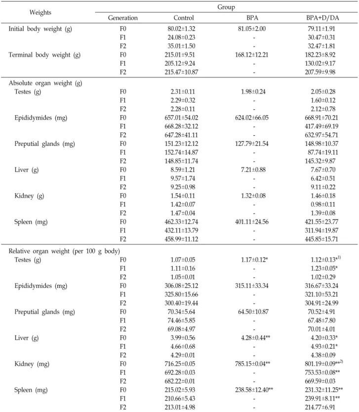
Table 1. Absolute and relative organ weights of males (F0, F1, and F2) were administrated for 30 days
Weights Group
Generation Control BPA BPA+D/DA
Initial body weight (g)
Terminal body weight (g)
F0 F1 F2 F0 F1 F2
80.02±1.32 24.08±0.23 35.01±1.50 215.01±9.51 205.12±9.24 215.47±10.87
81.05±2.00 - - 168.12±12.21 - -
79.11±1.91 30.47±0.31 32.47±1.81 182.23±8.92 130.02±9.17 207.59±9.98 Absolute organ weight (g)
Testes (g)
Epididymides (mg)
Preputial glands (mg)
Liver (g)
Kidney (g)
Spleen (mg)
F0 F1 F2 F0 F1 F2 F0 F1 F2 F0 F1 F2 F0 F1 F2 F0 F1 F2
2.31±0.11 2.29±0.32 2.28±0.11 657.01±54.02 668.28±32.12 647.28±41.11 151.23±12.12 152.74±14.87 148.85±11.74 8.59±1.21 9.57±1.74 9.25±0.98 1.54±0.11 1.42±0.07 1.47±0.04 462.33±12.74 432.11±13.79 458.99±11.12
1.98±0.24 - - 624.02±66.05 - - 127.79±21.54 - - 7.21±0.88 - - 1.32±0.08 - - 401.11±24.56 - -
2.05±0.28 1.60±0.12 2.12±0.78 668.91±70.21 417.49±69.19 632.97±54.71 148.98±10.37 87.74±19.11 145.32±9.87 7.67±0.70 6.42±0.51 9.11±0.22 1.46±0.18 0.98±0.11 1.39±0.08 421.55±23.77 311.94±19.87 445.85±15.71 Relative organ weight (per 100
Testes (g)
Epididymides (mg)
Preputial glands (mg)
Liver (g)
Kidney (mg)
Spleen (mg)
g body) F0 F1 F2 F0 F1 F2 F0 F1 F2 F0 F1 F2 F0 F1 F2 F0 F1 F2
1.07±0.05 1.11±0.16 1.05±0.01 306.08±25.12 325.80±15.66 300.40±19.44 70.34±5.64 74.46±5.85 69.08±4.97 3.99±0.56 4.66±0.68 4.29±0.01 716.25±0.05 692.28±0.03 682.22±0.01 215.02±5.93 210.66±5.43 213.01±4.98
1.17±0.12*
- - 315.11±33.34 - - 64.50±10.87 - - 4.28±0.44**
- - 785.15±0.04**
- - 238.58±12.40**
- -
1.12±0.13*1) 1.23±0.05*
1.02±0.29 316.67±33.24 321.10±53.21 304.91±24.99 70.52±4.91 67.48±7.80 70.01±4.01 4.20±0.33*
4.93±0.21*
4.38±0.09 801.19±0.09**2) 753.53±0.08**
669.59±0.03 231.32±11.25**
239.91±8.11**
214.77±6.91 All values represent the means±S.D. (n=15). 1)2)Significantly different from the control.*p<0.05, **p<0.01.
Organ weights in F0, F1, and F2 generation Previously, we reported the effect of organ weights by BPA and D/DA [11]. A significantly decreased weight of the body weight, testes, liver, kidney and spleen was de-
tected in F0 and F1 males at 50 mg/kg/day of BPA+D/DA.
The organ weights of F0 and F1 males are presented in
Table 1. Although the absolute weights of the Epididymides
and the Preputial glands at BPA were significantly lower,
no significant differences in the relative weights between the control and BPA groups. However, there was con- sistently affected in F1 offspring in BPA group. In the BPA+
D/DA group, the terminal body weights of F0 (17.98%,
p<0.05) and F1 (57.76%, p<0.01) decreased and the weightsof the testes F0 (4.47%, p<0.05) and F1 (8.76%, p<0.05), the liver F0 (5.00%, p<0.05) and F1 (5.48%, p<0.05), the kidney F0 (10.61%, p<0.01) and F1 (8.13%, p<0.01), the spleen F0 (7.05%, p<0.01) and F1 (12.20%, p<0.01) increased sig- nificantly compared to the corresponding relative weights in the control group. Absolute and relative organ weight data for F1 weanling pups indicate that the absolute organ weights were reduced, and the relative weights were in- creased in BPA+ D/DA group. Some of the effects on weights in the F1 weanlings were also present in the adults.
These results may cause the effect by reduced terminal body weight at dietary dose. However, some of the effects on absolute weights in the F0 males were significantly re- duced at only BPA group compared to the BPA+D/DA group. Similar results were also recorded by Tyl et al. [21], who mentioned that the highest dose of BPA intakes ap- proximately 1.5 to 2 times in the food consumption com- pared to other doses. These data indicated that the weights of the kidney, liver, spleen and testes were significantly cor- related (p<0.05 and p<0.01) with body weight. The organ weight of F2 males are presented in Table 1. No significant differences in the weight of any organs between control and BPA+D/DA groups were detected in F2 males. These re- sults indicate that oral doses of 50 mg/kg/day of D/DA recover damaged reproductive parameters by BPA over two-generation. More importantly, although organ weights of F0 and F1 males in the BPA+D/DA and BPA groups were different from those in the control group, the evidence of organ weights by D/DA (F2 males) affected, in as much as the overall ANOVA showed significant differences (p<
0.05 and p<0.01) between the BPA+D/ DA and BPA groups.
Epididymal sperm analysis and reproductive effects It is known that administration of D/DA increases the epididymal sperm motility and sperm count by the anti- oxidant effect of lipid peroxidation when compared with the corresponding group of control animals [20]. The mean epididymal sperm number was decreased (64.70%, p<0.01) in the BPA group compared to that of the control group.
However, the mean epididymal sperm number was slightly decreased (7.69%, p<0.05) in the BPA+D/DA group com-
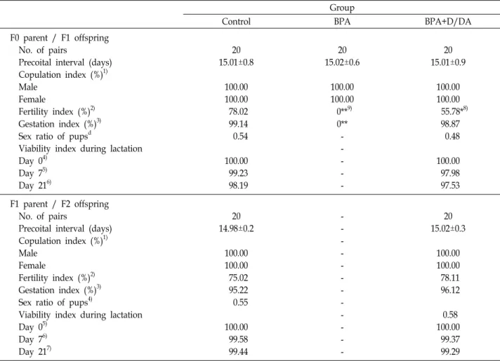
pared to that of the control group. These results match those observed in our earlier studies which D/DA recover sperm cell count against BPA damage [11]. The repro- ductive parameters for F0 parent/F1 offspring are shown Table 2. There were significant differences in the fertility in- dex and gestation index during lactation of BPA group.
Although the BPA group was observed copulation of males and females, no significantly pups were observed in BPA group. The majority of the effects observed for parental re- productive parameters (the epididymal sperm concentration of males) occurred only at the highest dose of BPA in BPA group. Tyl et al. [21] demonstrated that the highest dose of BPA was significantly reduced the number of implants, to- tal pups, and live pups per litter at birth. Biegel et al. [4]
and Liu et al. [15] also suggested that BPA may be reduced live litter sizes associated with reduced number of im- plantations per litter and a major contributor leading to male reproductive disorders. Reel et al. [18] also concluded that BPA was a reproductive toxicant that caused a reduc- tion in the number of live pups born in the F0 generation, reduced sperm motility and weight of some male re- productive organs in both the F0 and F1 generation, and reduced postnatal survival of the F1 generation. In the BPA+D/DA group, fertility index (55.78%, p<0.05) and ges- tation index (98.87%) restored significantly compared to the corresponding in the BPA group (Table 2). Previously, we reported that D/DA acts as protective agents against BPA-induced testicular toxicity. Administration of D/DA with BPA treatment also restored the testicular damage, and quality of sperm [11]. McLachlan et al. [16] suggested that a change of sperm count was directly affected to a spermatogenesis, including a level of testosterone. The lu- teinizing hormone of the pituitary gland regulated the se- cretion of testosterone, lead to a difference of spermatozoa [22]. Our results also showed a change of testosterone level (data not shown). As shown in Table 2, an increase in the level of fertility index, gestation index and viability index during lactation were observed in reproductive parameters of rats exposed to BPA+D/DA compared to BPA group.
The fertility index (22.24%) and gestation index (0.27%) in the BPA+D/DA group were a little lower when compared with control and BPA+D/DA groups. The reproductive pa- rameters for F1 parent/ F2 offspring are also shown in Table 2. The fertility index of control (75.02%) and BPA+D/
DA (78.11%) groups were no significant difference for two-
generation compared to one-generation. This result in-
Table 2. Reproductive data for F0 parent / F1 offspring and F1 parent / F2 offspring Group
Control BPA BPA+D/DA
F0 parent / F1 offspring No. of pairs
Precoital interval (days) Copulation index (%)1) Male
Female
Fertility index (%)2) Gestation index (%)3) Sex ratio of pupsd
Viability index during lactation Day 04)
Day 75) Day 216)
20 15.01±0.8
100.00 100.00 78.02 99.14 0.54
100.00 99.23 98.19
20 15.02±0.6
100.00 100.00 0**9)
0**
- - - - -
20 15.01±0.9
100.00 100.00 55.78*8)
98.87 0.48
100.00 97.98 97.53 F1 parent / F2 offspring
No. of pairs
Precoital interval (days) Copulation index (%)1) Male
Female
Fertility index (%)2) Gestation index (%)3) Sex ratio of pups4)
Viability index during lactation Day 05)
Day 76) Day 217)
20 14.98±0.2
100.00 100.00 75.02 95.22 0.55
100.00 99.58 99.44
- - - - - - - - - - - -
20 15.02±0.3
100.00 100.00 78.11 96.12
0.58 100.00 99.37 99.29
1)Copulation index (%)= (No. of rats copulated/no. paired) ×100.
2)Fertility index (%)= (No. of females pregnant/no. of females copulated) ×100.
3)Gestation index (%)= (No. of females with parturition/no. of females pregnant) ×100.
4)Sex ratio pups= No. of male pups/total no. of pups.
5)Viability index on day 0 of lactation (%)= (No. of live pups delivered/total no. of pups delivered) ×100.
6)Viability index on day 7 of lactation (%)= (No. of live pups delivered/total no. of pups delivered) ×100.
7)Viability index on day 21 of lactation (%)= (No. of live pups delivered/total no. of pups delivered) ×100.
Significantly different from the control.8)*p<0.05, 9)**p<0.001.
dicates that D/DA affect reproductive performance in gen- eration. No significant difference in the fertility index, ges- tation index, and viability index during lactation was also observed between control and BPA+D/DA groups. In our study, these findings suggest that D/DA effects on the number of pups per litter, sex ratio, fertility index, gestation index, and viability index in rats exposed to BPA (50 mg/
kg/day) during lactation. Animals treated with D/DA in- creased the number of mating and mounting comparing with animals treated with only BPA. Fertility index and body size of animals treated with D/DA were also normal- ized in two-generation. Though every baby was normalized in two-generation, further extensive research will be needed
for exacted evaluation in several conditions.
Acknowledgement
This research was (partially) supported by a Busan met- ropolitan city Grant (Busan Brain 21) in 2014.
References
1. Ahn, M. J., Lee, M. K., Kim, Y. C. and Sung, S. H. 2008.
The simultaneous determination of coumarins in Angelica gigas root by high performance liquid chromatography-di- ode array detector coupled with electrospray ioniza- tion/mass spectrometry. J. Pharmaceut. Biomed. 46, 258-266.
2. Ahn, K. S., Sim, W. S. and Kim, I. H. 1996. Decursin: a cyto- toxic agent and protein kinase C activator from the root of Angelica gigas. Planta. Med. 62, 7-9.
3. Akhtar, N., Srivastava, M. K. and Raizada, R. B. 2009.
Assessment of chlorpyrifos toxicity on certain organs in rat, Rattusnorvegicus. J. Environ. Biol. 30, 1047-1053.
4. Biegel, L. B., Cook, J. C., Hurtt, M. E. and O’Connor, J. C.
1998a. Effects of 17 β-estradiol on serum hormone concen- trations and estrous cycle in female Crl:CD BR rats: Effects on parental and first generation rats. Toxicol. Sci. 44, 143- 154.
5. Goodman, J. E., McConnell, E. E., Sipes, I. G., Witorsch, R.
J., Slayton, T. M., Yu, C. J., Lewis, A. S. and Rhomberq, L. R. 2006. An updated weight of the evidence evaluation of reproductive and developmental effects of low doses of bisphenol A. Crit. Rev. Toxicol. 36, 387-457.
6. Konoshima, M., Chi, H. J. and Hata, K. 1968. Coumarins from the root of Angelica gigas Nakai. Chem. Pharm. Bull.
16, 1139-1140.
7. Kang, S. Y., Lee, K. Y., Sung, S. H., Park, M. J. and Kim, Y. C. 2001. Coumarins isolated from Angelica gigas inhibit acetylcholinesterase structure-activity relationships. J. Nat.
Prod. 64, 683-685.
8. Kim, S. J., Kim, M. R., Hwang, S. Y., Bae, W. J., Kim, S., Hong, S. H., Lee, J. Y., Hwang, T. K., Wang, Z. and Kim, S. W. 2013. Preliminary Report on the safety of a new herb- al formula and its effect on sperm quality. World J. Mens Health 31, 254-261.
9. Kim, K. M., Kim, T. H., Park, Y. J., Kim, I. H. and Kang, J. S. 2009. Evaluation of the genotoxicity of decursin and decursinol angelate produced by Angelica gigas Nakai. Mol.
Cell. Toxicol. 5, 83-87.
10. Kim, K. M., Lee, Y. J., Hong, Y. G. and Kang, J. S. 2009.
Oral acute and subacute toxicity studies of decursin and decursinol angelate of Angelica gigas Nakai. Mol. Cell.
Toxicol. 5, 153-159.
11. Kim, K. M., Seo, J. L. and Kang, J. S. 2014. Decursin and decursinol angelate affect spermatogenesis in the adult rat at oral administration. Mol. Cell. Toxicol. 10, 83-89.
12. Kim, K. M., Jung, J. Y., Hwang, S. W., Kim, M. J. and Kang, J. S. 2009. Isolation and purification of decursin and de- cursinol angelate in Angelica gigas Nakai. J. Korean Soc. Food Sci. Nutr. 38, 653-656.
13. Lee, S. H., Lee, Y. S., Jung, S. H., Shin, K. H., Kim, B. K.
and Kang, S. S. 2003. Anti-tumor activities of decursinol an- gelate and decursin from Angelica gigas. Arch. Pharm. Res.
26, 727-730.
14. Lee, S. H., Shin, D. S., Kim, J. S., Oh, K. B. and Kang, S.
S. 2003. Antibacterial courmins from Angelica gigas roots.
Arch. Pharm. Res. 26, 449-452.
15. Liu, C., Duan, W., Li, R., Xu, S., Zhang, L., Chen, C., He, M., Lu, Y., Wu, H., Pi, H., Luo, X., Zhang, Y., Zhong, M., Yu, Z. and Zhou, Z. 2013. Exposure to bisphenol A dis- rupts meiotic progression during spermatogenesis in adult rats through estrogen-like activity. Cell Death Dis. 4, 1-10.
16. McLachlan, R. I., Wreford, N. G., O’Donnell, L., de Kretser, D. M. and Robertson, D. M. 1996. The endocrine regulation of spermatogenesis: independent roles for testosterone and FSH. J. Endocrinol. 148, 1-9.
17. Ryu, K. S., Hong, N. D., Kim, N. J. and Kong, Y. Y. 1990.
Studies on the coumarin constituents of the root of Angelica gigas Nakai. Isolation of decursinol angelate and assay of decursinol angelate and decursin. Kor. J. Pharmacogn. 21, 64-68.
18. Reel, J. R., George, J. D., Myers, C. B., Lawton, A. D. and Lamb, I. V. 1985. Bisphenol A: reproduction and fertility assessment in CD-1 mice when administered in the feed.
Final study report, NTP/NIEHS Contract No. N01-ES-2- 5014, National Technical Information Service (NTIS) Acces- sion No. PB86103207, May 3.
19. Šutiaková, I., Kovalkovičová, N., Tulenková, M. and Šutiak, V. 2012. Bisphenol A and its potential toxic effects on living organisms. J. Microb. Biotechnol. Food Sci. 2, 526-535.
20. Son, C. Y., Baek, I. H., Song, G. Y., Kang, J. S. and Kwon, K. I. 2009. Pharmacological effect of decursin and decursi- nol angelate from Angelica gigas Nakai. J. Kor. Pharm. Sci.
53, 303-313.
21. Tyl, R. W., Marr, M. C., Thomas, B. F., Keimowitz, A. R., Brine, D. R., Veselica, M. M., Fail, P. A., Chang, T. Y., Seely, J. C., Joiner, R. L., Butala, J. H., Dimond, S. S., Cagen, S.
Z., Shiotsuka, R. N., Stropp, G. D. and Waechter, J. M. 2002.
Three-generation reproductive toxicity study of dietary bi- sphenol Ain CD Sprague-Dawley rats. Toxicol. Sci. 68, 121- 146.
22. Uzun, F. G., Kalender, S. Durak, D., Demir, F. and Kalender, Y. 2009. Malathion-induced testicular toxicity in male rats and the protective effect of vitamins C and E.
Food Chem. Toxicol. 47, 1903-1908.
23. WHO, 1992. Research guidelines for evaluating the safety and efficacy or herbal medicines. Regional office for the western pacific working group on the safety and efficacy on herbal medicine. pp. 5-9. Manila, Philippines.
24. Yook, C. S. 1990. Coloured medicinal plants of Korea, pp. 390, Academy Book Co., Seoul, Korea,
25. Yim, D. S., Singh, R. P., Agarwal, C., Lee, S. Y. and Chi, H. J. 2005. A novel anticancer agent, decursin, induces G1
arrest and apoptosis in human prostate carcinoma cells.
Cancer Res. 65, 1035-1044.
초록:Decursin and decursinol angelate가 2세대 출산률 회복에 미치는 영향
김강민․옥 선․고윤석․강재선*
(경성대학교 약학과)