∙ Received: June 29, 2012. Accepted: September 28, 2012.
∙ Corresponding author : Chung Eun Mi
Department of Nuclear Medicine, Seoul Asan Medical Center 88, Olympic-ro 43-gil, Songpa-gu, Seoul 138-736, Korea Tel: +82-2-3010-4607, Fax: +82-2-3010-4588 E-mail: jjjeunmi@nate.com
Original Article
유방특이감마영상검사에서 액와부 영상 획득 방법에 대한 연구서울아산병원 핵의학과
장지연 ․ 정은미Technical Details Imaging Axillary Lymph Nodes in Breast-Specific Gamma Imaging
Ji Yeon Jang, Eun Mi Jung
Departmant of Nuclear Medicine, Asan Medical Center, Seoul, Korea
Purpose : The initial Breast-Specific Gamma Imaging (BSGI) protocol included bilateral breast imaging with 2 views of each breast-craniocaudal (CC) and mediolateral oblique (MLO). Furthermore, Axillary lymph nodes view can be acquired easily. The most meaningful prognosis factor for prediction of breast cancer is whether or not the breast cancer has metastasized to the lymph nodes. However, axillary view doesn't conduct in clinical.
This article collates a diverse data of BSGI and describes technical details to acquire optimal imaging. Materials and Methods : A retrospective review was performed on 343 patients who had undergone BSGI between May 2011 and March 2012. Patients who had undergone BSGI received intravenous injection of 740 MBq (20 mCi)
99m
Tc-sestamibi. Results : The following contents are the technical details for optimal axillary imaging.
99m
Tc-sestamibi should be administered using an indwelling venous catheter or scalp needle followed by 10 cc of saline to flush to reduce extravasation and vascular trapping. After administration, patients raise their arm over their head and exercise with stress ball for 1 full minute. A lead shield attached to the gamma camera is removed and patients axilla is placed as close as possible to the camera at a 90° angle. A lead apron is placed across the shoulder to reduce background from other organs. Acquisition time is enough for 120 sec~180 sec. Conclusion : If patients undergo bilateral axillary imaging as a standard with CC, MLO views, it could improve cancer treatment. Result of this study could maximize efficiency axillary imaging of breast cancer patients. (Korean J Nucl Med Technol 2012;16(2):115-119)
Key Words : Breast-Specific Gamma Imaging (BSGI), Axillary lymph node, Axillary imaging
INTRODUCTION
Westernized diets, increased obesity rates, and decreased birth rates and breastfeeding have increased breast cancer rates to such an extent that breast cancer is now the number one type of cancer in Korean women. Its early detection is now possible, however, using various methods such as mam-
mography, ultrasound, and magnetic resonance imaging; and its survival and recovery rates are also rising through devel- opments in chemotherapy and targeted therapy.
Early detection and accurate stage determination are essen-
tial in reducing mortality from breast cancer, and one of the
most important factors of the prediction of the relapse of and
survival from breast cancer is metastasis in the axillary lymph
nodes.
1-2)A number of studies have been conducted to inves-
tigate detection methods for axillary lymph node metastasis
that are less invasive and do not hinder the local regulation
of cancer cells. Breast ultrasound is currently considered the
easiest way to detect axillary lymph nodes, and it has several
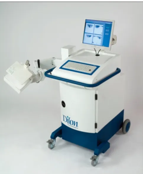
Fig. 1. Dilon 6800 (Dilon Technologies Inc, Newport News, USA).
benefits. For example, it can detect the longest diameter of a tumor at different angles without enlarging the tumor, and the patient does not have to be exposed to radiation.
15-16)Breast ultrasound is not objective, however, because its read- ing varies depending on the examiner and it is accurate only when the walls of the tumor are definite.
3)Recently, breast-specific gamma imaging (BSGI) using
99m
Tc-sestamibi and a high-resolution gamma camera was introduced. This is a method that uses the physiological char- acteristics of lesions in the breast more than their anatomy.
As the cancerous cells in the breast have increased mitochon- dria metabolic activity and more distributed blood vessels, they absorb more radiotracer than normal cells do.
4-5)Although, the most important prognosis factor for prediction of breast can- cer is axillary metastasis, axillary view on BSGI doesn’t con- duct in clinical. Our institution has incorporated axillary view at the time of BSGI to gain information on the status of the axillary lymph nodes to aid in surgical planning for these patients.
MATERIALS AND METHODS
BSGI was performed 3-12 days after the menstrual periods of selected women or on postmenopausal women. After the
99m
Tc-sestamibi injection, the images were obtained using a BSGI 6800 gamma camera manufactured by Dilon (Fig. 1).
The camera size and gantry design provided the flexibility re-
quired to emulate the same views obtained in mammography.
After acquiring bilateral craniocaudal (CC), mediolateral oblique (MLO) view, axillary imaging began. This hospital have included BSGI axillary lymph node imaging in its rou- tine procedures since May 2011. A retrospective review was performed on 343 patients who had undergone BSGI be- tween May 2011 and March 2012. And then, we studied about various axillary image data and suggested technical de- tails acquiring optimal axillary imaging.
RESULTS
Among the 343 patients, 77.8% patients had already un- dergone surgery or chemotheraphy, and 22.2% patients were waiting for surgery. Of the patients who were waiting for surgery, 47.4% had lymph node metastasis and in 36.1% of these patients, the uptake of the radiotracer in the lymph no- des was locally observed in their images. The patients who did not show any uptake in their lymph nodes either had no initial lymph node metastasis or undetected because of too small size tumor. The uptake was locally identified in the im- ages of 1.2% patients on follow up testing after surgical procedure. Whose uptake resulted from the extravasation.
Among all the patients, vascular trapping image was shown in the images of 9.6% patients.
Consequently, there are 2 complications with this techni- que that may cause artifacts-vascular trapping and subcuta- neous extravasation. To reduce these images and acquire opti- mal images, I suggest that we comply with the following contents.
1. Injection Technique
For breast imaging,
99mTc-sestamibi 740 MBq (20 mCi) is typically injected into the venous system of the upper limb contralateral to the breast of concern. Most of the time, it is injected through the anterior cardinal vein or the dorsum of the hand, but it can cause artifacts when the axillary image is obtained.
The first possible case is when the radiotracer is trapped
in the blood vessels.
99mTc-sestamibi mainly remains in the
walls of blood vessels and stays there more than 5 minutes
Fig. 2. Vascular trapping can be reduced as exercising anc
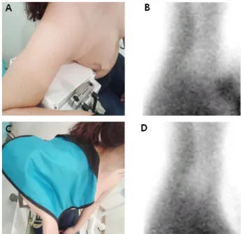
flushing with normal saline.Fig. 3. The image A is pre-shielding position and image B
shows artifacts due to the adjacent organs. The image C is post-shielding position and image D indicates that artifacts are reduced.after the injection. This trapping is shown as straight lines aligned with the cephalic vein and the basilica vein in the ax- illary images (Fig. 2).
Second, artifacts can be explained when
99mTc-sestamibi is not injected properly and extravasation occurs. This out- flowing radiotracer is carried in the lymphatic system and eventually accumulates in the axillary part and forms the fo- cal area. It has been reported that it was difficult to de- termine quantitatively.
6)To prevent the mentioned possible artifacts primarily, di- rect injection of radiotracer from a syringe should be avoided.
It is recommended that scalp needles be used first to find the blood vessel, before injecting
99mTc-sestamibi and after ad- ministering 10 cc of normal saline to confirm that there is no effluent. After the injection, the patient raises their arms than ear and exercises for a few minutes using a stress ball. By do- ing this, the artifacts can be significantly reduced.
2. Imaging and Acquisition Settings
The craniocaudal and mediolateral oblique images are tak- en within 10 minutes after the injection. As
99mTc-sestamibi is relatively less absorbed by fat tissues, more than 100k counts is used for 5-10 minutes. A paddle attached to a de- tector is used when such images are taken. As the paddle contains lead, it reduces the artifacts caused by the accumu- lated
99mTc-sestamibi in the surrounding organs. After the craniocaudal and mediolateral oblique images are obtained,
the axillary images are taken. To obtain the axillary images, the paddle is removed and a lead apron was draped across patient’s shoulder to shield the detector from uptake in the organs of the head (Fig. 3). In particular, the detector should be set at an appropriate angle by observing the images to minimize the shattered rays from the heart when the left axil- lary images are obtained. The detector should head toward the humeral head. As
99mTc-sestamibi is accumulated in the axillary area, more than 150k counts must be used for 1-2 minutes.
DISCUSSION AND CONCLUSION
BSGI is a useful supplementary method of diagnosing breast cancer when it is used with mammography or ultrasound.
BSGI is known to be 77.8% sensitive and 84.2% specific so far.
7-8)The usefulness of additional BSGI with traditional methods in detecting breast cancer has already been proven.
It has been reported to be 28-81% sensitive and 89-92% spe-
cific in detecting axillary lymph node metastasis.
9-14)Despite
such positive figures, however, axillary imaging using BSGI
is not a common procedure. Researchers believe additional
bilateral axillary imaging with the conventional imaging
would be helpful in determining the diagnosis and treatment plans for breast cancer patients.
In particular, the size and shape of a BSGI gamma camera is designed to allow its easy use to obtain images such as in mammography. As the standards for axillary lymph node imaging have not been revised despite the many studies on it in different study areas, more organized studies need to be conducted in various areas. In addition, a specific detector for axillary imaging must be developed, and further research on it is needed.
요 약
유방특이감마영상검사(Breast-specific Gamma Imaging, BSGI) 는
99mTc-sestamibi와 고해상도 감마카메라를 이용하여 영상 을 획득하는 방법으로 기본적으로 양측상하영상, 내외사방 향영상을 얻으며 추가로 액와 림프절 검사를 시행 할 수 있 다. 액와 림프절 전이 여부가 중요한 인자임에도 불구하고 아직까지 BSGI의 액와부 검사는 잘 시행되지 않고 있는 실 정이다 . 이에 본 연구에서는 2011년 5월부터 2012년 3월까지 본원에서 유방특이감마영상검사를 시행한 총 343명의 환자 를 대상으로 액와 림프절 검사 결과를 분석하고 올바른 검사 를 위한 기술적 사항을 연구하였다. 방사성의약품 주입 시
일어날 수 있는 혈관 포획 , 혈관 외 유출현상이 영상에 영향
을 끼치는 경우가 많았으며 , 이를 방지하는 방법과 영상의
획득 방법을 규정하였다 . 혈관 외 유출과 혈관의 포획현상을
최소화하기 위하여
99mTc-sestamibi를 주입 후, 10 cc의 생리
식염수를 주입한다 . 주사 후 팔을 귀 위로 올리고 공을 이용
하여 약 1분간 운동한다. 감마카메라의 납 차폐체를 제거하 고 검출기에 기울기를 주어 최대한 검출기에 액와부를 밀착
시키고 , 납 앞치마를 이용하여 촬영하려는 측의 어깨 부분을
가려주어 배후 방사능을 최소화 한다. 액와부 영상은 2-3분 획득한다 . BSGI 검사 시, 기존에 시행되던 양측 상하방향, 내 외사방향영상만을 얻는 방식에서 액와 림프절 영상을 함께 획득 한다면 유방암의 치료성적 향상에 도움이 되며, 본 연 구에서 얻은 기술적 사항을 검사에 적용한다면 유방암 환자 들의 액와 림프절 영상의 효과를 더욱 극대화 시킬 수 있을 것으로 사료된다.
REFERENCES
1. McCready DR, Hortobagyi GN, Kau SW, Smith TL, Buzdar
AU, Balch CM. The prognostic significance of lymph node metastases after preoperative chemotheraphy for locally ad- vanced breast cancer. Afch Surg 1989;124:21-5.
2. Fisher B, Bauer M, Wickerham DL, Redmond CK, Fisher ER.
Relation of the number of positive axillary nodes to the prog- nostic of patients with primary breast cancer. Cance 1983;52:
1551-7.
3. Nori J, Vanzi E, Bazzocchi M, Bufalini FN, Distante V, Branconi F, et al. Role of axillary ultrasound examination in the selection of breast cancer patients for sentinel node biopsy. Am J Surg 2007;193:16-20.
4. Sharma S, Sharma MC, Sarkar C. Morphology of angiogenesis in human cancer: a conceptual overview, Histopathology 2005;46:481-9.
5. Delmon-Moingeon LI, Piwnica-Worms D, Van den Abbeele AD, Holman BL, Davison A, Jones AG. Uptake of the cation hexakis(2-methoxyisobutylisonitrile)-technetium-99m by hu- man carcinoma cell lines in vitro. Cancer Res 1990;50:2198-202.
6. John Werner, MD, Jocelyn A. Rapelyea, MD, Kristen G. Yost, MS, et al. Quantification of radio-tracer uptake in axillary lymph nodes using breast-specific gamma imaging: Benign ra- dio-tracer extravasation versus uptake secondary to breast cancer. The Breast Journal, Volume15 Number 6 2009;579-582.
7. Brem RF, Petrovitch I, Rapelyea JA, Young H, Teal C, Kelly T.
Breast-specific gamma imaging with 99mTc-sestamibi and mag- netic resonance imaging in the diagnosis of breast cancer-a com- parative study. Breast J 2007;13:1465-9.
8. Bream RF, Floerke AC, Rapelyea JA, Teal C, Kelly T, Mathur V. Breast-specific gamma imaging as an adjunct imaging modal- ity for the diagnosis of breast cancer. Radiology 2008;2467:
651-7.
9. Tolmos J, Khalkhali I, Vargas H, Stuntz M, Cutrone J, Mishkin F, et al. Detection of axillary lymph node metastasis of breast carcinoma with technetium-99m-sestamibi scintimammography.
Am Surg 1997;63:850-3.
10. Taillefer R, Robidoux A, Turpin S, Lambert R, Cantin J, Leveille J. Metastatic axillary lymph node technetium-99m- MIBI imaging in primary breast cancer. J Nucl Med 1998;39:
459-64.
11. Lumachi F, Tregnaghi A, Ferretti G, Povolato M, Marzola MC, Zucchetta P, et al. Accuracy of ultrasonography and 99mTc- sestamibi scintimammography for assessing axillary lymph node status in breast cancer patients. A prospective study. Eur J Surg Oncol 2006;32:933-6.
12. Lumachi F, Ferretti G, Povolato M, Bui F, Cecchin D, Marzola MC, et al. Axillary lymph node metastasis detection with 99m-sestamibi scintimammography in patients with breast can- cer undergoing curative surgery. Anticancer Res 2007;27:2949- 52.
13. Elizabeth A, Jones, Trinh D. Phan, Nathalie M. Johnson, Deborah A. Blanchard. A protocol for imaging axillary lymph nodes in patients undergoing breast-specific Γ-imaging. J Nucl Med Technol 2010;38:28-31.
14. Bream RF, Rapelyea JA, Zisman G, Mothashemi K, Raub J,
Teal CB, et al. Occult breast cancer: scintimammography with high-resolution breast-specific gamma camera in women at high risk for breast cancer. Radiology 2005;237:274-80.
15. Rajesh YS, Ellenbogen S, Banerjee B. Preoperative axillary ul- trasound scan: Its accuracy in assessing the axillary nodal status
in carcinoma breast. Breast 2002;11:49-52.
16. Cowher MS, Erb KM, Poller W, Julian TB. Correlation of the use of axillary ultrasound and lymph node needle biopsy with surgical lymph node pathology in patients with invasive breast cancer. Am J Surg 2008;196:756-9.