R E S E A R C H Open Access
A clinical study of inferior alveolar nerve damage caused by Carnoy ’s solution used as a complementary therapeutic agent in a cystic lesion
Hyun-Jun Jo, Hee-Youl Kim, Dong-Cheol Kang, Dae-Ho Leem, Jin-A Baek and Seung-O Ko
*Abstract
Background: Cyst enucleation, which extracts only the tumor with the application of Carnoy ’s solution (CS), has been suggested as a conservative treatment with a low recurrence rate and morbidity. However, there has been a concern that CS ’s contact with inferior alveolar nerve (IAN) can cause neurons to degenerate and cause sensory dysfunction. The purpose of this retrospective cohort study aimed to investigate the neurosensory function after surgical treatment with or without the application of CS.
Methods: While controlling the effects of sex, age, follow-up period, and invasion size of the tumor, we performed the binary logistic regression analysis to examine whether or not the sensory function of the patients who were treated with CS (n = 19) for the cyst enucleation procedure was significantly different from those who were not treated with CS (n = 58) at the end of the follow-up period.
Results: The logistic regression result showed that the use of CS was not significantly related to the normalness of sensory function at the end of the follow-up period. Rather, the invasion size of the cyst was significantly associated with sensory dysfunction.
Conclusions: CS may be used for patients who are diagnosed with OKC and UAM without much fear of its impact on sensory dysfunction. However, a small number of patients who were treated with CS experienced severe sensory damage and did not recover at the end of the follow-up period, suggesting the need for further analysis of these patients.
Keywords: Carnoy ’s solution, Hypoesthesia, Paresthesia, Sensory dysfunction, Inferior alveolar nerve damage
Background
Carnoy ’s solution (CS) is a chemically cauterizing agent that has been used to treat various cysts and tumors of aggressive nature in the oral and maxillofacial surgery area. Although many studies indicated that the use of CS is effective to reduce the recurrence of various cysts and tumors in the oral and maxillofacial surgery area, such as odontogenic keratocyst (OKC), unicystic
ameloblastoma (UAM) [1 – 4], researchers have not ex- amined the effect of CS on the inferior alveolar nerve (IAN) and sensory dysfunction. Therefore, it is import- ant to research the relationship between the use of CS and sensory dysfunction.
In particular, CS has been commonly used for the treatment of odontogenic keratocyst (OKC) and unicys- tic ameloblastoma (UAM) and one of the three clinical variants of ameloblastoma (AM). CS composed of 60 ml (1 mol: 46.07 g) of absolute alcohol (ethanol), 30 ml (1 mol: 119.38 g) of chloroform, and 10 ml (1 mol: 60.05 g) of acetic acid. The penetration of this solution into
© The Author(s). 2020 Open Access This article is licensed under a Creative Commons Attribution 4.0 International License, which permits use, sharing, adaptation, distribution and reproduction in any medium or format, as long as you give appropriate credit to the original author(s) and the source, provide a link to the Creative Commons licence, and indicate if changes were made. The images or other third party material in this article are included in the article's Creative Commons licence, unless indicated otherwise in a credit line to the material. If material is not included in the article's Creative Commons licence and your intended use is not permitted by statutory regulation or exceeds the permitted use, you will need to obtain permission directly from the copyright holder. To view a copy of this licence, visithttp://creativecommons.org/licenses/by/4.0/.
* Correspondence:
[email protected]Department of Oral and Maxillofacial Surgery, School of Dentistry, Chonbuk
National University Dental Hospital, 20, Geonji-ro, Deokjin-gu, Jeonju-si,
Jeollabuk-do, Republic of Korea
tissues results in rapid local fixation and denaturation [5]. CS has been reported to have systemic toxicity that can cause local damage to anatomical structures, espe- cially nerves [6]. However, Blanas et al. [7] suggested that there would be no damage to the nerves if the infer- ior alveolar nerve (IAN) is not exposed to CS for more than 3 min [7].
The inferior alveolar nerve (IAN) is located on the mandible. If the sensory nerves are damaged, the hypoesthesia or paresthesia may occur. Therefore, IAN is a structure that should be preserved as much as pos- sible without much damage [8, 9]. It is reasonable for an oral surgeon to choose cyst enucleation with CS rather than extensive resection to preserve IAN and the sur- rounding bone tissue when the lesion contains IAN. The surgeon needs to remove the lesion carefully while pro- tecting the nerves. At this time, CS can be applied with chemical cauterization ability because OKC, UAM daughter’s cyst, and epithelial cell residue exist in the space adjacent to the nerve of the bone and cause recur- rence. However, CS may damage neurons beyond the peripheral sheath of the nerve and cause sensory dys- function when CS comes in contact with IAN. The pur- pose of this retrospective cohort study aimed to investigate the neurosensory function after surgical treatment with or without the application of CS.
Sample and methods Sample and study design
The current research project was reviewed and approved by the IRB at the Chonbuk National University before the project began. This study was a retrospective case- control one consisting of patients who underwent man- dibular cystic extraction. Radiologic and surgical records were reviewed for the patients who underwent mandibular cyst extraction for 5 years (2014–2018) at the Chonbuk National University Hospital in South Korea. For the pre- operative cone beam computed tomography (CBCT) examination, patients who had cystic lesions with the in- ferior alveolar nerve canal (IAC) invasion were selected.
The patients with normal sensation were included, while those who complained about abnormal sensory functions before the enucleation were excluded. Patients complain- ing of abnormal sensory function after the surgery were subjected to follow-up examinations of between 10 and 66 months.
The records of all patients who were treated for OKC and UAM for the 5 years between 2014 and 2018 were reviewed. Based on the pathology reports, all cases of cystic lesions of the mandible were screened for inclu- sion in the study. However, the patients who were diag- nosed with other subtypes of AM, such as solid/
multicystic (i.e., intraosseous) and peripheral ameloblas- tomas, were excluded. Out of a total of 248 patients with
a cystic lesion in mandible, 77 cases with IAC invasion, as shown by CBCT, were selected for further analysis. The analysis included scrutiny of intraoperative findings as documented in the surgical records and the histological diagnosis from incisional biopsies and final enucleation specimens. Oral pathologists reviewed histological sec- tions of all 77 cystic cases. The patients were divided into two groups according to the use (non-use) of CS.
Surgical procedure
During the 5 years (2014–2018), the treatment for the patients with OKC and UAM in the Department of Oral and Maxillofacial Surgery, the Chonbuk National Uni- versity Hospital, was consisted of the followings: a biopsy-confirmed diagnosis, enucleation of the lesion and application of CS to the bony cavity, and long-term clinical and radiological follow-up. In performing enu- cleation of the lesion, the teeth directly related to the periphery of the cyst were extracted before proceeding with the enucleation. If IAN was exposed during the enucleation procedure, the cyst was carefully stripped from the nerve. The bony cavity was then examined for any remaining cystic tissue. If a residue was found, it was removed. CS was applied to the bony cavity for 3 min using cotton applicators or soaked ribbon gauze.
However, the use of CS was minimized as much as pos- sible when the mandibular nerve was visible in the bony cavity. This procedure was followed by copious irrigation with normal saline, and clinical and radiological follow- up was carried out.
Data analyses
Data analyses were performed using SPSS version 25 (SPSS, Inc., Chicago, IL). Descriptive statistics of the sample and the comparison of means and the frequen- cies between the CS group and non-CS groups were per- formed by using one-way ANOVA and chi-square tests to assess the relationship between the use of CS and sen- sory function before the multivariate logistic regression analysis. The dependent variable for the logistic analysis was the subjective numeric rating scale (NRS), and the patient expressed the degree of his or her sensory func- tion in numbers. A value of 10 indicates a high-level sensory function and 0 for no sensation at all. A total of 77 patients were divided into two groups: one group that indicated their sensory scale of between 8 and 10, show- ing somewhat normal sensory function, and the other group which indicates below 8, showing non-normal sensory function. Therefore, we considered that a pa- tient’s sensory function was almost normal when NRS was eight or above.
On the other hand, the independent variables for the
logistic regression analysis included sex, age in years,
follow-up period, invasion size of the lesion, and the use
of CS. The invasion size had three groups according to the degree of cyst invasion of IAC: less than one third (1), between one third and two thirds (2), and over two thirds of the circumference of the canal (3). Given the nature of the dependent variable, binary logistic regres- sion was used and the invasion size was treated as an interval measure for logistic regression analysis, ranging from 1 to 3.
Results
Descriptive statistics
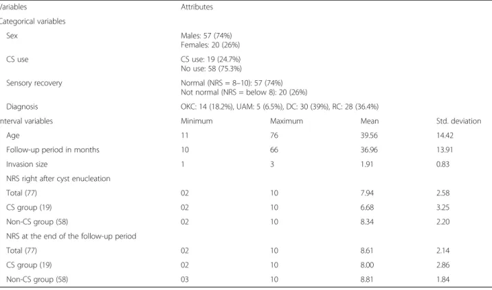
Table 1 shows the descriptive statistics. First of all, among a total of 77 patients in this study, 57 (74%) and 20 of them (26%) were males and females, respectively.
Next, among a total of 77 patients, a postoperative bi- opsy revealed OKC for 14 and UAM for five patients.
Out of a total of 77 patients, CS was used for a total of 19 (24.7%) patients who were diagnosed with either OKC or UAM. The postoperative biopsy also revealed dentigerous cysts (DC) for thirty patients (39%) and ra- dicular cysts (RC) for twenty-eight patients (36%). Fifty- seven patients (74%) indicated that their sensory func- tion, NRS, at the end of the follow-up period was be- tween 8 and 10 (normal) and 20 (26%) of them expressed below scale 8 (not normal).
The mean age of the patients was 39.56 and the mean follow-up period was 36.96 months. The average NRS right after the surgery was 7.94, and the one at the end
of the follow-up period was 8.61. Therefore, on average, there was only a 0.67 improvement of NRS at the end of the follow-up from the one right after the surgery. Fi- nally, the mean invasion size was 1.91.
General description of sensory function
Out of 14 OKC patients, the cyst recurred only for one patient (7.14%) during the 5-year follow-up period. For two patients with nevoid basal cell carcinoma (NBCC), multiple OKCs were found in various areas of the jaw, but no lesion recurred. Eight patients complained of ab- normal sensory function (NRS = below 8) immediately after the treatment with CS. However, four of them re- ported that they recovered their sensory function and expressed their sensory function of NRS at between 8 and 10 during the follow-up period. On the other hand, the other four OKC patients who were treated with CS (28.57%) still expressed abnormal sensory function dur- ing the follow-up period.
Among five patients in the UAM group, four patients complained of abnormal sensory function immediately after the treatment with CS. Three of them had almost recovered sensory function with NRS of from eight to ten during the follow-up period, and one of them still complained of discomfort due to sensory dysfunction at the end of the follow-up period. Therefore, five out of 19 patients (26%) who were treated with CS developed permanent abnormality of sensory function. Twenty out
Table 1 Descriptive statistics
Variables Attributes
Categorical variables
Sex Males: 57 (74%)
Females: 20 (26%)
CS use CS use: 19 (24.7%)
No use: 58 (75.3%)
Sensory recovery Normal (NRS = 8 –10): 57 (74%)
Not normal (NRS = below 8): 20 (26%)
Diagnosis OKC: 14 (18.2%), UAM: 5 (6.5%), DC: 30 (39%), RC: 28 (36.4%)
Interval variables Minimum Maximum Mean Std. deviation
Age 11 76 39.56 14.42
Follow-up period in months 10 66 36.96 13.91
Invasion size 1 3 1.91 0.83
NRS right after cyst enucleation
Total (77) 02 10 7.94 2.58
CS group (19) 02 10 6.68 3.25
Non-CS group (58) 02 10 8.34 2.20
NRS at the end of the follow-up period
Total (77) 02 10 8.61 2.14
CS group (19) 02 10 8.00 2.86
Non-CS group (58) 03 10 8.81 1.84
Note: OKC odontogenic keratocyst, UAM unicystic ameloblastoma, DC dentigerous cyst, RC radicular cyst, CS Carnoy’s solution, NRS numeric rating scale