Evaluation of Sealing Effect and Working Time of Root Canal Filling MTA Materials
Hyojin Kim, Youngjin Kim, Soonhyeun Nam, Kwon Taeyub, Hyunjung Kim
Department of Pediatric Dentistry, School of Dentisry, Kyungpook National University
The purpose of this study is to examine the sealing effect and efficiency of root canal filling MTA (Endoseal, Endoseal MTA).
A total of 106 extracted single rooted teeth were used and classified with group AH (AH-26), group PR (ProRoot MTA), group ES (Endoseal) and group EM (Endoseal MTA) depending on filled sealers. Time was measured in each group when sealers were filled. The groups were divided into subgroup A and subgroup B. The sealing of root canal walls and penetration of sealer in the dentinal tubule were evaluated, respectively.
According to the results, the sealing of root canal walls and dentinal tubule penetration of root canal filling MTA were inferior to AH-26 ( p < 0.05). When compared with ProRoot MTA, however, there was no significant difference in sealing of root canal walls (p > 0.05), but dentinal tubule penetration was high (p < 0.05). Working time was shorter in root canal filling MTA than ProRoot MTA and AH-26 (p < 0.05).
In conclusion, root canal filling MTA has lower root canal sealing effect than resin-based sealer, however, when in MTA needed root canal filling, it could be an effective alternative.
Key words : Mineral trioxide aggregate, Dye leakage, Sealer penetration, Time Abstract
Corresponding author : Hyunjung Kim
Department of Pediatric Dentistry, School of Dentistry, Kyungpook National University, 2177 Dalgubeol-daero, Jung-gu, 41904, Korea Tel: +82-53-600-7211 / Fax: +82-53-426-6608 / E-mail: jungkim@knu.ac.kr
Received September 2, 2015 / Revised October 30, 2015 / Accepted October 29, 2015
Ⅰ. Introduction
Complete sealing of the root canal is an important re- quirement for successful root canal treatment. Root canal lesions do not occur in dental pulp exposed to a sterile environment, but do occur in dental pulp exposed to bacteria
1). Therefore, if the root canal is not sealed properly, re-infection occurs via micro-leakage, which leads to treatment failure
2).
The most commonly used root canal filling material is the combination of a gutta percha (GP) cone and resin- based sealer. Bowman
3)first introduced the GP cone in 1867, and it has since been used as the standard filling material because of its stable volume, adhesion, thermo-
plasticity, antibiotic effects, and radio-opacity. The resin sealer is also radio-opaque, easy to mix, non-soluble in body fluids, and has excellent bonding capacity.
However, it causes tissue irritation until it hardens and it is difficult to use with a perforation or open apex
4,5).
Mineral trioxide aggregate (MTA) was first used in 1993
6). Given its stable volume, adhesion, thermoplastic- ity, antibiotic effects, and radio-opacity, MTA is used widely in apical barrier formation, pulp capping, pulpo- tomy, and sealing perforated dental roots with an open apex
7-9). However, it is difficult to handle, making it com- plicated to fill a narrow root canal with the material.
This problem can be partially overcome with the lentulo
spiral, MTA carrier, and other methods, but it is still
difficult to apply MTA to a narrow root apex. It is also difficult to remove MTA after it hardens, which makes retreatment difficult. These characteristics have limited the use of MTA as a direct filling material within the root canal
10).
Recently, two new silicate-based sealers (Endoseal and Endoseal MTA) have been developed and used as a root filling material because of their ease of use, remov- ability after hardening, and penetration into narrow root canals
11,12). The two have the same basic ingredient, sili- cate, although Endoseal is a powder and Endoseal MTA is pre-mixed. With both products, the root canal is filled using a new method: sonic vibration.
Many studies have examined the functions of MTA in apical barrier formation, pulp capping, pulpotomy, and sealing perforated dental roots and have demonstrated its stable volume, bioaffinity, antibiotic effects, and hard tissue formation
13-15). However, only a few studies have examined MTA for filling root canals.
A root canal filler must not only seal all root canals in three dimensions but also block the re-entry of bacteria by penetrating the dentinal tubules
16,17). Various MTAs are currently being tested for filling root canals because of their material characteristics. Clinically, however, it is only possible to check the seal of the root canal using pe- riapical radiographs, which are not precise enough to evaluate micro-leakage and can observe only two-dimen- sional filling. Consequently, more research is needed to check if these MTAs are suitable for filling root canals.
Therefore, this study evaluated the suitability of MTA as root canal filler by comparing the sealing of root canal walls and penetration into the dentinal tubule of various MTAs (ProRoot MTA, Endoseal, and Endoseal MTA) with a proven epoxy resin-based sealer (AH-26).
Ⅱ. Materials and Methods
The study used 106 single-root teeth kept in physio- logical saline after extraction. Organic debris on the
tooth surface was removed by immersion in 1% NaOCl solution for 4 days. The crown of the tooth was removed to the cement-enamel junction with a high-speed fissure bur. Then, the dental pulp was removed using a #10 K- file (MANI, Tochigi, Japan) and the working length was measured by pulling it 1 mm out of the root apex.
The root canal was enlarged to #40/0.04 taper with the Crown Down method using Profile (Dentsply, Ballaigues, Switzerland). In this process, the root canal was disinfected with 5.25% NaOCl before moving on to the next larger profile. After the root canal was shaped, 18% ethylenediaminetetraacetic acid (EDTA) was ap- plied for 3 minutes and then the canal was irrigated again with 5.25% NaOCl. Moisture was removed with paper points and patency was confirmed by passing #10 K-file. The teeth were divided randomly into four groups of 25 according to the filling material used (Table 1).
In group AH, the root canal was filled using lateral condensation with an epoxy-resin-based sealer (AH-26, DeTrey/Dentsply, Germany) and a #40/0.04 taper GP cone as the master cone. Then, the upper GP cone was removed with B&L-α(B&L Biotech, Ansan, Korea) and the cavity entrance was sealed with Cavit (3M ESPE, Seefeld, Germany). In group PR, the toot canal was filled using ProRoot MTA (Dentsply, Switzerland). A va- riety of methods were attempted because the manufac- turer does not specify a method for filling a root canal.
After mixing 1 g ProRoot MTA powder and 0.35 g Repair Material Water, MTA carrier (Dentsply, Swiss) was applied to the apical 1/3 of the root and compressed using a plugger. Then, the cavity entrance was sealed with Cavit. In group ES, the filling materials were Endoseal (Maruchi, Wonju, Korea) and a #30/0.04 taper GP cone. After mixing 300 mg powder and 0.14 mL dis- tilled water, the mixture was put in the cap and Endoseal was applied to the root canal using a centrix gun (Shinwoo Dental, Korea). Then, the Endoseal was induced to penetrate the canal by rotating a #15 K-file.
Insertion was carried out to the root canal using the GP
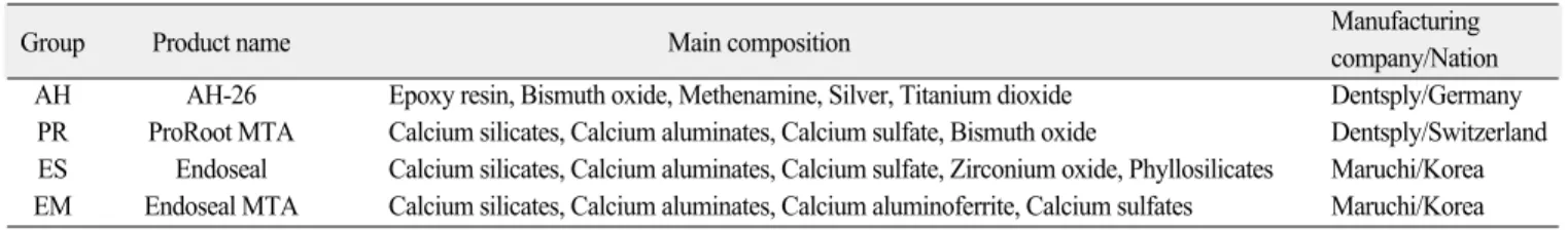
Table 1. Materials used for the experiment
Group Product name Main composition Manufacturing
company/Nation
AH AH-26 Epoxy resin, Bismuth oxide, Methenamine, Silver, Titanium dioxide Dentsply/Germany
PR ProRoot MTA Calcium silicates, Calcium aluminates, Calcium sulfate, Bismuth oxide Dentsply/Switzerland
ES Endoseal Calcium silicates, Calcium aluminates, Calcium sulfate, Zirconium oxide, Phyllosilicates Maruchi/Korea
EM Endoseal MTA Calcium silicates, Calcium aluminates, Calcium aluminoferrite, Calcium sulfates Maruchi/Korea
cone and then ultrasonic vibration was applied to the master cone for 5 seconds using a pincette. After remov- ing the upper GP cone with B&L-α , the cavity entrance was sealed with Cavit. In group EM, the filling materials were Endoseal MTA (Maruchi, Wonju, Korea) and a
#30/0.04 taper GP cone. As per the manufacturer’ s in- structions, using a 23 gauge needle, the Endoseal MTA was injected until it tightly filed the root apex. Then, a
#30/0.04 taper GP cone was inserted and pumped light- ly. Ultrasonic vibration was applied to the master cone for 5 seconds using a pincette. Then, the upper GP cone was removed with B&L-αand the cavity entrance was sealed with Cavit. The roots of all group were then stored at 37℃ and 100% humidity for 48 hours.
The time spent on each filling procedure until the Cavit was used was recorded and the filling shape was evaluated with periapical radiographs. All of the filling processes and time measurements were performed by one clinician.
Within each group, the samples were randomly divided into subgroups A and B. Subgroup A was used to evalu- ate the root apex seal based on the penetration of a dye.
Three positive and negative controls were also tested. In the positive controls, only a #40 GP cone was inserted into the root canal and all of the outer surfaces of the teeth were covered with two layers of nail varnish except a 1 mm region at the root tip. In the negative controls, the entire outer surfaces of the teeth, including the root tip, were covered with two layers of nail varnish. In the experimental group, which comprised 60 samples, 15 from each group, the outer surfaces of the teeth except a 1 mm region at the root tip were coated with two layers of nail varnish and stored at room temperature for 24 hours. Then, after confirming that the nail varnish had hardened, they were immersed in 1% methylene blue solution for 72 hours. Finally, the dental root was washed with running tap water, dried for 24 hours, and cut into longitudinal sections with diamond disk under sprinkled water.
To quantify dye leakage, the maximum distance that the dye moved from the anatomical apex toward the crown was measured under an illuminated microscope (SZ61, Olympus, Tokyo, Japan) at X 12 magnification with an objective micrometer (Union, Tokyo, Japan).
Subgroup B was used to check the degree of penetra- tion of the sealer within the dentinal tubules using scan- ning electron microscopy (SEM). In the experimental group, which comprised 40 samples, 10 from each group,
the samples were cut with a diamond disk under sprin- kled water, leaving the lower 6 mm of the cervical area, and a groove was cut in the lower part using a fissure bur. Then, the specimen was dehydrated for 10 minutes each in a series of solutions with ethanol concentrations increasing from 50% to 100% in 10% increments and dried for 24 hours at room temperature in a closed con- tainer containing silica gel. Cross-sections were cut us- ing a chisel and hammer in the upper notch and these were coated with Pt via ion sputtering (E-1030, Hitachi High Technologies, Tokyo, Japan).
Using SEM (SU-8220, Hitachi High Technologies, Tokyo, Japan), images of the 4 mm of root apex were obtained at ×500, ×1000, and ×1500 magnification de- pending on the degree of sealer penetration. In the im- ages, the maximum penetration of the sealer was mea- sured using Adobe Photoshop 7.0. The data were ana- lyzed using one-way analysis of variance (ANOVA) and Dunnett’ s T3 post hoc test. The confidence level was p <
0.05.
Ⅲ. Results 1. Dye penetration assay
In the three positive controls, the dye penetrated all of the root canals, while in the three negative controls, no dye penetration was observed. Except for two samples in group AH, dye penetration was observed in all of the ex- perimental samples.
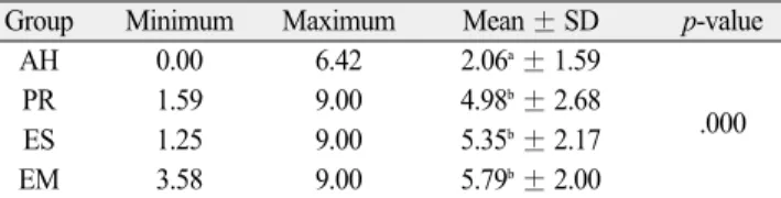
Table 2 summarizes the micro-leakage in the experi- mental groups. The micro-leakage was the lowest in group AH, followed in order by groups ES, EM, and PR. The dif- ferences between group AH and the other experimental groups were significant (p < 0.05) in one-way ANOVA and Dunnett’ s T3, while there were no significant differ- ences (p > 0.05) among groups ES, EM, and PR.
Table 2. Extent of apical dye penetration (mm)
Group Minimum Maximum Mean ± SD p-value
AH 0.00 6.42 2.06
a± 1.59
PR 1.59 9.00 4.98
b± 2.68
ES 1.25 9.00 5.35
b± 2.17 .000
EM 3.589.00 5.79
b± 2.00
SD indicates standard deviation
One-way ANOVA test followed by Dunnett ' s T3 post hoc analysis
a,b: Dunnett ' s T3 grouping, which means the same letter are not signifi-
cantly different
2. Depth of sealer penetration in dentinal tubules
Table 3 shows the penetration depth of the sealer within the dentinal tubules. In group AH and the con- trols, penetration to the maximum depth was observed at ×500 magnification in all samples. In the experimen- tal groups, the maximum penetration was in the order group EM, ES, and PR (Fig. 1). There were significant differences among all of the groups (p < 0.05).
3. Time required for root canal filling
Table 4 summarizes the time spent filling the canals in each group when canal enlargement was completed with a #40/0.04 taper. group EM required the least time (44.70 seconds), followed by groups ES (74.97 seconds), PR (280.39 seconds), and AH (328.32 seconds). All of the differences were significant (p < 0.05) and the time differences between groups EM and ES and groups PR and GP were particularly large.
Table 3. Sealer penetration depth into dentinal tubules at the apical section (㎛)
Group Minimum Maximum Mean ± SD p-value
AH 329.27 636.36 483.57 ± 108.12
PR 0.00 37.81 16.20 ± 13.16
ES 21.37 62.05 35.18 ± 14.03 .000
EM 44.3876.44 66.99 ± 8.88
SD indicates standard deviation
One-way ANOVA test followed by Dunnett ' s T3 post hoc analysis There are significant differences among all of the groups
Table 4. Time required for root canal filling (second)
Group Minimum Maximum Mean ± SD p-value
AH 293.58395.91 328 .32 ± 28.98
PR 245.36 300.52 280.39 ± 16.06
ES 64.36 91.47 74.97 ± 8.19 .000
EM 35.46 53.82 44.70 ± 5.49
SD indicates standard deviation
One-way ANOVA test followed by Dunnett ' s T3 post hoc analysis There are significant differences among all of the groups
Fig. 1. Longitudinal view of specimens. (A) Group AH. All dentinal tubules are filled with AH-26 (magnification: ×500). (B) Group PR. Dentinal tubules
nearby are empty (magnification: ×1500). (C) Group ES. Partial dentinal tubules are filled with Endoseal (magnification: ×1500). (D) Group EM. Most
dentinal tubules are filled with Endoseal MTA (magnification: ×1500).
Ⅳ. Discussion
This study evaluated whether MTA can be used as a root canal filling material. In this in vitro study, the MTA groups took less time than AH-26, both sealing of the root canal walls and penetration within the dentinal tubules were inferior with MTA. Comparing ProRoot MTA, Endoseal, and Endoseal MTA within the MTA groups, there were no significant differences in sealing the root canal walls, but Endoseal MTA had the best re- sults in terms of dentinal tubule penetration and work- ing time.
Following root canal sealing, leakage occurs mainly be- tween the filling material and root wall. The degree of leakage can be reduced using root filling materials that fit within the canal snuggly
20,21). Several methods have been used to evaluate the seal between these filling ma- terials and the root wall, such as dye penetration, bacte- rial penetration and fluid filtration. The methylene blue penetration method we used is relatively inexpensive, safe and results in high penetration because the dye is a very small molecule
22,23). Therefore, if the methylene blue molecules do not pass the filling material, the micro- leakage of bacteria and their byproducts should also be prevented
24).
In this study, the extracted teeth were immersed in methylene blue solution for 3 days to ensure full pene- tration. A vacuum device was not used because it has been reported that the amount of micro-leakage can be over-evaluated in vivo using such a device
25).
The root sealing effect of AH-26 was significantly bet- ter than that of the MTA groups. For a sealer to pene- trate the narrow root canal, its flowability should be good. However, the flowability of MTA made from pow- der is inevitably inferior to that of the paste type AH-26.
MTA expands while hardening and moisture is required for hardening. There may be insufficient moisture in the prepared root canal for complete hardening of the MTA.
The penetration of the sealer within dentinal tubule improves the sealing ability by increasing the contact surface between the filling material and dentin
18,19). Chemical bonding between the sealer and dentin does not occur, except with GI sealer, but the mechanical bonding increases the retention of the filling material in the root canal
26-28). MTA sealers with antibiotics are more effective at preventing bacteria from penetrating the dentinal tubules
18,29).
Many factors affect dentinal tubule penetration by a
sealer, such as the presence or absence of a smear layer, the clinician’ s technique, the diameter of the dentinal tubule, the sealer application method, and the physical and chemical characteristics of the sealer
30-32). After root canal preparation, we immersed the samples in 17%
EDTA for 3 minutes to remove the smear layer. To re- duce the variability in dentinal tubule diameter, we studied only single-rooted tooth and examined only the apical 4 mm. In addition, because one clinician per- formed all of these procedures, interobserver errors were eliminated. Therefore, the significant differences seen in the dentinal tubule penetration of each sealer in this study resulted from the physical and chemical character- istics of the sealers.
For a sealer to penetrate the dentinal tubules, it should have sufficient flowability and low surface activity
33). There was a clear difference between AH-26 and the MTA groups: AH-26 is a liquid sealer, while the MTA groups are particle types, as seen in the SEM im- ages. This difference affects the flowability and surface activity of each sealer. Within the MTA groups, the Endoseal MTA particles were the smallest and ProRoot MTA particles were the largest. This was reflected in the differences in penetration of the dentinal tubules.
Therefore, both the type of material and size of particles affect the dentinal tubule penetration depth.
Compared to the resin-based sealer, the working times were faster for the MTA groups. Endoseal and Endoseal MTA were faster to use than ProRoot MTA because the latter must be applied in layers in the root canal using MTA carrier after mixing. This process is time-consum- ing and working with the plugger increases hand fatigue.
In comparison, Endoseal and Endoseal MTA are injected into the root canal, which is filled using sonic vibration.
As Endoseal MTA is pre-mixed, the time required is re- duced further.
Studies that have compared root canal filling with a variety of MTA products have concluded that MTA is an effective root apex filling material
11,34,35), which is the op- posite to our conclusion. Nevertheless, we found that Endoseal and Endoseal MTA were better than ProRoot MTA. Therefore, when treating root canals that are diffi- cult to fill with AH-26, such as an open apex or dental root perforation, Endoseal or Endoseal MTA might be useful. However, when filling the root canals of mature permanent teeth, we recommend using a GP cone and epoxy resin-based sealer.
The results in an actual oral environment might differ
from our in vitro findings. Although the sealing effect of a sealer should last for a long time, we did not consider changes in the physical properties of the sealers over time. Therefore, long-term studies should analyze the root canal filling effect of MTA in vivo.
Ⅴ. Conclusion
In our study, Endoseal and Endoseal MTA, new MTA products, were faster to use than AH-26, but did not seal the root canal walls or penetrate the dentinal tubules as well. However, both were better than ProRoot MTA as root canal filling materials, with better dentinal tubule penetration and shorter times required.
Therefore, AH-26 and GP cones are recommended as root canal filling material for mature permanent teeth, while Endoseal and Endoseal MTA may be useful when treating the root canals with an open.
References
1. Kakehashi S, Stanley HR, Fitzgerald RJ : The effects of surgical exposures of dental pulps in germ- free and conventional laboratory rats. Oral Surg Oral Med Oral Pathol, 20:340-349, 1965.
2. Holland R, Murata SS, DezanJunior E, et al. : Apical seal of root canals with gutta-percha calcium hydroxide. Braz Dent J, 15:26-29, 2004.
3. Bowman GA : In history of Dentistry in Missouri.
Ovid Bell Press 423,1938.
4. Bergdahl M, Wennberg A, Sp å ngberg L : Biologic effect of polyisobutylene on bony tissue in guinea pigs, Scand J Dent Res, 82:618-621, 1974.
5. Feldmann G, Nyborg H : Tissue reactions to root filling materials. II. A comparison of implants of sil- ver and root filling material AH26 in rabbits' jaws.
Odontol Revy, 15:33-40, 1964.
6. Lee SJ, Monsef M, Torabinejad M : Sealing ability of a mineral trioxide aggregate for repair of lateral root perforations. J Endod, 19:541-544, 1993.
7. Torabinejad M, Watson TF, Pitt Ford TR : Sealing ability of a mineral trioxide aggregate when used as a root end filling material. J Endod, 19:591-595, 1993.
8. Li Z, Cao L, Fan M, Xu Q : Direct Pulp Capping with Calcium Hydroxide or Mineral Trioxide Aggregate: A Meta-analysis. J Endod, 15:381-387, 2015.
9. Dorileo MC, Pedro FL, Borges AH, et al. : Comparative analysis of physicochemical properties of root perforation sealer materials. Restor Dent Endod, 39:201-209, 2014.
10. S ö nmez IS, Oba AA, S ö nmez D, Almaz ME : In vit- ro evaluation of apical microleakage of a new MTA- based sealer. Eur Arch Paediatr Dent, 13:252-5, 2012.
11. Gomes-Filho JE, Moreira JV, Otoboni Filho JA, et al. : Sealability of MTA and calcium hydroxide con- taining sealers. J Appl Oral Sci, 20:347-351, 2012.
12. Vizgirda PJ, Liewehr FR, Buxton TB, et al. : A comparison of laterally condensed gutta-percha, thermoplasticized gutta-percha, and mineral trioxide aggregate as root canal filling materials. J Endod, 30:103-106, 2004.
13. Park DS, Sohn SJ, Kye SB, et al. : An electrochemi- cal study of the sealing ability of three retrofilling materials. J Kor Acad Cons Dent, 29:365-369, 2004.
14. Antonopoulos KG, Attin T, Hellwig E. : Evaluation of the apical seal of root canal fillings with different methods. J Endod, 24:655-658, 1998.
15. Hashem AA, Hassanien EE : Proroot MTA, MTA- Angelus and IRM used to repair large furcation per- forations: Sealability Study. J Endod, 34:59-61, 2008.
16. O ¨zcan E, Eldeniz AU, Ari H : Bacterial killing by several root filling materials and methods in an ex vivo infected root canal model. Int Endod J, 44:
1102-1109, 2011.
17. Cergneux M, Ciucchi B, Dietschi JM, Holz J : The influence of the smear layer on the sealing ability of canal obturation. Int Endod J, 20:228-232, 1987.
18. Kokkas AB, Boutsioukis ACh, Vassiliadis LP, Stavrianos CK : The influence of the smear layer on dentinal tubule penetration depth by three different root canal sealers: An in vitro study. J Endod, 30:
100-102, 2004.
19. Moradi S, Ghoddusi J, Forghani M : Evaluation of dentinal tubule penetration after the use of dentin bonding agent as a root canal sealer. J Endod, 35:
1563-1566, 2009.
20. Ingle JI, Newton CW, West JD, et al. : Obturation of the radicular space. Endodontics 5th edition, p.571, 2002.
21. Sagsen B, Er O, Kahraman Y, Orucoglu H. :
Evaluation of microleakage of roots filled with differ-
ent techniques with a computerized fluid filtration technique, J Endod, 32:1168-1170, 2006.
22. Asgary S, Eghbal MJ, Parirokh M : Sealing ability of a novel endodontic cement as a root-end filling material. J Biomed Mater Res A, 87:706-709, 2008.
23. Boussetta F, Bal S, Farge P, et al. : In vitro evalua- tion of apical microleakage following canal filling with a coated carrier system compared with lateral and thermomechanical Gutta-percha condensation techniques. Int Endod J, 36:367-371, 2003.
24. Kubo CH, Gomes AP, Mancini MN : In vitro evalua- tion of apical sealing in root apex treated with dem- ineralization agents and retrofiled with mineral tri- oxide aggregate through marginal dye leakage. Braz Dent J, 16:187-191, 2005.
25. Ha
¨lkel Y, Wittenmeyer W, Allemann C : A new method for the quantitative analysis of endodontic microleakage. J Endod, 25:172-177, 1999.
26. White RR, Goldman M, Lin PS : The influence of the smeared layer upon dentinal tubule penetration by plastic filling materials. J Endod, 10:558-62, 1984.
27. Saunders WP, Saunders EM : The effect of smear layer upon the coronal leakage of gutta-percha root fillings and a glass ionomer sealer. Int Endod J, 25:
245-259, 1992.
28. Kennedy WA, Walker WA, Gough RW : Smear layer removal effects on apical leakage. J Endod, 12:21-
27, 1986.
29. Heling I, Chandler NP : The antimicrobial effect within dentinal tubules of four root canal sealers. J Endod, 22:257-259, 1996.
30. Vassiliadis LP, Sklavounos SA, Stavrianos CK : Depth of penetration and appearance of grossman sealer in the dentinal tubules: An in vivo study. J Endod, 20:373-376, 1994.
31. Calt S, Serper A : Dentinal tubule penetration of root canal sealers after root canal dressing with cal- cium hydroxide. J Endod, 25:431-433, 1999.
32. Kara Tuncer A, Tuncer S : Effect of different final irrigation solutions on dentinal tubule penetration depth and percentage of root canal sealer. J Endod, 38:860-3, 2012.
33. Aktener BO, Cengiz T, Pi kin B : The penetration of smear material into dentinal tubules during instrumentation with surface active reagents. J Endod, 15:588-90, 1989.
34. Torabinejad M, Rastegar AF, Kettering JD, Pitt Ford TR : Bacterial leakage of mineral trioxide aggregate as a root-end filling material. J Endod, 21:109-12, 1995.
35. Hwang JH, Chung J, Kim HC, et al. : Comparison
of bacterial leakage resistance of various root canal
filling materials and methods: confocal laser-scan-
ning microscope study. Scanning, 37:422-428, 2015.
근관 충전용 MTA의 밀폐 효과와 작업 시간 평가