• 검색 결과가 없습니다.

Characterization of Lactobacillus reuteri BCLR-42 and Lactobacillus plantarum BCLP-51 as novel dog probiotics with innate immune enhancing properties

N/A
N/A
Protected

Academic year: 2021

Share "Characterization of Lactobacillus reuteri BCLR-42 and Lactobacillus plantarum BCLP-51 as novel dog probiotics with innate immune enhancing properties"

Copied!
10
0
0

로드 중.... (전체 텍스트 보기)

전체 글

(1)

Korean J Vet Res(2016) 56(2) : 75~84 http://dx.doi.org/10.14405/kjvr.2016.56.2.75

75

<Original Article>

Characterization of Lactobacillus reuteri BCLR-42 and Lactobacillus plantarum BCLP-51 as novel dog probiotics with innate immune enhancing properties

Eun Jin Kim

, Yeong Im Kang

, Tae Il Bang, Myoung Han Lee, Sang Won Lee, In Soo Choi, Chang Seon Song, Joong Bok Lee, Seung Yong Park*

Laboratory of Veterinary Immunology, College of Veterinary Medicine, Konkuk University, Seoul 05029, Korea (Received: January 15, 2016; Revised: April 19, 2016; Accepted: April 26, 2016)

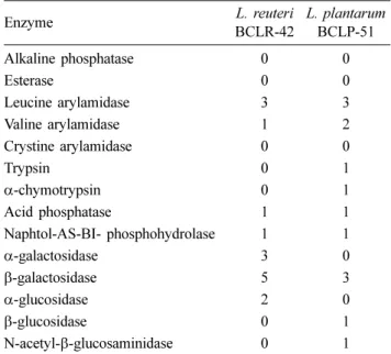
Abstract : Probiotics that are able to provide beneficial effects on animal health have become important ingredients of dog foods. This study was conducted to characterize the probiotic potentials of two strains, Lactobacillus reuteri BCLR-42 and Lactobacillus plantarum BCLP-51, that were derived from feces of healthy dogs and evaluated based on tolerance to low pH and bile acid, antimicrobial activities, enzyme profiles, sensitivity to antibiotics, and innate immune enhancing potentials. Both strains showed survival of more than 90% at pH 3 and 0.2% bile acid and exhibited broad antimicrobial activities against indicator bacteria. Moreover, both strains showed high sensitivity to antibiotics, except vancomycin, metronidazole, and gentamicin. The alkaline phosphatase was negligible (score 0), whereas they showed strong beta galactosidase activity (score range 5 or 3, respectively). The phagocytosis and oxidative burst activities of canine granulocytes were significantly enhanced in response to both strains. These results show that both strains have the capability to act as probiotics and the potential for application as ingredients in dog foods.

Keywords : dog, feces, in vitro innate immune activity, lactic acid bacteria, probiotics

Introduction

The maintenance of good health of companion animals including dogs is the major concern of the owners. In addi- tion to proper vaccination and regular checkups, the supply of the good quality of foods, which should be consumed every day, is the critical thing in keeping health of dogs. The importance of dog foods is reflected by the dramatic increases of pet food market. According to Transparency Market Research, the global market for food for companion animals in 2011 was 58.6 billion United States dollars (USD), and it is expected to be grown to 74.8 USD in 2017. As the inter- est in functional foods for humans with a variety of potential positive effects on health beyond basic nutrition has increased, so has the need for the development and commer- cialization of functional foods for dogs, and one of the mostly used ingredients in such foods is probiotics.

The WHO defines probiotics as live microorganisms which, when administered in adequate numbers, improve host health [27], and effects of probiotics have been implicated in curing and preventing various diseases such as allergy [6], inflam- mations [35], some cancers [41], metabolic diseases [18], and even mental disorders [17]. The utilization of probiotics has

been expanded to animals, as such, probiotics for animals including chicken [6], cattle [21], pigs [15], and fishes [23]

have been developed. The critical beneficial aspect of probi- otics for these livestock is to replace antibiotics, which have long been used as growth promoters for industrial animals, but its use for such purpose has recently been banned in a global way due to the issue of public health [35]. In sharp contrast to livestock, the application of probiotics for dogs as companion animals should be rather similar to that for humans [12].

While the interest in the use of probiotics for dogs has increased sharply, the related studies are scarce, and it is apparent that the probiotics developed for humans are used for dogs without verifying its effect on the host. Although argues in the significance of host species-specific probiotics remain, considering the fact that one critical aspect of probi- otics is to colonize sufficiently to host intestine, the develop- ment of probiotics derived from the same species should be important. Indeed, McCoy et al. [27] reported that a success- ful probiotics for dogs should be of canine intestinal origin since these species exhibit host specificity.

In order to develop probiotics for dogs, we have cultured the lactic acid bacteria (LAB) from feces from healthy dogs.

*Corresponding author

Tel: +82-2-450-3713, Fax: +82-2-3437-1941 E-mail: [email protected]

The first two authors equally contributed to this work.

(2)

Although non-LAB such as bacillus and yeasts also can be functional as probiotics, the major bacterial species for probi- otics are LAB including Lactobacillus, Lactococcus, Entero- coccus, Streptococcus and Bifidobacterium. LAB produce lactic acid from glucose source, and do not make harmful factors including skatole, phenol, and ammonia, thereby pre- venting putrefaction, all of which confers the beneficial effects to the host. In this study, we have chosen and charac- terized two strains through screening more than 50 different strains to develop probiotics that could be available for the ingredients of the dog foods.

Materials and Methods

Isolation and identification of bacterial strains Fecal samples were obtained from 5 healthy female beagle dogs (age, 1 to 3 years; body weight, 12 to 15 kg). One gram of feces from healthy dogs was suspended in 10 mL phos- phate-buffered saline (PBS), and 0.1 mL of 10-fold serially diluted samples with PBS were inoculated onto de Man-Rog- osa-Sharpe (MRS; Merck, Germany) agar plate and incu- bated for 36 h at 37

o

C anaerobically using a GasPak (Becton, Dickinson and Company, USA). The identification of result- ing colonies was determined by sequencing 16S ribosomal RNA PCR product with the primer set 357F-926Rb (357F;

CCTACGGGAGGCAG CAG, 926Rb; CCGTCAATTYMTT TRAGT). As a reference strain, we used Lactococcus lactis isolated from a commercial probiotic product (Activia; Pul- muon, Korea).

Low pH and bile resistance

Resistance of the isolates to low pH and bile salt was determined by the method reported by Brink et al. [3] with minor modifications. Briefly, the pH of MRS broth was adjusted to 2, 3, 4, or 5 with 1 M HCl, and each was dis- pensed into a 96-well plate (Nunc V bottom; Thermo Fisher Scientific, USA) at a volume of 250 µL per well. Twenty five µL of overnight culture of each isolate, the cell density of which was normalized to an optical density at 600 nm of 0.5, was inoculated into the MRS broth, and incubated at 37

o

C for 3 h under anaerobic condition. The number of viable bac- teria as colony forming units was determined by incubation the bacteria on MRS agar plates at 37

o

C for 36 h under anaerobic condition. Resistance to bile salt was determined by the same method as for pH resistance, except that the bile concentration was 0.05, 0.1, 0.2, and 0.4%.

Antimicrobial activity

The antimicrobial activity of the isolated LAB was evalu- ated according to Strompfova and Laukova [40]. Three µL of an overnight culture was spotted onto a MRS agar plate, and incubated for 36 h at 37

o

C under anaerobic condition. The spot was then covered with TSB soft agar (0.8% w/v) inocu- lated with the indicator bacteria (Salmonella typhimurium, Escherichia [E.] coli, Citrobacter freundii, Enterococcus

faecium, Staphylococcus aureus, Staphylococcus interme- dius, Listeria monocytogenes, Corynebacterium auriscanis, and Bacillus circulans), and the plate was incubated aerobi- cally at 37

o

C for 24 h. The antimicrobial activity of each strain was determined by the inhibition zone surrounding the spot. This test was performed in triplicate.

Enzyme activities

The enzyme activities of the selected strains were evalu- ated using API ZYM system (Analytab Products, USA) fol- lowing the manufacturer’s instructions. Briefly, overnight cultured bacteria were centrifuged at 2,500 × g for 10 min, and diluted with PBS to adjust the cell density to a McFar- land standard 1.0 (SD2301; Pro-Lab Diagnostics, USA). An aliquot of the cell suspension was inoculated into each well of the kit, and after addition of the reagents (Zym A and Zym B), the kit was incubated for 4 h at 37

o

C under anaerobic condition. The evaluation of enzyme activity was carried out depending on the color intensity ranged from 0 to 5, which was determined by using a color code supplied by the manu- facturers.

Antibiotic susceptibility

To determine antibiotic susceptibility of selected probiot- ics, wide classes of antibiotics including erythromycin, oxacillin, vancomycin, gentamicin, ampicillin, cefotaxime, metronidazole, and tetracycline were used. Strains were grown overnight in MRS broth under anaerobic conditions at 37

o

C. Bacterial cul- tures were then inoculated on MRS agar plates with appro- priate minimum inhibitory concentration (MIC) evaluator strip (Oxoid, UK) and incubated for 36 h under the same conditions. The MIC was determined as the lowest concen- tration of antimicrobial agent at which no visible growth of the isolate was observed.

Preparation of samples for evaluating immune func- tions of LABs

Selected strains were grown in MRS broth for 16 h at 37

o

C under anaerobic condition, and the cell density was adjusted to optical density of 0.8 at 560 nm, which was used for sam- ples of live bacteria. Inactivated bacteria were prepared by incubating the cells in the PBS containing 6 mM binary eth- ylenmine and 0.06% formaldehyde overnight at 37

o

C, when the reaction was stopped by adding same of volume of steril- ized 1 M sodium thiosulfate. After 30 min, the cells were centrifuged at 2,500 × g for 10 min, and finally resuspended in RMPI 1640. To obtain metabolites, the overnight cultured LABs in MRS broth were centrifuged at 2,500 × g for 10 min, and the volume of supernatant was 10 times concen- trated in a freeze-dryer, which was finally resuspended in RPMI 1640 medium (Invitrogen, USA). The cell walls of LABs were obtained as reported by Camacho et al. [5].

Briefly, overnight-cultured LABs were centrifuged and resus-

pended in 4% sodium dodecyl sulfate in PBS, and were

boiled for 30 min. After washing the cells with PBS, the pel-

(3)

lets were treated with PBS containing 5 µg/mL DNAse I at 37

o

C for 2 h, which were then incubated in PBS containing 200 µg/mL trypsin and 400 µg/mL lysozyme at 37

o

C for 3 h.

The remaining pellets were extensively washed with PBS, and finally resuspended in PBS. The protein content in the solution was determined by the Bradford assay [2]. All reagents in this assay were purchased from Sigma-Aldrich.

Phagocytosis

Evaluation of phagocytic activity of granulocytes was per- formed as described by Maeda et al. [22] with minor modifi- cation. Briefly, heparinized canine whole blood was diluted with same volume of RPMI 1640 without antibiotics and fetal bovine serum (FBS), and 100 µL of diluted whole blood was stimulated with the same volume of samples in the pres- ence of same volume of fluorescence beads (Fluoresbrite car- boxy YG 1.0 micron microspheres; Polysciences, USA) solution for 30 min at 37

o

C. The beads solution was pre- pared by washing twice with PBS, and resuspended in RPMI 1640 at a density of 1 × 10

9

beads/mL. After incubation, the cells were washed with FACS buffer (PBS with 1% FBS and 0.1% sodium azide), and red blood cells were removed by treatment with the RBC Lysis Buffer (eBioscience, USA).

The cells were finally suspended in FACS buffer, and ana- lyzed by a flow cytometer (FACSCalibur; BD Biosciences, USA) with the FlowJo software (FlowJo, USA). Phagocytic activity was expressed as percentage of the granulocyte pop- ulations, which was gated based on forward and side scatter, emitting green fluorescence.

Oxidative burst

The production of reactive oxygen species (ROS) by gran- ulocytes was measured by flow cytometry as previously described [16]. Briefly, 100 µL diluted canine whole blood cells were incubated with 25 µL samples for 30 min at 37

o

C in a microcentrifuge tube, when 1 µM dihydrorhodamine 123 (DHR; Sigma-Aldrich, USA) was added and incubated for

15 min at 37

o

C. These cells were then treated with 1 mL RBC lysis buffer for 10 min at room temperature to remove erythrocytes while terminating the reaction. After washing the cells three times with PBS containing 3% FBS, the cells were analyzed within 30 min using a flow cytometer. The mean green fluorescence intensity of the granulocytes that were obtained by gating based on the forward and side scat- ter plot was analyzed to evaluate the oxidative burst activity.

Statistics

Statistical analysis was performed using a SPSS statistical package (ver. 22; IBM, USA). We determined significance by one-way ANOVA with the significance level of p value less than 0.05.

Results

Selection of two strains for dog probiotics

To develop homologous probiotics for dogs, we isolated LAB by culturing the fecal supernatant from healthy dogs on MRS selective media. We obtained 124 strains, which were composed of Lactobacillus (L.) animalis (52%), L. reuteri (20%), L. johnsonii (14%), L. acidophilus (12%), L. plan- tarum (1%), and other Lactobacillus spp. (3%). Unexpect- edly, Bifidobacteria were not isolated in our hands. These strains except L. animalis were screened for their potentials as probiotics. The reason we excluded the L. animalis was that these strains are not legally allowed for food yet. Based on the screening results including resistance to low pH and phagocytic activities, we choose two strains, which were denoted as L. reuteri BCLR-42 and L. plantarum BCLP-51.

Resistance to low pH and bile acid

The resistance activities against low pH and bile salt are important factors that determine the beneficial properties of probiotics that should be consumed by oral administration.

The number of bacteria after incubation in MRS broth

Fig. 1. Resistance to low pH and bile acid. The selected strains were anaerobically incubated in the presence of difference levels of

pH and bile acid at 37

o

C for 3 h, when they were incubated on Man-Rogosa-Sharpe agar plates at 37

o

C for 36 h under anaerobic con-

dition, and the colony forming units were determined.

(4)

adjusted to different pH values is shown in Figure 1A. Both strains exhibited resistance to pH 3, while half of L. reuteri BCLR-42 survived even at pH 2. The number of colony- forming unit (CFU)/mL after incubation with different levels of bile acid is shown in Figure 1B. At 0.2% bile acid concen- tration, both strains showed considerable resistance to bile acid, while the CFUs at 0.4% bile acid were decreased to three orders of magnitude when compared to the start of inoculum.

Antimicrobial activity

Antimicrobial activity is one of the desirable properties of probiotics, because it may help maintain gut commensal microorganisms by suppressing the growth of pathogenic bacteria. As shown in Table 1, both strains showed broad spectrum antimicrobial activities in which significant inhibi- tion zones against Gram negative bacteria (Salmonella, E.

coli, and Citrobacter) and Gram positive bacteria (Enterococ- cus, Staphylococcus, Listeria, Corynebacterium, and Bacil- lus) were observed, while the activities of L. plantarum BCLP-51were superior to those of L. reuteri BCLR-42.

Enzyme activities

Enzyme profiles of the LAB could be a good indication of the bacteria to demonstrate their beneficial or detrimental effects on the host, which means that the enzyme profiles can be utilized for the in vitro assessment of both functions and safety. Moreover, the enzyme activities of the bacteria can affect the tastes of the food that composed of the probiotics [36], which is also an important factor for commercialization of the probiotic products. The API-ZYM system showed that there were almost same but some difference in the enzyme activities of two selected strains (Table 2). Both strains com- monly had no or low (score 1) activities of alkaline phos- phatase, esterase, crystine acrlyamidase, trypsin, α-chymotrypsin, acid phosphatase, naphtol-AS-BI-phosphydrolase, β-glucosi-

dase, and N-acetyl- β-glucosaminidase. In contrast, both strains showed strong activity of β-galactosidase (5 for L.reuteri BCLR-42 and 3 for L.planatarum BCLP-51).

Sensitivity to antimicrobial agents

The sensitivity of the probiotics to antimicrobial agents should be considered in the context of the safe and efficacy for host animals. The high sensitivity to antimicrobial agents indicates that the bacteria have no acquired antimicrobial resistance, which implies them to be safe for host animals.

On the other hand, the extreme sensitivity to the antimicro- bial agents of the usual clinical use in veterinary medicine harbors the weakness of the probiotics to be low survival rate in the gut, which may dampen the effects of the probiotics.

As shown in Table 3, the results of the sensitivity test dem- onstrated that these selected strains could be grouped into three categories based on the MIC values; the group of high sensitivity with less than 1 µg/mL of MIC, moderate sensitiv- ity group with less than 16 µg/mL of MIC, and resistant group with more than 128 µg/mL of MIC. Both L. reuteri BCLR-42 and L. plantarum BCLP-51 were susceptible to erythromycin, ampicillin and cefotaxime, while they were moderately sensitive to oxacillin and tetracycline. The met- ronidazole, vancomycin and gentamycin were included in the resistant group in which the MIC values were 128, > 256, and > 256, respectively.

Phagocytosis

Phagocytosis of pathogenic microorganisms is one of innate immune defense mechanisms, and should be an important parameter to demonstrate the functions of immune status of animals. In addition to live bacteria, we examined Table 1. Antimicrobial activity of selected strains

Indicator bacteria

Lactobacillus (L.) L. reuteri

BCLR-42

L. plantarum BCLP-51

Salmonella typhimurium n.i. 6.0 ± 0.1

Escherichia coli 0.5 ± 0.7 5.0 ± 0.1

Citrobacter freundii 7.0 ± 1.4 5.7 ± 0.3 Enterococcus faecium 0.2 ± 0.3 2.7 ± 0.3 Staphylococcus aureus 7.0 ± 1.4 7.0 ± 2.8 Staphylococcus intermedius 3.5 ± 2.1 9.0 ± 1.4 Listeria monocytogenes 5.2 ± 1.0 12.0 ± 1.4 Corynebacterium auriscarnis 2.0 ± 2.8 11.0 ± 0.1 Bacillus circulans 2.0 ± 0.1 10.0 ± 1.4 Numbers indicate the diameter (mm) of the inhibition zones around the bacterial spots. Data represent the mean ± SD for triplicate experiments. n.i., no inhibition.

Table 2. Enzymatic profiles of selected strains

Enzyme L. reuteri

BCLR-42

L. plantarum BCLP-51

Alkaline phosphatase 0 0

Esterase 0 0

Leucine arylamidase 3 3

Valine arylamidase 1 2

Crystine arylamidase 0 0

Trypsin 0 1

α-chymotrypsin 0 1

Acid phosphatase 1 1

Naphtol-AS-BI- phosphohydrolase 1 1

α-galactosidase 3 0

β-galactosidase 5 3

α-glucosidase 2 0

β-glucosidase 0 1

N-acetyl-β-glucosaminidase 0 1

Numbers indicate the enzyme activities ranging from 0 (no activ-

ity) to 5 (strong activity), which was determined by color intensities.

(5)

the effects of the inactivated bacteria, metabolites, and cell walls from them. As shown in Figure 2, the granulocytes in the presence of samples increased phagocytosis, while it was dependent on the preparations. Live L. reuteri BCLR 42 and L. plantarum BCLP 51 increased phagocytosis by 31% and 36% respectively, while the increased phagocytic activity by live L. lactis, a reference strain, was superior to them.

Whereas the inactivated L. reuteri BCLR 42 did not affect the phagocytosis, inactivated L. plantarum BCLP 51 and L.

lactis increased phagocytosis by 50% and 52% respectively.

The cell wall fraction from L. plantarum BCLP 51 increased the phagocytic granulocytes up to 62%, whereas the granulo- cyte populations that were involved in phagocytosis in the presence of cell wall fractions from both L. reuteri BCLR 42 and L. lactis were less than 40%. As for metabolite, that from

L. reuteri BCLR 42 only significantly increased the phagocy- tosis of granulocytes compared to those of media.

Oxidative burst

The production of ROS by immune cells through respira- tory burst is another critical function in innate immune defense system, where ROS kills the invading microbes as well as induces inflammatory responses which lead to the clearance of pathogens from the host. To investigate whether the live tested strains or the preparations derived from the strains enhance the capacity of the granulocytes in involving respiratory burst, we loaded the blood cells deprived erythro- cytes with DHR substrate that emits green fluorescence when reacted with ROS. The reaction of granulocytes that were obtained by gating based on forward and side scatter plot Table 3. Minimum inhibitory concentration (MIC) of selected strains to antimicrobial agents

Strain MIC (µg/mL)

E OX VA CN AMP CTX MTZ TE

L. reuteri BCLR-42 0.5 8 > 256 128 0.55 0.12 > 256 52

L. plantarum BCLP-51 1.5 8 > 256 128 0.06 0.03 > 256 16

E, erythromycin; OX, oxacillin; VA, vancomycin; CN, gentamycin; AMP, ampicillin; CTX, cefotaxime; MTZ, metronidazole; TE, tetracy- cline.

Fig. 2. Phagocytic activities of dog granulocytes. Whole blood cells deprived of erythrocytes were incubated for the fluorescent beads

in the presence of various samples from the tested strains for 30 min at 37

o

C, when the percentage of granulocytes involved in phago-

cytosis was determined by a flow cytometry. Each bar represents mean ± SD of triplicate experiments. The different numbers on each

bar indicate the statistical significance between groups (p < 0.05).

(6)

was demonstrated as mean fluorescence intensity (MFI) of green fluorescence. As shown in Figure 3, regardless whether the samples are in the form of live and inactivated, cell wall, and metabolites, the capabilities of L. plantarum BCLP 51 in the production of ROS was superior to those of L. reuteri BCLR 42 and L. lactis. The MFIs by live, inactivated, cell walls, and metabolites from the L. plantarum BCLP 52 were 2200, 3200, 2500, and 2800 respectively, which were much higher than those from L. reuteri BCLR 42 and L.lactis. The MFIs by L. reuteri BCLR 42 in the form of live, inactivated and cell wall were lower than those by L. lactis, metabolites from L. reuteri BCLR 42 generated significantly higher MFI than L. lactis.

Discussion

The importance of intestinal commensals in various health conditions including gastrointestinal, respiratory, nervous, and immune systems has been dramatically accumulated recently. The removal of intestinal commensals by oral administration of antibiotics increases the incidence and sever-

ity of diseases in these diverse systems, and supply of suffi- cient number of beneficial bacteria recovers the host from diseases [8]. These studies have highlighted the importance of probiotics that function as live beneficial bacteria in the host in maintaining health of dogs. To develop probiotics for dogs, we selected two strains of LABs, and their probiotic potentials were investigated.

It is generally recognized that the beneficial effects of pro- biotics are dependent on the survival of the bacteria during transit through the stomach and upper small intestine. The orally administered bacteria should overcome the harsh con- ditions of the stomach with low pH as low as 2 [11]. When L. reuteri BCLR 42 and L. plantarum BCLP 51 each were exposed to various pH levels for 3 h, they could survive at pH 3, while half of L. reuteri BCLR 42 survived at pH 2.

Although this result shows that both strains' resistance capa- bility against low pH is not so strong, we considered the thing that these strains will be utilized as food ingredients.

When diets incorporated with probiotics are entered the stomach, its pH rises to more than 5 [8], where these strains could tolerate such pH levels, because the viability at pH 4 Fig. 3. Oxidative burst activities of dog granulocytes. Diluted whole blood cells were stimulated with various samples from the selected strains, followed by dihydrorhodamine (DHR) loading. After removing the erythrocytes, the cells were analyzed with a flow cytometer.

The mean fluorescence intensities (MFI) from granulocytes were determined to evaluate the oxidative burst activity. Each bar represents

mean ± SD of triplicate experiments. The different numbers on each bar indicate the statistical significance between groups (p < 0.05).

(7)

and pH 5 were not diminished (Fig. 1A). After the bacteria pass through the stomach, they enter the intestinal tract, where they are exposed to bile acid. Because there was no information about the concentration of bile acid in the small intestine of dogs, we set tested bile acid concentrations based on human data, the mean level of which is 0.3% (w/v) [10].

As shown in Figure 1B, the both strains were survived at 0.2

% bile acid concentration, which indicates that both strains are resistance to bile acid.

One of underlying mechanisms by which probiotics exert their effects is to exclude gastrointestinal pathogens both by competitive binding for adhesion sites and nutrition and by secreting antimicrobial agents, and these potentials are known to be strain-dependent [24]. The antimicrobial activities of L.

reuteri BCLR 42 and L. plantarum BCLP 51 against several Gram-positive and Gram-negative bacteria were examined by an agar spot test. As for the E. coli and Salmonella, the most-representative Gram-negative bacteria, L. plantarum BCLP 51 but not L. reuteri BCLR 42 exhibited antimicro- bial activity, as determined by a clear zone of inhibition. The mean diameters (mm) of inhibition zone of L. plantarum BCLP 51 against E. coli and Salmonella were 6 and 5 respectively, while those of L. reuteri BCLR 42 were negligi- ble. Similar antimicrobial activity against Gram-negative bacteria were reported by Perelmuter et al. [33] where L.

murinus isolated from dogs demonstrated inhibition zone against E. coli up to 15 mm. Whether the antimicrobial activ- ity of L. plantarum BCLP 51 against Gram-negative bacteria was due to bacteriocins or metabolic products was not clear in this study, although it has a high possibility that metabolic products such as lactic acid should be main factors demon- strating antimicrobial activity. Such understanding is based on the fact that Gram-negative bacteria with outer-mem- brane are more resistant to bacteriocins from LABs [15]. The antagonistic activities of L. reuteri BCLR 42 and L. plan- tarum BCLP 51 against Staphylococcus were similar to the mean diameters of inhibition zone that were the same as 7.

However, as for other Gram-positive bacteria the activities of L. plantarum BCLP 51 were superior to L. reuteri BCLR 42.

While the range of inhibition zones of L. reuteri BCLR 42 were 0.2 to 7, those of L. plantarum BCLP 51 were 2.7 to 12, showing that L. plantarum BCLP 51 exhibits much stron- ger antimicrobial activities against Gram-positive bacteria.

The factors involved in antimicrobial activities of LAB may include metabolites of oxygen such as hydrogen peroxide and oxygen radicals, acetaldehyde, diacetyl, lipophilic fac- tors, and bacteriocins. It remains to be clarified which fac- tors function as main antimicrobial agents of both strains.

The enzymes that probiotics harbor should be important aspects of probiotics, since diverse beneficial or detrimental effects of bacteria are derived from such enzyme activities.

For example, alkaline phosphatase has been implicated in the induction of intestinal inflammation [37]. As shown in Table 2, both selected strains showed negligible activities of alka- line phosphatase, while this result is in agreement with a pre-

vious report with L. plantarum [39]. Both strains exhibited no esterase and crystine acrylamidase, either. Another impor- tant harmful enzyme is the β-glucosidase which hydrolyzes glycoside to aglycones, because algycones have been sus- pected to cause the colon cancer due to their carcinogenic properties [14]. The activities of β-glucosidase in L. reuteri BCLR 42 and L. plantarum BCLP 51 were 0 and 1, respec- tively, demonstrating that both strains exhibited low to negli- gible activities. In contrast, β-galactosidase which acts on the β-galactosidic bond is reported from Bifidobacterium [34], and has been implicated in diminishing the side effects due to lactose maldigestion [9]. The scores of β-galactosidase of L.

reuteri BCLR 42 and L. plantarum BCLP 51 were 5 and 3, respectively, demonstrated that both strains have strong β- galactosidase activities.

The susceptibility of a bacterial strain against commer- cially-used antibiotics is another key prerequisite to be probi- otic strains. It is believed that antibiotics used for industrial animals can increase the emergence of antibiotics-resistant strains, and when these strains enter humans, they can trans- fer resistance gene to other pathogens by way of plasmids or transposons [41]. As shown in Table 3, L. reuteri BCLR 42 and L. plantarum BCLP 51 showed strong resistance against vancomycin and metronidazole (MICs > 256 µg/mL). Partic- ularly, the resistance against vancomycin was in agreement with the study by Handwerger et al. [13], where most Lacto- bacillus showed resistance to vancomycin, and this resis- tance should be due to intrinsic interactions between Lactobacillus and vancomycin. Lactobacillus has D-Ala-D- lactate in their peptidoglycan, and vancomycin cannot dis- rupt this kind of structure [13]. So it should be reasonable to consider that this resistance to vancomycin was not acquired due to previous exposure to vancomycin. In contrast, both strains showed high susceptibility to erythromycin, ampicil- lin, and cefotaxime and the MICs of which were less than 1 µg/mL. Some researchers also argue that resistance to antibi- otics should not be considered as only low side of probiotics, because some resistance to antibiotics should be important to keep balance of intestinal commensal during and after antibi- otics therapy [40]. One more thing we should consider was that companion animals such as dogs are not animals in food-chain. So even the strains acquired resistance by pre- exposure to antibiotics, there should no opportunity for such strains to be transferred to humans. Moreover, Oh et al. [31]

reported that oral microbiomes in the dogs and humans are different, indicating no exchange of microbiomes between two parties occurred.

The beneficial effects of probiotics are known to derive from various functions of probiotics, among which immuno- potentiation is recognized as one of important characters.

Such effects are usually dependent on strains with different

glycolipid and protein composition in their cell walls and dif-

ferent CpG content of their DNA [22]. In this study, we eval-

uated the in vitro immunomodulatory effects of selected

strains by using erythrocyte-deprived whole blood cells from

(8)

dogs rather than peripheral blood mononuclear cells (PMBC).

The reason we chose to use whole blood cells rather than PBMC is based on the fact that the immune cells in the blood should be exposed to live bacteria or molecules derived from the bacteria in the presence of other blood cells. Indeed, Schmitz et al. [38] revealed that appropriate responses for TLR ligands were well recognized by whole blood cells rather than PBMCs, and they concluded that the stimulation of whole blood cells could be a useful diagnostic tool in a clinical setting, where the available blood volume is fre- quently limited. In determining the immunoenhancing effects of the selected strains, we used, in addition to the live bacte- ria, inactivated bacteria, cell walls fractions and metabolites thereof as the immunomodulatory activities by LAB have been attributed to peptidoglycan, teichoic acid, and exopolysac- charide, which are generated when the LABs are undergoing lysis in the body [4]. Moreover, the metabolites from LABs are also known to affect the host immune response [1].

The neutrophils which are composed of 60 to 70% of canine white blood cells are the most essential immune cells that are involved in phagocytosis, by which the invading pathogenic bacteria are removed and adaptive immune response are triggered by presenting antigens to lympho- cytes [16]. In addition, the capacity of probiotics to stimulate phagocytic leukocytes has been reported to be a critical parameter in establishing immunity against bacterial infec- tion [24]. As shown in Figure 2, the selected strains as vari- ous forms of preparations including live bacteria, inactivated bacteria, cell wall fractions, and metabolites increased the neutrophil populations engaged in phagocytosis as compared to control (media). The mechanisms by which these samples stimulate the neutrophils to be involved in phagocytosis are not clear in this study, but direct contact of samples to neu- trophils may be necessary for this action. For example, the binding of molecules derived from probiotics to pattern-rec- ognition receptors on neutrophils should be one of possible mechanisms [29]. Otherwise, other factors such as cytokines that are produced by stimulation of leukocytes by samples might be implicated in enhancement of phagocytosis. For example, IFN- γ which is known to be produced by stimula- tion with probiotics is a potent promoter for phagocytosis [32]. The granulocytes engaged in phagocytosis also pro- duce a variety of toxic products that help to destroy the engulfed microbes. One of the important antimicrobial agents is ROS, which are generated by the nicotinamide adenine dinucleotide phosphate oxidase reaction or the respiratory burst. Previous study has shown that oral administration of probiotics in mice leads to increases in the activities of enzymes associated with the respiratory bursts in neutro- phils, indicating that probiotics can potentiate the neutro- phils in producing ROS [20]. As shown in Figure 3, the MFIs of neutrophils stimulated with L. plantarum BCLP 51 in any preparation were significantly higher than those of neutrophils with L. reuteri BCLR 42 and L. lactis. Interest- ingly, the metabolites, the culture supernatants from L. reu-

teri BCLR 42 or L. plantarum BCLP 51 enhanced the production of ROS by neutrophils significantly, whereas they did not affect the phagocytosis by neutrophils (Fig. 2), which suggest that the phagocytosis and the oxidative burst are closely associated reactions but are not always activated in a coupled way, as demonstrated by Nilsson et al. [30]. As a polysaccharide peptidoglycan complex from L. casei has been known the major component in modulating immune response [26], identifying the active factors from the metab- olites of the selected strains should be a valuable study.

In conclusion, this study shows that L. reuteri BCLR 42 and L. plantarum BCLP 51, which were isolated from dogs, can function as probiotics for dogs. Several studies have reported potential properties of L. reuteri and L. plantarum for dogs. For instance, Strompfova et al. [40] reported that they isolated L. reuteri and L. plantarum from dog feces, and that both strains could be a candidate probiotics for dogs. In addition, L. plantarum VET14A is already recognized probi- otics for dogs [12]. These results deserve further investiga- tion to apply these strains as probiotics for dog foods.

Acknowledgments

This work was funded by Binggrae Co. Ltd. in Gyeonggi- do, Korea.

References

1. Adams CA. The probiotic paradox: live and dead cells are biological response modifiers. Nutr Res Rev 2010, 23, 37-46.

2. Bradford MM. A rapid and sensitive method for the quantitation of microgram quantities of protein utilizing the principle of protein-dye binding. Anal Biochem 1976, 72, 248-254.

3. Brink M, Todorov SD, Martin JH, Senekal M, Dicks LM.

The effect of prebiotics on production of antimicrobial com- pounds, resistance to growth at low pH and in the presence of bile, and adhesion of probiotic cells to intestinal mucus. J Appl Microbiol 2006, 100, 813-820.

4. Bron PA, Tomita S, Mercenier A, Kleerebezem M. Cell surface-associated compounds of probiotic lactobacilli sustain the strain-specificity dogma. Curr Opin Microbiol 2013, 16, 262-269.

5. Camacho AI, Souza-Rebouças J, Irache JM, Gamazo C.

Towards a non-living vaccine against Shigella flexneri: from the inactivation procedure to protection studies. Methods 2013, 60, 264-268.

6. Cisek AA, Binek M. Chicken intestinal microbiota function with a special emphasis on the role of probiotic bacteria. Pol J Vet Sci 2014, 17, 385-394.

7. Cuello-Garcia CA, Bro ek JL, Fiocchi A, Pawankar R, Yepes-Nuñez JJ, Terracciano L, Gandhi S, Agarwal A, Zhang Y, Schünemann HJ. Probiotics for the prevention of allergy: a systematic review and meta-analysis of randomized controlled trials. J Allergy Clin Immunol 2015, 136, 952-961.

8. Dembinski A, Konturek SJ, Thor P. Gastric and pancreatic responses to meals varying in pH. J Physiol 1974, 243, 115- 128.

(9)

9. Di Pierro F, Bertuccioli A, Marini E, Ivaldi L. A pilot trial on subjects with lactose and/or oligosaccharides intolerance treated with a fixed mixture of pure and enteric-coated α- and β-galactosidase. Clin Exp Gastroenterol 2015, 8, 95-100.

10. Gilliland SE, Staley TE, Bush LJ. Importance of bile tolerance of Lactobacillus acidophilus used as a dietary adjunct.

J Dairy Sci 1984, 67, 3045-3051.

11. Grazul H, Kanda LL, Gondek D. Impact of probiotic supplements on microbiome diversity following antibiotic treatment of mice. Gut Microbes 2016, 7, 101-114.

12. Grze kowiak , Endo A, Beasley S, Salminen S. Microbiota and probiotics in canine and feline welfare. Anaerobe 2015, 34, 14-23.

13. Handwerger S, Pucci MJ, Volk KJ, Liu J, Lee MS.

Vancomycin-resistant Leuconostoc mesenteroides and Lacto- bacillus casei synthesize cytoplasmic peptidoglycan precursors that terminate in lactate. J Bacteriol 1994, 176, 260-264.

14. Heavey PM, Rowland IR. Microbial-gut interactions in health and disease. Gastrointestinal cancer. Best Pract Res Clin Gastroenterol 2004, 18, 323-336.

15. Helander IM, Mattila-Sandholm T. Permeability barrier of the Gram-negative bacterial outer membrane with special reference to nisin. Int J Food Microbiol 2000, 60, 153-161.

16. Holst BS, Gustavsson MH, Lilliehöök I, Morrison D, Johannisson A. Leucocyte phagocytosis during the luteal phase in bitches. Vet Immunol Immunopathol 2013, 153, 77- 82.

17. Hou C, Zeng X, Yang F, Liu H, Qiao S. Study and use of the probiotic Lactobacillus reuteri in pigs: a review. J Anim Sci Biotechnol 2015, 9, 6-14.

18. Hsiao EY, McBride SW, Hsien S, Sharon G, Hyde ER, McCue T, Codelli JA, Chow J, Reisman SE, Petrosino JF, Patterson PH, Mazmanian SK. Microbiota modulate behavioral and physiological abnormalities associated with neurodevelopmental disorders. Cell 2013, 155, 1451-1463.

19. Hulston CJ, Churnside AA, Venables MC. Probiotic supplementation prevents high-fat, overfeeding-induced insulin resistance in human subjects. Br J Nutr 2015, 113, 596-602.

20. Kapila R, Sebastian R, Varma D VP, Sharma R, Kapasiya M, Salingati V, Kapila S, Dang AK. Comparison of innate immune activation after prolonged feeding of milk fermented with three species of Lactobacilli. Microbiol Immunol 2013, 57, 778-784.

21. Klaenhammer TR, Azcarate-Peril MA, Altermann E, Barrangou R. Influence of the dairy environment on gene expression and substrate utilization in lactic acid bacteria. J Nutr 2007, 137 (3 Suppl 2), 748S-750S.

22. Lammers KM, Brigidi P, Vitali B, Gionchetti P, Rizzello F, Caramelli E, Matteuzzi D, Campieri M. Immunomodulatory effects of probiotic bacteria DNA: IL-1 and IL-10 response in human peripheral blood mononuclear cells. FEMS Immunol Med Microbiol 2003, 38, 165-172.

23. Lazado CC, Caipang CM. Mucosal immunity and probiotics in fish. Fish Shellfish Immunol 2014, 39, 78-89.

24. Lee Y, Lee TS. Enhancement in ex vivo phagocytic capacity of peritoneal leukocytes in mice by oral delivery of various lactic-acid-producing bacteria. Curr Microbiol 2005, 50, 24- 27.

25. Maeda K, Sakonju I, Kanda A, Suzuki T, Kakuta T, Shimamura S, Okano S, Takase K. Priming effects of

lipopolysaccharide and inflammatory cytokines on canine granulocytes. J Vet Med Sci 2010, 72, 55-60.

26. Matsumoto S, Hara T, Hori T, Mitsuyama K, Nagaoka M, Tomiyasu N, Suzuki A, Sata M. Probiotic Lactobacillus- induced improvement in murine chronic inflammatory bowel disease is associated with the down-regulation of pro- inflammatory cytokines in lamina propria mononuclear cells.

Clin Exp Immunol 2005, 140, 417-426.

27. McCoy S, Gilliland SE. Isolation and characterization of Lactobacillus species having potential for use as probiotic cultures for dogs. J Food Sci 2007, 72, M94-97.

28. Morelli L, Capurso L. FAO/WHO guidelines on probiotics:

10 years later. J Clin Gastroenterol 2012, 46 (Suppl), S1-2.

29. Moretti J, Blander JM. Insights into phagocytosis-coupled activation of pattern recognition receptors and inflammasomes.

Curr Opin Immunol 2014, 26, 100-110.

30. Nilsson M, Weineisen M, Andersson T, Truedsson L, Sjöbring U. Critical role for complement receptor 3 (CD11b/

CD18), but not for Fc receptors, in killing of Streptococcus pyogenes by neutrophils in human immune serum. Eur J Immunol 2005, 35, 1472-1481.

31. Oh C, Lee K, Cheong Y, Lee SW, Park SY, Song CS, Choi IS, Lee JB. Comparison of the oral microbiomes of canines and their owners using next-generation sequencing.

PLoS One 2015, 10, e0131468.

32. Paturi G, Phillips M, Kailasapathy K. Effect of probiotic strains Lactobacillus acidophilus LAFTI L10 and Lactobacillus paracasei LAFTI L26 on systemic immune functions and bacterial translocation in mice. J Food Prot 2008, 71, 796- 801.

33. Perelmuter K, Fraga M, Zunino P. In vitro activity of potential probiotic Lactobacillus murinus isolated from the dog. J Appl Microbiol 2008, 104, 1718-1725.

34. Rabiu BA, Jay AJ, Gibson GR, Rastall RA. Synthesis and fermentation properties of novel galacto-oligosaccharides by β-galactosidases from Bifidobacterium species. Appl Environ Microbiol 2001, 67, 2526-2530.

35. Reid G. Friendship R. Alternatives to antibiotic use:

probiotics for the gut. Anim Biotechnol 2002, 13, 97-112.

36. Saez-Lara MJ, Gomez-Llorente C, Plaza-Diaz J, Gil A.

The role of probiotic lactic acid bacteria and bifidobacteria in the prevention and treatment of inflammatory bowel disease and other related diseases: a systematic review of randomized human clinical trials. Biomed Res Int 2015. Epub ahead of print. doi: 10.1155/2015/505878.

37. Sánchez de Medina F, Martínez-Augustin O, González R, Ballester I, Nieto A, Gálvez J, Zarzuelo A. Induction of alkaline phosphatase in the inflamed intestine: a novel pharmacological target for inflammatory bowel disease. Biochem Pharmacol 2004, 68, 2317-2326.

38. Schmitz S, Henrich M, Neiger R, Werling D, Allenspach K. Comparison of TNFα responses induced by Toll-like receptor ligands and probiotics Enterococcus faecium in whole blood and peripheral blood mononuclear cells of healthy dogs. Vet Immunol Immunopathol 2013, 153, 170- 174.

39. Stoyanovski S, Gacovski Z, Antonova-Nikolova S, Kirilov N, Ivanova I, Tenev T, Hadjinesheva V. API ZYM enzymatic profile of lactic acid bacteria isolated from traditional Bulgarian meat product "Lukanka". Bulg J Agric Sci 2013,

s í L

(10)

19 (Suppl 2), 86-89.

40. Strompfová V, Lauková A. Isolation and characterization of faecal bifidobacteria and lactobacilli isolated from dogs and primates. Anaerobe 2014, 29, 108-112.

41. Teuber M, Meile L, Schwarz F. Acquired antibiotic resistance

in lactic acid bacteria from food. Antonie Van Leeuwenhoek 1999, 76, 115-137.

42. Zhong L, Zhang X, Covasa M. Emerging roles of lactic

acid bacteria in protection against colorectal cancer. World J

Gastroenterol 2014, 20, 7878-7886.

수치

Fig. 1. Resistance to low pH and bile acid. The selected strains were anaerobically incubated in the presence of difference levels of pH and bile acid at 37 o C for 3 h, when they were incubated on Man-Rogosa-Sharpe agar plates at 37 o C for 36 h under ana
Table 2. Enzymatic profiles of selected strains
Fig. 2. Phagocytic activities of dog granulocytes. Whole blood cells deprived of erythrocytes were incubated for the fluorescent beads in the presence of various samples from the tested strains for 30 min at 37 o C, when the percentage of granulocytes invo

참조

관련 문서