| Abstract |
Purpose: This study aimed to adjust the craniovertebral angle and shoulder alignment through shoulder and abdominal stabilization exercises in adults with a forward head posture.
Methods: The study participants were 29 adults with a forward head posture, and they were randomly divided into the following groups: 14 participants in a combined exercise group that used shoulder and abdominal stabilization exercises and 15 participants in a shoulder exercise group that used just shoulder stabilization exercises. The participants performed the stabilization exercises for 30 minutes per day, three times a week for five weeks.
Results: There were significant differences in the craniovertebral angle after intervention in the shoulder stabilization exercise group (p < 0.05). There were significant differences in the craniovertebral angle and location of the right root of the spine and both inferior angles before and after intervention in the shoulder and abdominal stabilization exercise group (p < 0.05). There was a significant difference in the location of the right root of the spine and the left inferior angle between the groups at the post-test (p < 0.05), and there was a larger change in the shoulder and abdominal stabilization exercise group.
Conclusion: There was a significant difference in the craniovertebral angle and a partially significant difference in shoulder alignment before and after intervention in both groups.
Key Words: Forward head posture, Shoulder stabilization exercise, Abdominal stabilization exercise, Craniovertebral angle
†Corresponding Author : Min-Hyung Rhee ([email protected])
Original Article Open Access
어깨 안정화운동과 복부 안정화 운동에 의한 전방머리자세 성인의 머리척추각도와 어깨뼈 정렬 변화
김재현⋅이민형1†11)
부산가톨릭대학교 일반대학원 물리치료학과, 1부산대학교병원 재활의학팀
Change of Craniovertebral Angle and Scapula Alignment in Adults with Forward Head Posture by Shoulder and Abdominal Stabilization Exercise
Jae-Hyun Kim, P.T., M.S.⋅Min-Hyung Rhee, P.T., Ph.D.1†
Department of Physical Therapy, Graduate School, Catholic University of Pusan
1Department of Rehabilitation Medicine, Pusan National University Hospital Received: August 1, 2021 / Revised: August 10, 2021 / Accepted: August 13, 2021
ⓒ 2021 Journal of Korea Proprioceptive Neuromuscular Facilitation Association
This is an Open Access article distributed under the terms of the Creative Commons Attribution Non-Commercial License (http://creativecommons.org/licenses/by-nc/3.0) which permits unrestricted non-commercial use, distribution, and reproduction in any medium, provided the original work is properly cited.
& Koo, 2016). 지속적인 전자 장비의 사용은 시선을 아래로 향하게 되고 구부정한 자세가 유발되며 이러 한 자세의 변화는 각 관절에 가해지는 근육의 활동이 나 부하를 변화시켜 정렬이 변화될 수 있다(Greig et al., 2005; Janwantanakul et al., 2012; Kang et al., 2015).
전방머리자세(forward head posture)는 깊은목폄근 (deep neck extensors)과 목굽힘근(cervical flexors)의 길 이가 짧아지고, 깊은목굽힘근(deep neck flexors)과 목 폄근(cervical extensors)의 근 길이가 늘어나서 1번째에 서 3번째 목뼈는 뒤로 펴지게 되고 4번째에서 7번째 목뼈는 앞으로 굽히게 되어 머리는 전방으로 향하게 된다(Khayatzadeh et al., 2017; Sheikhhoseini et al., 2018). 7번째 목뼈의 가시돌기에서 전방으로 향하는 수평선과 7번째 목뼈의 가시돌기에서 귀구슬을 잇는 선 사이의 각도인 머리척추각도(craniovertebral angle, CVA)가 48º 미만일 시 전방머리자세라 한다(Diab &
Moustafa, 2011; Fard et al., 2016).
전방머리자세가 장기간 지속될 경우 어깨뼈의 안 정성을 제공하는 앞톱니근(serratus anterior)의 근력이 감소하게 되어 어깨뼈 주변의 근육과 조직에 변화가 일어나게 된다(Park et al., 2010; Weon et al., 2010).
그러하여 어깨뼈는 앞쪽경사(anterior tilt), 아래돌림 (downward rotation), 안쪽돌림(internal rotation), 올림 (elevation)되어 둥근어깨(round shoulder) 또는 날개어 깨뼈(winging scapula)처럼 정렬의 변화가 나타나게 된 다(Singla & Veqar, 2017).
배곧은근(rectus abdominis), 배바깥빗근(external oblique), 배속빗근(internal oblique), 배가로근(transverse abdominis)으로 구성된 복부근육(abdominal muscle)은 몸의 중심부 쪽 뿐만 아니라 먼 쪽의 움직임에도 관여 하여 영향을 미치게 된다(Fard et al., 2016; Kudo et al., 2019). 특히 배가로근은 골반과 척추의 안정성을 제공하는 등허리근막(thoracolumbar fascia)의 장력
화 운동을 병합하여 실시한 군의 어깨뼈 위돌림근의 근활성도와 근육의 두께가 유의하게 증가되었다고 보 고하였다. 결과적으로 복부와 어깨는 서로 연관성이 있고, 어깨와 목도 서로 연관성이 있으므로, 전방머리 자세는 복부에도 영향을 미치게 된다.
어깨뼈의 안정성 개선으로 인한 전방머리자세의 변화를 분석한 연구는 있었으나(Ahn et al., 2020; Song et al., 2020), 복부의 안정성 개선으로 인한 전방머리자 세의 변화와 관련된 연구는 미흡한 실정이다. 이에 본 연구는 복부 안정화 운동에 의한 머리척추각도와 어깨뼈 정렬의 변화를 분석하고 전방머리자세에 어떠 한 변화가 나타나는지 연구하고자 하였다.
Ⅱ. 연구 방법 1. 연구설계 및 대상
본 연구의 대상자는 B시의 K병원에서 근무하는 성 인을 대상으로 남녀 나이 구분없이 29명을 선정하였 다. 머리척추각도가 48º 미만인 자(Fard et al., 2016)를 대상으로 선정하였으며, 본 연구에 대한 목적과 절차 를 충분히 설명을 들은 후 자발적으로 동의한 자를 대상으로 연구를 진행하였다. 신경학적 질환 및 과거 력이 있는 자, 팔, 다리를 포함하여 인체의 어느 한곳이 라도 정형외과적인 수술의 과거력이 있는 자, 인지 장애 및 의사 전달에 장애가 있는 자는 제외하였다.
2. 측정 방법 1) 머리척추각도
연구자는 대상자의 가쪽면에서부터 1.5m 거리에
카메라를 설치하고 렌즈의 높이는 대상자의 어깨 봉 우리에 위치시켰다(Kocur et al., 2019). 대상자는 바로 선 자세에서 대상자의 귀구슬(tragus)과 7번째 목뼈의 가시돌기(spinous process)에 스티커를 부착한 후 사진 을 촬영하였다(Fig. 1).
2) 어깨뼈 정렬
Lennie 검사를 통하여 어깨뼈 정렬을 측정하였다 (Fig. 2). 대상자는 선 자세에서 연구자는 대상자의 뒤 에서 7번째 목뼈의 가시돌기에 스티커를 부착하였다.
그 후 대상자의 등뼈 12개의 가시돌기와 양측 어깨뼈 의 위각(superior angle), 가시 뿌리(root of spine), 아래각 (inferior angle)에 스티커를 부착하였다. 위각, 가시 뿌 리, 아래각을 기준으로 수평선을 그린 후 수평선에서
가까운 등뼈로 위치를 지정하였다. 수평선이 등뼈 사 이의 중간일 경우 두 등뼈 사이의 중간으로 지정하였 다(Sobush et al., 1996).
3. 중재 방법
운동 시 시작 동작에서 끝 범위로 가서 유지 후 돌아오기까지의 시간을 총 15초로 설정하였다. 4분 운동 후 1분 휴식을 1세트로 설정하였고, 30분간 총 6세트, 주 3회, 5주에 걸쳐 실시하였다. 복합운동군은 어깨와 복부 안정화 운동을 각각 3세트씩 실시하였고, 어깨운동군은 어깨 안정화 운동만 6세트를 실시하였 다. 각 군별 중재 방법은 다음과 같다.
Fig. 1. Head and spine angle measurement.
Fig. 2. Scapular alignment measurement.
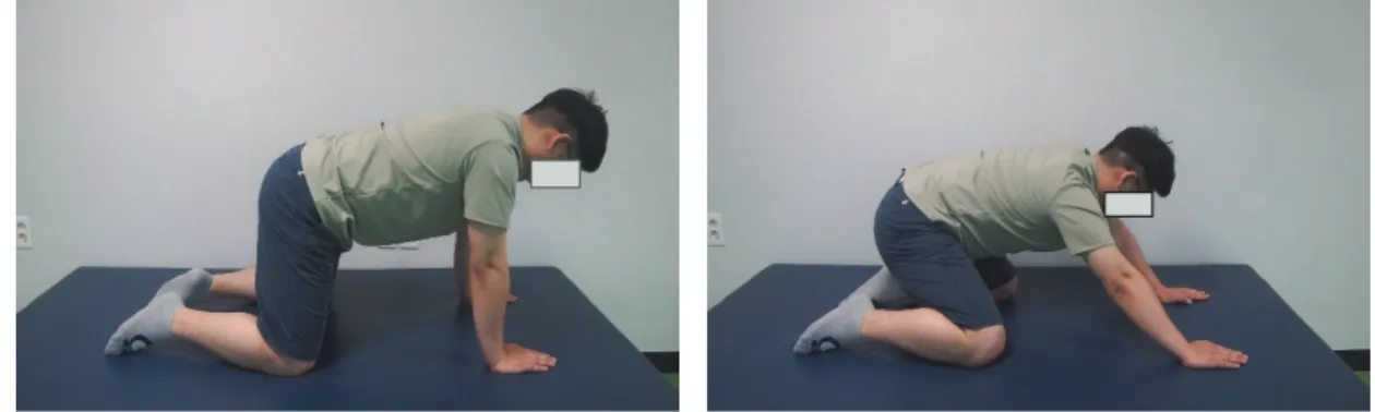
Fig. 3. shoulder stabilization exercises.
반대측 발뒤꿈치의 대각선 방향으로 내려가면서 궁둥 뼈결절측 팔의 동작은 위 가쪽 대각선 방향으로 뻗게 하였다(Park & Lee, 2013a). 1세트의 16회중 8회는 오른 쪽 어깨, 8회는 왼쪽 어깨에 운동을 실시하였다(Fig. 3).
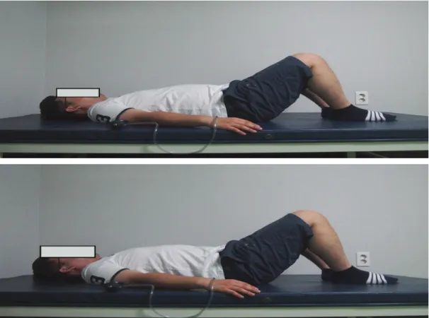
2) 복부 안정화 운동
대상자는 누운 자세에서 양 발을 어깨 넓이만큼 벌리고, 엉덩관절 45º, 무릎관절 90º 굽힌 자세에서 복 부 드로잉-인 기법(abdominal drawing-in maneuver)을 실시하였다(Fig. 4). 연구자는 대상자의 5번째 허리뼈
이때 압력게이지가 0∼2mmHg만큼 증가하도록 조절 하였다(Park et al., 2013; Park & Lee, 2013b).
4. 자료 분석
수집된 자료는 윈도우 용 SPSS version 18.0을 이용 하여 분석하였으며 각 군의 정규성을 확인하기 위하 여 Shapiro-Wilk 검정을 실시하였다. 각 군의 중재 전⋅
후 비교는 대응표본 t-검정(paired t-test)을 실시하였고, 각 군 간 비교는 독립표본 t-검정(independent t-test)을 실시하였으며, 유의수준(α)은 0.05로 설정하였다.
Fig. 4. abdominal stabilization exercise.
Ⅲ. 결 과 1. 연구 대상자의 일반적인 특성
본 연구에 참여한 대상자 29명 중에서 복합운동군 은 14명(남자1명, 여자13명)이었고, 평균 연령은 58.79±11.13세, 키는 158.71±6.27㎝, 체중은 63.43±10.01㎏, BMI는 24.76±3.05㎏/㎡이였다. 다른 나 머지 인원인 어깨운동군 15명(남자2명, 여자13명)의 평균 연령은 60.47±8.96세, 키는 159.00±4.39㎝, 체중은 58.73±8.66㎏, BMI는 23.15±2.53㎏/㎡이였다. 대상자 의 일반적인 특성에서 두 군 간의 유의한 차이가 없었 다(p>0.05).
2. 머리척추각도 측정 결과
1) 중재 전⋅후 군 내 머리척추각도 비교
본 연구의 실험에 참여한 복합운동군과 어깨운동 군은 중재 전⋅후 모두에서 머리척추각도에 대한 유 의한 차이가 있었다(p<0.05)(Table 1).
2) 중재 전⋅후 군 간 머리척추각도 비교
본 연구의 실험에 참여한 복합운동군과 어깨운동 군은 중재 전⋅후 머리척추각도에 대한 군 간의 유의 한 차이는 없었다(p>0.05)(Table 2).
3. 어깨뼈 정렬 측정 결과
1) 중재 전⋅후 군 내 어깨뼈 정렬 비교
본 연구의 실험에 참여한 복합운동군과 어깨운동 군의 어깨뼈 정렬은 중재 전⋅후 복합운동군에서의 오른쪽 가시 뿌리와 양측 아래각에서 유의한 위치 변 화가 있었지만(p<0.05) 다른 부위에서의 유의한 위치 변화는 없었다(p>0.05). 어깨 운동군에서는 어깨뼈 정렬에 대한 유의한 위치 변화가 없었다(p>0.05) (Table 3).
2) 중재 전⋅후 군 간 어깨뼈 정렬 비교 본 연구의 실험에 참여한 복합운동군과 어깨운동
Group Before intervention After intervention t p
SASE 42.50±3.80 50.57±3.98 -9.82 0.01*
SSE 42.67±3.52 50.47±3.85 -6.65 0.01*
*: significant difference (p<0.05) Unit: º
SASE: shoulder and abdominal stabilization exercises group SSE: shoulder stabilization exercises group
Table 1. Comparison of craniovertebral angles within groups before and after intervention (n=29)
CVA SASE SSE t P
Before intervention 42.50±3.80 42.67±3.52 -0.12 0.90
After intervention 50.57±3.9850.47±3.85 0.07 0.94
*: significant difference (p<0.05) Unit: º
CVA: craniovertebral angles
SASE: shoulder and abdominal stabilization exercises group SSE: shoulder stabilization exercises group
Table 2. Comparison of craniovertebral angles between two groups before and after intervention (n=29)
가 유의하게 컸다. 는 변화를 알아보기 위해 본 연구를 실시하였다. 본
Before intervention After intervention t p Right SASE
SA 2.68±0.61 2.41±0.33 1.68 0.12
RS 4.36±0.53 4.00±0.482.69 0.02*
IA 9.54±0.91 8.86±0.79 2.85 0.01*
Left SASE
SA 2.50±0.44 2.32±0.37 1.59 0.14
RS 4.29±0.43 3.93±0.55 2.11 0.06
IA 9.57±0.76 8.71±0.83 3.38 0.01*
Right SSE
SA 3.10±0.71 2.83±0.75 1.84 0.09
RS 4.63±0.55 4.57±0.65 0.62 0.55
IA 9.40±0.47 9.33±0.56 0.380.71
Left SSE
SA 2.87±0.72 2.63±0.77 1.45 0.17
RS 4.50±0.63 4.37±0.61 1.00 0.33
IA 9.13±0.40 9.33±0.62 -1.10 0.29
*: significant difference (p<0.05) Unit: thoracic level
SASE: shoulder and abdominal stabilization exercises group SSE: shoulder stabilization exercises group
SA: superior angle RS: root of spine IA: inferior angle
Table 3. Comparison of scapular alignment measurements within groups before and after intervention (n=29)
Distance SASE SSE t P
Right
SA 2.68±0.61 3.10±0.71 -1.71 0.10
RS 4.36±0.53 4.63±0.55 -1.37 0.18
IA 9.54±0.91 9.40±0.47 0.50 0.62
Left
SA 2.50±0.44 2.87±0.72 -1.64 0.11
RS 4.29±0.43 4.50±0.63 -1.07 0.30
IA 9.57±0.76 9.13±0.40 1.97 0.06
*: significant difference (p<0.05) Unit: thoracic level
SASE: shoulder and abdominal stabilization exercises group SSE: shoulder stabilization exercises group
SA: superior angle RS: root of spine IA: inferior angle
Table 4. Comparison of scapular alignment measurements between two groups before intervention (n=29)
연구에서 시행한 어깨 안정화 운동은 Park과 Lee (2013a)의 연구에서 시행된 운동으로 중재 전⋅후 앞 톱니근의 근활성도가 증가되었다고 보고하였다. Kim 등(2016b)은 깊은목굽힘근의 근활성도가 증가되면 앞 톱니근의 근활성도 또한 증가된다고 보고하였으며, 이는 앞톱니근의 근활성도 증가는 깊은목굽힘근의 근 활성도 또한 같이 증가될 것으로 사료된다. 따라서 본 연구에서도 대상자들의 앞톱니근의 근활성도가 증 가되고, 그로 인하여 깊은목굽힘근의 근활성도가 증 가되어 머리척추각도가 증가됨으로써 전방머리자세 가 개선된 것으로 사료된다.
앞톱니근의 근활성도 감소는 어깨뼈의 운동장애가 유발되고, 위등세모근의 근활성도를 증가시켜 전방머 리자세가 나타나게 된다(Khan et al., 2020). Kim 등 (2016a)은 앞톱니근의 근활성도가 증가되면 아래등세 모근의 근활성도가 증가되고, 위등세모근의 근활성도 가 감소되어 머리척추각도가 증가된다고 보고하였다.
앞톱니근의 정지점은 어깨뼈의 척추모서리(Vertebral border of scapula)이며, 아래등세모근의 정지점은 어깨 뼈의 척추모서리에 있는 가시 뿌리이다. 앞톱니근과 아래등세모근의 정지점은 상호 연결이 되어 있으므 로, 앞톱니근의 근활성도 변화는 아래등세모근의 근 활성도에 영향을 주어 어깨뼈의 정렬이 변화되었고, 그로 인하여 위등세모근의 근활성도가 감소된 것으로
사료된다. 본 연구에서 시행한 어깨 안정화 운동은 아래앞톱니근과 아래등세모근의 근활성도를 증가시 키고, 위등세모근의 근활성도를 감소시켜 어깨의 안 정성을 증진시켰다고 보고된 운동(Kim & Park, 2015;
Park & Lee, 2013a)이며 Kim 등(2016a)의 연구 결과와 일치한다. IJspeert 등(2019)은 4번째에서 9번째 갈비뼈 에서 어깨뼈의 척추모서리에 붙어 있는 아래앞톱니근 은 어깨뼈 아래각의 움직임에 영향을 준다고 보고하 였다. 따라서 본 연구에서는 가시 뿌리와 아래각에 유의한 변화가 나타났는데, 이는 아래앞톱니근의 근 활성도 증가로 인하여 아래등세모근의 근활성도 또한 증가되었고, 위등세모근의 근활성도가 감소되어 전방 머리자세가 개선된 것으로 사료된다.
Kim 등(2013)은 Lennie 검사처럼 줄자를 이용하는 측정방법은 다양한 연령대이거나 비만인 사람들에게 일반화하기에는 제한이 있을 것이라 보고하였다. 본 연구에 참여한 대상자의 평균연령은 59.66±9.92세이 고, BMI는 23.93±2.86㎏/㎡이며 대한비만학회의 기준 (Korean Society For The Study Of Obesity, 2020)에 의하 면 과체중 또는 비만에 속한다. Ko (2005)와 Kim (2007) 은 여성이 남성보다 체지방율이 높고, 연령이 높을수 록 증가된다고 보고하였다. 본 연구에 참여한 대상자 중에서 여성의 비율이 89.66%로 남성보다 더 많았다.
이러한 제한점을 고려한다면 Lennie 검사에 대한 정확
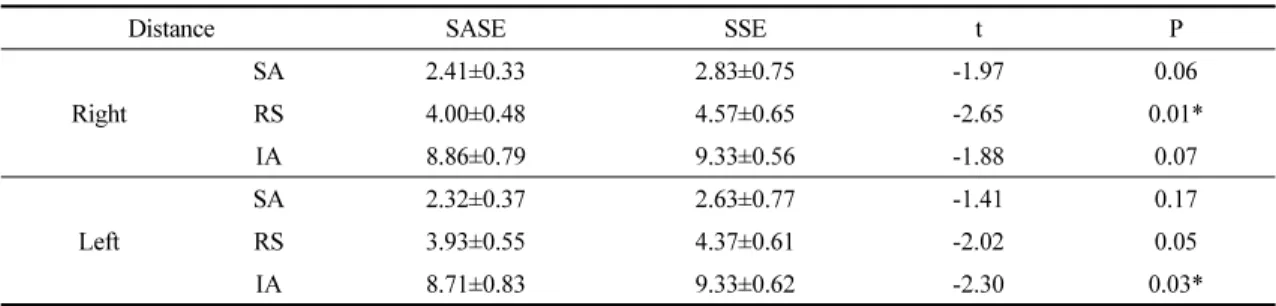
Distance SASE SSE t P
Right
SA 2.41±0.33 2.83±0.75 -1.97 0.06
RS 4.00±0.484.57±0.65 -2.65 0.01*
IA 8.86±0.79 9.33±0.56 -1.88 0.07
Left
SA 2.32±0.37 2.63±0.77 -1.41 0.17
RS 3.93±0.55 4.37±0.61 -2.02 0.05
IA 8.71±0.83 9.33±0.62 -2.30 0.03*
*: significant difference (p<0.05) Unit: thoracic level
SASE: shoulder and abdominal stabilization exercises group SSE: shoulder stabilization exercises group
SA: superior angle RS: root of spine IA: inferior angle
Table 5. Comparison of scapular alignment measurements between two groups after intervention (n =29)
기 때문에 어깨뼈 정렬의 변화가 크지 않은 것으로 사료된다. 따라서 앞톱니근의 활성화가 어깨의 운동장 애를 개선하고, 결과적으로 전방머리자세에도 유의한 영향을 미쳐 머리척추각도가 개선된 것으로 사료된다.
Shiravi 등(2019)은 젊은 여성을 대상으로 어깨 안정 화 운동과 복부 조절피드백을 동시에 시행하는 군과 어깨 안정화 운동만 시행하는 군을 대상으로 6주간 중재를 하였고, 두 군 모두 위등세모근의 근활성도가 감소되었고, 앞톱니근의 근활성도가 증가되어 전방머 리자세가 개선되었는데, 어깨 안정화 운동과 복부 조 절피드백을 동시에 시행하는 군이 유의하게 개선되었 다고 보고하였다. 이는 복부의 안정성이 어깨의 정렬 에 영향을 주어 앞톱니근의 근활성도가 증가되어 머 리척추각도가 증가되었다고 보고하였다(Shiravi et al., 2019). 하지만 본 연구에서는 복합운동군과 어깨운동 군 간의 차이는 없었다.
앞톱니근의 근막과 힘줄은 부분적으로 배바깥빗근 의 힘줄이 연결되어 있어 두 근육 중 한 근육이 활성화 할 시 다른 근육도 활성화하게 된다(Kaur et al., 2014;
Neumann & Camargo, 2019; Toro et al., 2016). 본 연구에 서 시행한 복부 드로잉-인 기법은 배바깥빗근의 활동 을 최소화하면서 배속빗근과 배가로근을 수축하는 운 동 기법이다(Madokoro et al., 2020). 그러하여 앞톱니 근의 근활성도를 증가시키기에는 제한이 있을 것이 며, 복부 드로잉-인 기법 보다는 배바깥빗근을 활성화 시킬 수 있는 복부 브레이싱(Lee, 2014)기법이 더 효과 적일 것으로 사료된다.
이상의 내용을 종합해 보면 복부 안정화 운동은 배바깥빗근의 근활성도 증가에 영향을 미치지 않아 머리척추각도에 영향을 미치지 않았을 것으로 사료된 다. 따라서 배바깥빗근의 근활성도를 증가시킬 수 있 는 운동에 초점을 두어 시행하면 전방머리자세의 개 선에 긍정적인 효과가 나타날 것으로 사료된다.
군 모두에서 부분적으로 유의한 변화가 나타났다. 그 러므로 전방머리자세의 개선은 복부의 안정성 보단 어깨의 안정성에 초점을 맞추는 것이 긍정적인 효과 가 나타날 것으로 사료된다.
Acknowledgement
본 논문은 김재현의 석사학위논문의 일부를 발취 한 것입니다.
References
Ahn SH, Yang JH, Lee SK, et al. Effect of chin tuck exercises on various postures and muscle activity of the neck and shoulder. PNF and Movement. 2020;18(3):
403-414.
Barry CM, Kestell G, Gillan M, et al. Sensory nerve fibers containing calcitonin gene-related peptide in gastrocnemius, latissimus dorsi and erector spinae muscles and thoracolumbar fascia in mice.
Neuroscience. 2015;219:106-117.
Diab AA, Moustafa IM. The efficacy of forward head correction on nerve root function and pain in cervical spondylotic radiculopathy: a randomized trial. Clinical Rehabilitation. 2011;26(4):351-361.
Fard BS, Ahmadi A, Maroufi N, et al. Evaluation of forward head posture in sitting and standing positions.
European Spine Journal. 2016;25(11):3577-3582.
Greig AM, Straker LM, Briggs AM. Cervical erector spinae and upper trapezius muscle activity in children using different information technologies. Physiotherapy.
2005;91(2):119-126.
IJspeert J, Kerstens HCJW, Janssen RMJ, et al. Validity and reliability of serratus anterior hand held dynamometry.
BMC Musculoskeletal Disorders. 2019;20(1):360.
Janwantanakul P, Sitthipornvorakul E, Paksaichol A. Risk factors for the onset of nonspecific low back pain in office works: a systematic review of prospective cohort studies. Journal of Manipulative and Physiological Therapeutics. 2012;35(7):568-577.
Kaur N, Bhanot K, Brody LT, et al. Effects of lower extremity and trunk muscles recruitment on serratus anterior muscle activation in healthy male adults. The International Journal of Sports Physical Therapy.
2014;9(7):924-937.
Kang KW, Kang DW, Kwon GY, et al. The impact of head repositioning accuracy and proprioception on cervical stabilization exercise in healthy adults. Physical Therapy Rehabilitation Science. 2015;4 (1): 49-54.
Khan A, Khan Z, Bhati P, et al. Influence of forward head posture on cervicocephalic kinesthesia and electromyographic activity of neck musculature in asymptomatic individuals. Journal of Chiropractic Medicine. 2020;19(4):230-240.
Khayatzadeh S, Kalmanson OA, Schuit D, et al. Cervical spine muscle-tendon unit length differences between neutral and forward head postures: biomechanical study using human cadaveric specimens. Physical Therapy.
2017;97(7):756-766.
Kim BB, Lee JH, Jeong HJ, et al. Can suboccipital release followed by cranio -cervical flexion exercise improve shoulder range of motion, pain, and muscle activity of scapular upward rotators in subjects with forward head posture? Physical Therapy Korea. 2016a;23 (2):57-66.
Kim BB, Lee JH, Jeong HJ, et al. Effects of suboccipital release with craniocervical flexion exercise on craniocervical alignment and extrinsic cervical muscle activity in subjects with forward head posture. Journal
of Electromyography and Kinesiology. 2016b;
30:31-37.
Kim BY, Kang SM, Kang JH, et al. 2020 Korean society for the study of obesity guidelines for the management of obesity in Korea. Seoul. Journal of Obesity &
Metabolic Syndrome. 2020.
Kim HW, Yun SJ, Ha SM. A reliability study of tape and photography measurement techniques for scapular position. Physical Therapy Korea. 2013;20(3):74-80.
Kim JH, Yoon HB, Park JH, et al. Comparative effect of modified shrug exercises with and without trunk stabilization exercise on scapular upward rotator EMG and thickness in subjects with scapular downward rotation syndrome. Physical Therapy Korea.
2017;24(4):60-67.
Kim MH. The effect of traditional Korean breathing exercises on body composition basic fitness and stress hormone levels in the elderly women. Korea Sport Research.
2007;18(5):91-102.
Kim SH, Park DJ. Effects of diagonal shoulder training in a closed kinematic chain for secondary impingement syndrome: a case study. Journal of Physical Therapy Science. 2015;27(6):2019-2020.
Kim SY, Koo SJ. Effect of duration of smartphone use on muscle fatigue and pain caused by forward head posture in adults. Journal of Physical Therapy Science.
2016;28(6):1669-1672.
Ko KH. A study on correlational relationship between percent body fat (%BF) and body mass index (BMI). Korea Sport Research. 2005;16(5):619-626.
Kocur P, Wilski M, Goliwas M, et al. Influence of forward head posture on myotonometric measurements of superficial neck muscle tone, elasticity, and stiffness in asymptomatic individuals with sedentary jobs.
Journal of Manipulative and Physiological Therapeutics. 2019;42(3):195-202.
Kudo S, Mastuda Y, Yanai T, et al. Contribution of upper
abdominal bracing, and dynamic neuromuscular stabilization on core stability and motor control in adults with core instability. Yonsei University.
Dissertation of Master’s Degree. 2014.
Madokoro S, Yokogawa M, Miaki H. Effect of the abdominal draw-in maneuver and bracing on abdominal muscle thickness and the associated subjective difficulty in healthy individuals. Healthcare. 2020;8(4):496.
Neumann DA, Camargo PR. Kinesiologic considerations for targeting activation of scapulothoracic muscles - part 1: serratus anterior. Brazilian Journal of Physical Therapy. 2019;23(6):459-466.
Park DJ, Lee HO. The intramuscular activation of scapular stabilizing muscles during push-up plus and PNF exercises in a quadruped position. Journal of Physical Therapy Science. 2013a;25(4):371-374.
Park DJ, Lee SK. What is a suitable pressure for the abdominal drawing-in maneuver in the supine position using a pressure biofeedback unit? Journal of the Physical Therapy Science. 2013b;25(5):527-530.
Park SK, Kim YN, Jung EY, et al. Abdominal draw in maneuver with shoulder isometric contractions on abdominal muscles thickness in healthy person. Physical Therapy Korea. 2013;20(2):38-45.
Park SK, Park JM, Lee JH. Effects of a push-up plus exercise program on scapular position and muscle activity in individuals with rounded shoulder posture. The Journal Korean Society of Physical Therapy.
2010;22(5):1-8.
Sheikhhoseini R, Shahrbanian S, Sayyadi P, et al. Effectiveness of therapeutic exercise on forward head posture: a
control feedback and scapula stabilization exercises in participants with forward head, round shoulder postures and neck movement impairment. Sports Health. 2019;11(3):272-279.
Singla D, Veqar Z. Association between forward head, rounded shoulders, and increased thoracic kyphosis: a review of the literature. Journal of Chiropractic Medicine.
2017;16(3):220-229.
Sobush DC, Simoneau GG, Dietz KE, et al. The lennie test for measuring scapular position in healthy young adult females: a reliability and validity study. The Journal of Orthopaedic and Sports Physical Therapy.
1996;23(1):39-50.
Song GB, Kim JJ, Kim KR, et al. The effects of neck stabilization exercise and proprioceptive neuromuscular facilitation on neck alignment, NDI, and static balance in adults with forward-head posture in a sitting position. PNF and Movement. 2020;18(1):11-22.
Toro ASV, Cools AMJ, Oliveira ASD. Instruction and feedback for conscious contraction of the abdominal muscles increase the scapular muscles activation during shoulder exercises. Manual Therapy. 2016;25:11-18.
Weon JH, Oh JS, Cynn HS, et al. Influence of forward head posture on scapular upward rotators during isometric shoulder flexion. Journal of Bodywork & Movement Therapies. 2010;14(4):367-374.
Wong KK, Chai HM, Chen YJ, et al. Mechanical deformation of posterior thoracolumbar fascia after myofascial release in healthy men: a study of dynamic ultrasound imaging. Musculoskeletal Science and Practice.
2017;27:124-130.