심한 유리체출혈에서 수술 중 빛간섭단층촬영의 유용성
Utility of the Intraoperative Optical Coherence Tomography for Dense Vitreous Hemorrhage
양영성1, 손준홍1, 박지인2, 황덕진1,3
Young Seong Yang1, Joonhong Sohn1, Ji In Park2, Duck Jin Hwang1,3
1한길안과병원, 2강원대학교병원 내과, 3가톨릭관동대학교 의과대학 안과학교실
1HanGil Eye Hospital, Incheon, Korea
2Department of Internal Medicine, Kangwon National University Hospital, Chuncheon, Korea
3Department of Ophthalmology, Catholic Kwandong University College of Medicine, Gangneung, Korea
Purpose: To evaluate the utility of intraoperative optical coherence tomography (iOCT) during pars plana vitrectomy surgery (PPV) for dense vitreous hemorrhage (VH).
Methods: The authors retrospectively reviewed medical records of 30 eyes from 30 patients in which iOCT was evaluated during PPV for dense VH that precluded preoperative OCT assessment. iOCT images were qualitatively evaluated for retinal abnormalities that might impact intraoperative or perioperative management.
Results: There were 18 males (60%) and 12 females (40%). The mean age of patients in the study was 56.0 ± 14.5 years (range: 34-86 years). The etiology for VH was proliferative diabetic retinopathy (18 eyes, 60.0%), retinal vein occlusion with neovascularization (5 eyes, 16.7%), age-related macular degeneration (2 eyes, 6.7%), retinal tear (2 eyes, 6.7%), posterior vitreous detachment (1 eye, 3.3%), retinal arterial macroaneurysm (1 eye, 3.3%) and Eale’s disease (1 eye, 3.3%). iOCT revealed epiretinal membrane (7 eyes, 23.3%), tractional retinal detachment (6 eyes, 20.0%), rhegmatogenous retinal detachment (2 eyes, 6.7%) and macular edema (1 eye, 3.3%). The median number of scan sessions per case was 2.87 ± 1.61 (range: 1-8 sessions) with 4.70 ± 2.20 (range: 3-11 sessions) median total scans. The mean time at which surgery was paused to perform iOCT imaging was 3.18 ± 1.53 minutes (range: 1.70-7.17 minutes).
Conclusions: iOCT during PPV for dense VH may provide surgeons with clinically relevant information that may influence perioperative management.
Keywords: Dense vitreous hemorrhage; Intraoperative optical coherence tomography; Pars plana vitrectomy
서론
유리체출혈은 망막 혈관질환에서 가역적인 시력저하를 일으키 는 흔한 질환이다[1]. 유리체출혈을 일으키는 흔한 원인들로는 증식당뇨망막병증, 망막열공, 후유리체박리, 망막정맥폐쇄, 연령 관련황반변성 등이 있다[1-5]. 유리체출혈이 지속될 경우 유리
체절제술이 첫 번째 치료방법으로 알려져있다[6,7].
빛간섭단층촬영(optical coherence tomography, OCT)은 비접 촉성, 비침습성 영상기술로 초음파 B-scan과 유사하나 음파 대 신 적외선 계열의 광선을 사용하며 안조직에서 반사되어 나오 는 반향반사지연시간을 통해 망막의 두께를 정량적으로 측정할 수 있는 진단도구이다[8]. 하지만 광학적 방법을 통한 검사이므
Address reprint requests to Duck Jin Hwang, MD
HanGil Eye Hospital, #35 Bupyeong-daero, Bupyeong-gu, Incheon 21388, Korea Tel: 82-32-503-3322, Fax: 82-32-504-3322
E-mail: [email protected]
Received: 2017. 9. 4 Revised: 2017. 10. 19 Accepted: 2017. 10. 20
로 유리체출혈과 같은 매체의 혼탁이 심한 경우 빛의 투사나 반 사가 간섭을 받아 측정이 제한될 수 있다[9,10]. 그래서 황반부 를 가리는 중심부에 출혈이 있는 유리체출혈에서 수술 전 OCT 를 포함한 여러 검사들이 제한을 받아 수술 계획을 세우는 데 어려움이 있다[11].
최근 몇 년 동안 수술 중 OCT (intraoperative OCT, iOCT)가 안과 수술의 보조장치로 소개되었고 유리체망막수술 중에 황 반원공, 유리체황반부견인, 망막박리, 망막앞막 등 다양한 망 막질환을 알아낼 수 있다고 보고하였다[12-15]. 이에 저자들은 수술 전 OCT 촬영에 제한이 있을 정도의 유리체출혈이 있어 출혈의 정확한 원인을 알 수 없는 유리체출혈환자의 수술을 시 행하면서 유리체출혈 외 어떤 유리체망막 소견이 동반되어 있 는지 알아보고, 수술 중 빛간섭단층촬영의 유용성을 알아보고 자 하였다.
대상과 방법
2016년 9월부터 2017년 3월까지 본원 안과에 내원하여 수술 당 시에 안저검사상 안저반사가 소실되고 시신경 및 주변부 망막 혈관이 보이지 않을 정도의 유리체출혈로 인해 유리체절제술 을 시행하면서 iOCT를 촬영한 30명 30안을 대상으로 후향적 으로 분석하였다. 본 연구는 한길안과병원 연구시험윤리위원회 (institutional review board, IRB)의 승인을 받았고 의학연구윤 리강령인 헬싱키 선언(Declaration of Helsinki)을 준수하였다.
모든 환자를 대상으로 나이, 성별, 동반 질환, 이전 유리체절 제술 시행 여부, 수술 전 시력, 수술 전 안압을 파악하였다. 모 든 수술은 25게이지 유리체절제술을 RESCAN 700 (Carl Zeiss Meditec, Oberkochen, Germany) 수술현미경을 이용하여 시행 하였으며, 한 명의 수술자가 수술을 집도하였다. 수정체혼탁이 있는 경우 유리체절제술과 함께 수정체초음파유화술 및 인공 수정체삽입술을 같이 시행하였다. 수술자는 유리체절제술 중 iOCT 영상을 활용하여 수술자의 판단하에 추가적인 치료를 시 행하였으며 iOCT 장비를 이용한 session 횟수(술 중 OCT 화면 을 켜고 volume scan으로 이미지를 구성하여 review 화면을 본 횟수)와 iOCT를 이용한 scan 횟수(술 중 OCT 화면을 켜서 망막 상태를 확인한 횟수) 및 수술 중 iOCT를 사용하기 위해 중단된 시간을 함께 기록하였다. 통계학적 분석은 SPSS ver. 21.0 (IBM Corp., Armonk, NY, USA)를 사용하였다.
결과
총 30명의 환자 중 남자가 18명(60%), 여자가 12명(40%)이었 고 평균 나이는 56.0 ± 14.5세(34-86세)였다. 당뇨가 있는 환자
가 20명, 고혈압이 있는 환자가 14명, 심장질환이 있는 환자가 2명, 뇌혈관질환이 있는 환자가 1명이 있었다(Table 1). 22안에 서는 유리체절제술과 함께 수정체초음파유화술 및 인공수정체 삽입술을 같이 시행하였다.
수술 중 알아낸 유리체출혈을 일으킨 원인으로 증식성당뇨 망막병증(18안, 60.0%), 신생혈관이 동반된 망막정맥폐쇄(5안, 16.7%), 나이관련황반변성(2안, 6.7%), 망막열공(2안, 6.7%), 후 유리체박리(1안, 3.3%), 망막동맥대혈관류(1안, 3.3%). 일스병 (1안, 3.3%)으로 확인되었다(Table 2).
수술 중 iOCT를 이용하여 망막앞막(7안, 23.3%), 견인성망 막박리(6안, 20.0%), 열공성망막박리(2안, 6.7%), 황반부종(1안, 3.3%)을 확인하였다(Table 3). 이 중 망막앞막 4안과 황반부종 1안은 수술자가 인지하지 못하고 iOCT 상으로만 확인이 가능하 였다. 수술자의 판단하에 유리체강내 항혈관내피세포성장인자 주입술(19안, 63.3%), 망막앞막제거술(7안, 23.3%), 실리콘기름 주입술(3안, 9.9%), 과불화프로판(C3F8) 가스주입술(1안, 3.3%)
Table 1. Baseline characteristics of the patients
Charicteristics Value
Number of eyes (patients) 30 (30)
Age (years) 56.0 ± 14.5
Sex (male/female) 18/12
Diabetes mellitus 20 (66.7)
Hypertension 14 (46.7)
Cardiovascular disease 2 (6.7)
Cerebrovascular disease 1 (3.3)
Number of eyes with previous vitrectomy Hx. 2 (6.7)
Preoprerative BCVA (logMAR) 1.88 ± 0.90
Preoprerative IOP (mmHg) 15.70 ± 3.57
Duration from diagnosis to surgery (days) 26.30 ± 23.27 Values are presented as number (%) or mean ± standard deviation.
Hx. = history; BCVA = best corrected visual acuity; logMAR = loga- rithm of minimal angle of resolution; IOP = intraocular pressure.
Table 2. Intraoperative diagnosis of dense vitreous hemorrhage
Final diagnosis Number of eyes
Proliferative diabetic retinopathy 18 (60.0) Retinal vein occlusion with neovascularization 5 (16.7) Age-related macular degeneration 2 (6.7)
Retinal tear 2 (6.7)
Posterior vitreous detachment 1 (3.3)
Retinal arterial macroaneurysm 1 (3.3)
Eale’s disease 1 (3.3)
Values are presented as n (%).
등을 시행하였다.
수술 중 iOCT를 이용한 평균 session 횟수는 2.87 ± 1.61회 (1-8회)였으며, 수술 중 빛간섭단층촬영 장비를 이용한 평균 scan 횟수는 4.70 ± 2.20회(3-11회)였다. 수술 중 빛간섭단층촬 영을 하기 위해 중단된 평균 시간은 3.18 ± 1.53분(1.70-7.17분) 이었다(Table 4).
대표 증례
증례 1
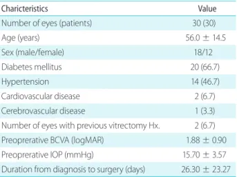
59세 여자로 수술 직전 시행한 검사에서 좌안 교정시력 안전수 지였다. 수술 전 안저사진에서 유리체출혈로 인하여 망막이 관 찰되지 않았다(Fig. 1A). 수술로 유리체출혈을 제거하고 난 후 망막분지정맥폐쇄를 진단하였고 iOCT를 이용하여 낭포황반부 종을 발견하였으며 망막전막은 관찰되지 않았다(Fig. 1B). 수술 중 유리체강내 항혈관내피세포성장인자 주입술과 산발레이저 광응고술(scatter laser photocoagulation)을 시행하였고 추가적 인 막제거술은 시행하지 않았다.
증례 2
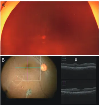
68세 여자로 내원 당시 시행한 검사에서 우안 교정시력 안전수 지였다. 수술 전 안저사진에서 유리체출혈로 인하여 망막이 관 찰되지 않았다(Fig. 2A). 진단 3달 후에 수술을 시행하였고 수 술 중 오래된 유리체출혈을 제거하였다. 수술로 유리체출혈을 제거하고 난 후 iOCT를 통하여 맥락막신생혈관막과 함께 망막
하액이 관찰되어 나이관련황반변성으로 인한 유리체출혈로 진 단하고 수술 중 유리체강내 항혈관내피세포성장인자 주입술을 시행하였다(Fig. 2B).
고찰
심한 유리체출혈이 있는 경우 유리체출혈의 원인을 알아내기가 힘들며 원인을 추정하기 위해서는 정밀한 안과적 검사뿐만 아 니라 환자의 나이와 성별, 기저 질환, 약물 복용력, 외상 과거 력, 혈액 검사 및 과거 안과 진료 기록 등이 이용된다[1,5]. 그래 서 유리체출혈이 있는 경우 수술 전 정확한 안과 검사가 힘들 기 때문에 어떠한 유리체망막질환을 동반하고 있는지 알기 어 려워 수술 계획을 구상하는 데 한계가 있고 수술 중에도 망막 막제거술이나 유리체강내 주사치료 등 추가적인 치료를 결정하 는 것 역시 어렵다[11].
본 연구에서는 유리체출혈을 일으킨 원인질환으로 증식성당 뇨망막병증이 30안 중 18안으로 가장 많았으며 신생혈관이 동 반된 망막정맥폐쇄, 나이관련황반변성, 망막열공 등이 뒤를 이 었는데 이는 기존의 연구들과 비슷한 결과였다[1-5]. 그리고 수 술 중 iOCT를 이용하여 망막앞막, 견인성망막박리, 열공성망막 박리, 망막부종 등을 확인할 수 있었는데 수술 중 소견을 토대 Figure 1. Case 1. (A) Fundus photograph of non-clearing vitreous hemorrhage. (B) Branched retinal vessel obstruction was diagnosed after removal of vitreous hemorrhage. Intraoperative optical coher- ence tomography revealed cystoid macular edema (white arrow) and no epiretinal membrane.
A
B
Table 3. Diagnosis of comorbid retinal disorder during vitrectomy by iOCT
Comorbid retinal disorder Number of eyes
Epiretinal membrane 7 (23.3)
Tractional retinal detachment 6 (20.0) Rhegmatogenous retinal detachment 2 (6.7)
Macular edema 1 (3.3)
Values are presented as n (%).
iOCT = intraoperative optical coherence tomography.
Table 4. Time implication for performing iOCT during surgery Value
Scan sessions 2.87 ± 1.61 (1-8)
Total scans 4.70 ± 2.20 (3-11)
Total duration surgery paused for iOCT imaging (minutes)
3.18 ± 1.53 (1.70-7.17) Values are presented as mean ± standard deviation (range).
iOCT = intraoperative optical coherence tomography.
로 추가적인 막제거술, 유리체강내 항혈관내피세포성장인자 주 입술 등을 시행할 수 있었다.
OCT를 수술 중 사용하는 데 있어서 처음으로 직면한 문제는 휴대화할 수 있게 경량화하는 것과 누워있는 환자로부터 이미 지를 얻어낼 수 있는 기술이었는데, 경량화된 휴대용 OCT가 개 발되어서 2000년대 중반부터 수술 중에 사용되기 시작하였다 [16,17]. 휴대용 OCT가 수술방에서의 진단의 용이함을 제공하 였지만 이미지의 정밀도와 재현성이 떨어지는 단점이 보고되었 다[12,15,18-21]. 그 뒤로 현미경에 휴대용 OCT를 장착하고 현 미경 발판을 이용하여 초점과 측면 및 수직 편차를 제어할 수 있는 장치가 개발되었고 이것은 휴대용 OCT보다 향상된 이미 지의 안정성과 재현성을 보고하였다[15,18]. 최근에는 Lumera 700 (Carl Zeiss Meditec, Oberkochen, Germany) 수술 현미경에 OCT가 결합된 RESCAN 700이 개발되었는데, 이것은 수술자가 수술 현미경을 보는 상태에서 ‘real-time’ 이미지를 통해 수술 중 기구의 조작으로 인한 조직의 변화까지 헤드업 디스플레이로 동 시에 이미지를 확인할 수 있는 장점이 있다[22,23]. iOCT 사용 에 관한 기존 연구에서 PIONEER 연구의 2년 결과는 수술 중 현미경에 장착된 OCT의 실행과 유용성에 관하여 보고하였고, iOCT에서 수집한 정보가 수술자의 의사 결정에 어떻게 도움이
되는지 발표하였다[19]. 연속적으로 DISCOVER 연구에서 수술 현미경과 결합된 OCT의 유용성에 대한 결과를 발표하였다[23].
본 연구는 iOCT를 사용한 소수의 증례를 후향적으로 분석하 였다는 제한점이 있으나, 수술 전 정확한 안과검사를 할 수 없 는 유리체출혈 환자에서 iOCT가 유용할 수 있음을 국내 최초 로 고찰해 보았다는 데에 의의가 있다. 대상안 중 망막앞막이나 망막부종이 동반돼 있어 수술 중 iOCT 검사가 도움이 될 수 있 는 경우가 전체의 26.6%로 나타났는데, 망막앞막이 있을 경우 망막앞막제거술을 추가로 시행하고 망막부종이 있을 경우 유 리체강내 항혈관내피세포성장인자 주입술 등을 추가로 시행할 수 있어 수술 중 추가적인 시술을 결정하는 데 도움을 주었다.
그리고 황반부뿐만 아니라 주변부에 있는 국소적인 열공성망막 박리나 견인성망막박리의 진단에도 도움이 되었다. iOCT의 사 용은 수술자에게 수술 중 망막의 구조에 대한 정보를 주기 때 문에 수술 전 안과검사가 어려운 심한 유리체출혈 환자의 수술 에 있어서 수술자의 판단하에 추가적인 술기 선택에 도움을 줄 수 있다고 생각된다.
Conflicts of interest
The authors have no conflicts to disclose.
References
1. Spraul CW, Grossniklaus HE. Vitreous hemorrhage. Surv Oph- thalmol 1997;42:3-39.
2. Butner RW, McPherson AR. Spontaneous vitreous hemorrhage.
Ann Ophthalmol 1982;14:268-70.
3. Dana MR, Werner MS, Viana MA, Shapiro MJ. Spontaneous and traumatic vitreous hemorrhage. Ophthalmology 1993;100:1377- 83.
4. Lean JS, Gregor Z. The acute vitreous hemorrhage. Br J Ophthal- mol 1980;64:469-71.
5. Lindgren G, Sjödell L, Lindblom B. A prospective study of dense spontaneous vitreous hemorrhage. Am J Ophthalmol 1995;119:458-65.
6. Early vitrectomy for severe vitreous hemorrhage in diabetic retinopathy. Two-year results of a randomized trial. Diabetic Retinopathy Vitrectomy Study report 2. The Diabetic Reti- nopathy Vitrectomy Study Research Group. Arch Ophthalmol 1985;103:1644-52.
7. Diabetic Retinopathy Clinical Research Network. Randomized clinical trial evaluating intravitreal ranibizumab or saline for vit- reous hemorrhage from proliferative diabetic retinopathy. JAMA Ophthalmol 2013;131:283-93.
Figure 2. Case 2. (A) Fundus photographs of non-clearing vitreous hemorrhage. (B) Intraoperative optical coherence tomography re- vealed choroidal neovascular membrane with subretinal fluid (white arrows) after removal of vitreous hemorrhage. Vitreous hemorrhage associated with age-related macular degeneration was diagnosed and intravitreal anti-vascular endothelial growth factor injection was performed during surgery.
A
B
8. Massin P, Vicaut E, Haouchine B, et al. Reproducibility of retinal mapping using optical coherence tomography. Arch Ophthal- mol 2001;119:1135-42.
9. Huang D, Swanson EA, Lin CP, et al. Optical coherence tomogra- phy. Science 1991;254:1178-81.
10. Kholodnykh AI, Petrova IY, Larin KV, et al. Precision of measure- ment of tissue optical properties with optical coherence to- mography. Appl Opt 2003;42:3027-37.
11. Ehlers JP, Griffith JF, Srivastava SK. Intraoperative optical coher- ence tomography during vitreoretinal surgery for dense vitre- ous hemorrhage in the pioneer study. Retina 2015;35:2537-42.
12. Ehlers JP, Ohr MP, Kaiser PK, Srivastava SK. Novel microarchitec- tural dynamics in rhegmatogenous retinal detachments identi- fied with intraoperative optical coherence tomography. Retina 2013;33:1428-34.
13. Binder S, Falkner-Radler CI, Hauger C, et al. Feasibility of intrasur- gical spectral-domain optical coherence tomography. Retina 2011;31:1332-6.
14. Dayani PN, Maldonado R, Farsiu S, Toth CA. Intraoperative use of handheld spectral domain optical coherence tomography imaging in macular surgery. Retina 2009;29:1457-68.
15. Ray R, Barañano DE, Fortun JA, et al. Intraoperative micro- scope-mounted spectral domain optical coherence tomogra- phy for evaluation of retinal anatomy during macular surgery.
Ophthalmology 2011;118:2212-7.
16. Scott AW, Farsiu S, Enyedi LB, et al. Imaging the infant retina with a hand-held spectral-domain optical coherence tomography
device. Am J Ophthalmol 2009;147:364-73.e2.
17. Chavala SH, Farsiu S, Maldonado R, et al. Insights into advanced retinopathy of prematurity using handheld spectral domain optical coherence tomography imaging. Ophthalmology 2009;116:2448-56.
18. Ehlers JP, Dupps WJ, Kaiser PK, et al. The prospective intraop- erative and perioperative ophthalmic imagiNg with Optical CoherEncE TomogRaphy (PIONEER) study: 2-year results. Am J Ophthalmol 2014;158:999-1007.
19. Ehlers JP, Petkovsek DS, Yuan A, et al. Intrasurgical assessment of subretinal tPA injection for submacular hemorrhage in the PIO- NEER study utilizing intraoperative OCT. Ophthalmic Surg Lasers Imaging Retina 2015;46:327-32.
20. Ehlers JP, Tam T, Kaiser PK, et al. Utility of intraoperative optical coherence tomography during vitrectomy surgery for vitreo- macular traction syndrome. Retina 2014;34:1341-6.
21. Ehlers JP, Xu D, Kaiser PK, et al. Intrasurgical dynamics of macular hole surgery: an assessment of surgery-induced ultrastructural alterations with intraoperative optical coherence tomography.
Retina 2014;34:213-21.
22. Ehlers JP, Kaiser PK, Srivastava SK. Intraoperative optical coher- ence tomography using the RESCAN 700: preliminary results from the DISCOVER study. Br J Ophthalmol 2014;98:1329-32.
23. Ehlers JP, Goshe J, Dupps WJ, et al. Determination of feasibility and utility of microscope-integrated optical coherence tomog- raphy during ophthalmic surgery: the DISCOVER Study RESCAN Results. JAMA Ophthalmol 2015;133:1124-32.
심한 유리체출혈에서 수술 중 빛간섭단층촬영의 유용성
목적: 심한 유리체출혈 환자의 유리체절제술 중 빛간섭단층촬영의 유용성에 대해서 알아보고자 하였다.
대상과 방법: 수술 전 빛간섭단층촬영이 어려운 유리체출혈로 유리체절제술을 받은 환자에서 수술 중 빛간섭단층촬영을 시행한 30명 30안을 대상으로 수술 중 유리체출혈 외 어떤 유리체망막 소견이 동반되어 있는지 알아보고 그 유용성을 알아보았다.
결과: 남자 18명(60%), 여자 12명(40%)이었고 평균 나이는 56.0 ± 14.5세(34-86세)였다. 유리체출혈을 일으킨 원인으로 증식성당 뇨망막병증(18안, 60.0%), 신생혈관이 동반된 망막정맥폐쇄(5안, 16.7%), 나이관련황반변성(2안, 6.7%), 망막열공(2안, 6.7%), 후 유리체박리(1안, 3.3%), 망막동맥대혈관류(1안, 3.3%), 일스병(1안, 3.3%)으로 확인되었다. 유리체절제술 중 빛간섭단층촬영으로 망 막앞막(7안, 23.3%), 견인성망막박리(6안, 20.0%), 열공성망막박리(2안, 6.7%), 황반부종(1안, 3.3%)을 확인할 수 있었다. 수술 중 빛간섭단층촬영 장비를 이용한 평균 session 횟수는 2.87 ± 1.61회(1-8회)였으며, 수술 중 빛간섭단층촬영 장비를 이용한 평균 scan 횟수는 4.70 ± 2.20회(3-11회)였다. 수술 중 빛간섭단층촬영을 하기 위해 중단된 평균 시간은 3.18 ± 1.53분(1.70-7.17분)이었다.
결론: 심한 유리체출혈로 유리체절제술을 할 경우 수술 중 빛간섭단층촬영이 수술 중 정보획득에 도움을 주고, 수술 중 치료 선택에 도 움을 줄 수 있다.
국문초록