Small Bowel Perforation Caused by Taenia Solium Infection
4
0
0
전체 글
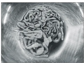
(2) 476. 대한외과학회지:제 71 권 제 6 호 2006. ꠏꠏꠏꠏꠏꠏꠏꠏꠏꠏꠏꠏꠏꠏꠏꠏꠏꠏꠏꠏꠏꠏꠏꠏꠏꠏꠏꠏꠏꠏꠏꠏꠏꠏꠏꠏꠏꠏꠏꠏꠏꠏꠏꠏꠏꠏꠏꠏꠏꠏꠏꠏꠏꠏꠏꠏꠏꠏꠏꠏꠏꠏꠏꠏꠏꠏꠏꠏꠏꠏꠏꠏꠏꠏꠏꠏꠏꠏꠏꠏꠏꠏꠏꠏꠏꠏꠏꠏꠏꠏꠏꠏꠏꠏꠏꠏꠏꠏꠏꠏꠏꠏꠏꠏꠏꠏꠏꠏꠏꠏꠏꠏꠏꠏꠏ. Fig. 1. Abdominal CT shows inhomogeneous attenuation of mesentery and omental fat with edematous change of small bowel, suggesting perforation site (arrow)(Case 1).. Fig. 2. Operation findings show ileal perforation with ongoing migration of tape-warm (arrow) at 120 cm proximal from I∼C valve (Case 1).. 충이 탈출하고 있었으며 천공 부위의 인접한 복강 내에 수 개의 기생충이 관찰되었다(Fig. 2). 천공 부위는 경화가 심 하지 않아 단순봉합을 실시하였다. 기생충학적으로 Taenia solium으로 보고되었고(Fig. 3), Praziquantel 10 mg/kg 복용 후 7병일째에 퇴원하였다. 증 례 2 88세 남자로 3일 전부터 발생된 갑작스런 복통으로 응급 실에 내원하였다. 이학적 검사에서 내원 시 혈압은 70/40 o mmHg, 맥박수 124회/분, 호흡수 20회/분, 체온은 36.8 C였 다. 복부는 전반적으로 압통 및 반발통이 심하게 있었다. 혈 3 액검사상 혈색소 16.3 g/dl, 백혈구 9,950/mm (호중구: 95%),. Fig. 3. Parasitological identification revealed double crowns of hooks containing scolex is found which is typical characteristic of Taenia solium species (Case 1).. Fig. 4. Abdominal CT shows a free air (arrow head) and loculated fluid collection with inflammatory changes on cecal area (white arrow), suggesting peritonitis and and periappendiceal abscess (Case 2).. 혈소판 102×103/ul, Na/K: 128/4.5 mmol/L, BUN/Cr: 63/3.3 mg/dl, 알부민: 2.8 g/dl, AST/ALT: 60/45 IU/L, Bilirubin (T/D): 3.7/2.0 mg/dl, PT/aPTT: 15.2/64 sec로 패혈증 소견이 보였다. 단순 흉부 X-선에서 유리공기는 보이지 않았고 복 부 전산화 단층 촬영에서 유리공기 소견과 비정상적인 복 강 내 저류액 및 전반적으로 작은창자가 팽창되어 보였으 며 충수주위 농양소견이 동반되었다(Fig. 4). 수술 소견에서 충수주위 농양 소견과 함께 공장 말단부에 기생충 감염에 의한 1 cm가량의 천공과 천공 부위와 인접한 복강내에 수 개의 기생충이 관찰되었다(Fig. 5). 천공 부위의 단순봉합과 충수절제술 및 배액술을 실시하였으나 패혈증으로 인한 합 병증으로 수술 다음날 사망하였으며 기생충학적으로 Taenia solium으로 보고되었다(Fig. 6)..
(3) 강동백 외:유구조충 감염에 의한 소장 천공. 477. ꠏꠏꠏꠏꠏꠏꠏꠏꠏꠏꠏꠏꠏꠏꠏꠏꠏꠏꠏꠏꠏꠏꠏꠏꠏꠏꠏꠏꠏꠏꠏꠏꠏꠏꠏꠏꠏꠏꠏꠏꠏꠏꠏꠏꠏꠏꠏꠏꠏꠏꠏꠏꠏꠏꠏꠏꠏꠏꠏꠏꠏꠏꠏꠏꠏꠏꠏꠏꠏꠏꠏꠏꠏꠏꠏꠏꠏꠏꠏꠏꠏꠏꠏꠏꠏꠏꠏꠏꠏꠏꠏꠏꠏꠏꠏꠏꠏꠏꠏꠏꠏꠏꠏꠏꠏꠏꠏꠏꠏꠏꠏꠏꠏꠏꠏ. Fig. 5. Operation findings show distal jejunal perforation (arrow) (Case 2).. 고. 찰. 유구조충(Taenia solium) 감염은 성서의 시대부터 기록되 어 있으며 1850년대 중반경에 생활사가 기술되었다.(11) 세 계적으로 산발적으로 많은 곳에서 발견되었고, 돼지고기를 생으로 먹거나 덜 익혀 먹는 지역에서 감염되는 기생충으 로 여겨지고 있으며(1-3) 우리나라에서는 특히 제주도에서 많은 감염보고가 있다.(12) 성충의 크기는 2∼7 m이고 두절은 사각형이며 직경은 1 mm정도로 두개의 갈고리(hook)줄을 지닌 둥근 두절돌출부 (rostellum)에 네 개의 큰 흡반(sucker)을 가지고 있다. 경부 부분은 짧고 두절 넓이의 반 정도 된다. 편절의 수는 1,000 개보다 적으며 미성숙 편절은 길이보다 폭이 크고 성숙 편 절은 대략 정사각형 모양이며 수태 편절은 폭보다 길이가 길다.(3,6,13) 형태학적으로 조충감별은 어려운 점이 있으나 본 증례는 사각형 모양의 두절과 두개의 갈고리 및 흡반을 가지고 있어 무구조충과 감별할 수 있었다.(6,13) 난자에 오염된 풀을 돼지가 먹으면 소장에서 부화한 유 충들이 장벽의 혈류를 따라 피하, 근육 등에 이르러 약 2개 월 후 유구낭충(Cysticercus cellulosae)이 된다. 이 낭충이 인 체에 생기는 경로는 음식물과 같이 충란이 섭취 되었을 때 항문 주위에 부착된 난자가 손가락 등을 통하여 구강에 들 어올 때(self infection), 그리고 소장에서 완숙한 마디가 위 에 역행되어 위액의 영향을 받았다가 다시 내려와 부화하 는 경우(internal autoinfection) 등이 있다.(14) 인체에 들어온 유구낭충은 소장에서 약 2개월 내에 성충이 되어 장 점막 내에 깊이 쳐박고 기생하므로 임상적으로 소화불량, 식욕 부진, 두통, 변비, 설사, 영양불량 등을 호소하며 migrating proglottids에 의해 급성충수염, 담관염이 발생 할 수 있다고 하나 정확한 기전은 밝혀지지 않고 있다. 대장에 기생한 무. Fig. 6. Parasitological identification revealed Taenia solium species (Case 2).. 구조충의 제거나(15) 무구조충에 의한 장천공(9) 이소성 폐 흡충증의 발현(16) 여러 기생충 등에 의한 장천공(17-19)이 드물게 보고되고 있으나 본 증례처럼 유구조충에 의한 장 천공은 문헌상 찾기 힘들다. 유구조충의 낭미충이 뇌로 퍼져 뇌낭미충증(neurocysticercosis)을 일으키며 이는 뇌에 있는 인간의 신경계 기생충 감염 중에 제일 흔한 형태이다. 이유는 알려지지 않았지만 라틴 아메리카에서는 같은 환자에게 뇌와 근육에서 동시에 낭미충이 6% 이하로 드물게 나타나지만 세계의 다른 장소 에서는 유구조충의 낭미충의 피하 침입은 뇌낭미충증 환자 중 78.5% 정도로 많이 보고되었다.(20) 이렇게 차이가 나게 되는 이유로는 환자의 면역상태, 인간의 백혈구 항원 상태, 환자의 영양상태, 환자에게 감염된 충란의 수, 그리고 유구 조충의 종의 차이 등이 있다.(3,20) 뇌에 위치한 낭충에 따 라 비정형적인 행동, 두통, 어지러움, 소뇌성실조, 지속적 구토, 시각적인 문제 등이 발생할 수 있어 일부 보고에서 가족력이 없는 간질 환자나 어렸을 때 걸리지 않았던 간질 환자의 모든 경우에서 뇌낭미충증의 가능성을 고려해야 한 다고 주장하였다.(21,22) 유구조충과 무구조충의 감별 및 진단은 외형상 똑같이 생겼기 때문에 충란만 가지고 구분하는 것은 힘들다. 형태 학적으로 보통 배출된 수태편절의 자궁촉지의 수, 두절의 검사로 감별진단을 확실하게 한다. 무구조충은 흡반 및 갈 고리가 없으며 본 증례처럼 유규조충은 네 개의 흡반과 갈 고리의 두절형태 존재로 다른 조충과 구분할 수 있 다.(3,14,23) 또한 Indian ink로 염색을 하거나 다른 투명화 기술을 이용하여 주요 자궁측지(uterine lateral branch)의 수 를 세면 유구조충에서는 7∼13개, 무구조충에서는 15∼20 개로 구분할 수 있으며 본 증례에서는 8∼9개로 유구조충 에 합당한 소견이다.(3,14,23) 또한 유구조충과 무구조충의 대변항원을 찾기 위한 예비.
(4) 478. 대한외과학회지:제 71 권 제 6 호 2006. ꠏꠏꠏꠏꠏꠏꠏꠏꠏꠏꠏꠏꠏꠏꠏꠏꠏꠏꠏꠏꠏꠏꠏꠏꠏꠏꠏꠏꠏꠏꠏꠏꠏꠏꠏꠏꠏꠏꠏꠏꠏꠏꠏꠏꠏꠏꠏꠏꠏꠏꠏꠏꠏꠏꠏꠏꠏꠏꠏꠏꠏꠏꠏꠏꠏꠏꠏꠏꠏꠏꠏꠏꠏꠏꠏꠏꠏꠏꠏꠏꠏꠏꠏꠏꠏꠏꠏꠏꠏꠏꠏꠏꠏꠏꠏꠏꠏꠏꠏꠏꠏꠏꠏꠏꠏꠏꠏꠏꠏꠏꠏꠏꠏꠏꠏ. 적인 ELISA는 진단적으로 매우 가치가 있어 대변에 알이 없어도 ELISA로 사람의 조충을 찾아내어 감염이 풍토성인 지역에서 조충의 보균자를 찾아내는 역학연구에 도움을 주 고 있다.(3,6,24) 최근에는 DNA와 PCR를 통한 조충의 구분, 각각의 종의 분류, 유전자 다양성 및 지역적 차이에 따른 유전자 변이 등의 분자 역학적인 접근이 시도되고 있다.(25) 치료로는 Praziquantel 5∼10 mg/kg 1회 복용 또는 Niclosamide 2 g 복용을 하며 치료 1달 뒤 변충란 검사를 하도록 권장하고 있다.(3) 예방은 감염경로의 자각과 개인적으로 청결한 위생상태 를 유지하는 것으로 돼지고기와 돼지와 관련된 제품을 o 65 C에서 요리하여 낭미충을 죽이는 것이 가장 중요하다. 또한 백신을 통한 유구낭미충증의 효과적인 예방법이 유구 조충의 치료하는데 있어 보고되고 있다.(26) 기생충 감염에 의한 장천공은 아주 드물어 장천공과 더 불어 기생충 감염이 동시에 발생할 수 있는 가능성을 배제 할 수 없지만 증례 1에서 정확한 외상의 현병력이 없으며 수술 소견상 천공 부위가 깨끗한 변연의 자연적인 천공소 견으로 외상과는 관계없는 기생충 자체에 의한 천공으로 생각되어지며 증례 2에서도 충수주위 농양에 의한 장벽의 자극증상으로 이차적인 소장천공 가능성을 배제할 수 없지 만 좌하복부의 공장 발단부에서 깨끗한 변연의 천공 부위 가 발생하여 충수주위 농양과 관계없이 발생한 기생충 감 염에 의한 천공으로 생각한다. 기생충 감염에 의한 증상은 아주 다양하여 기생충이 유 행하는 지역, 환자의 식습관 등 그 지역에 살고 있는 사람들 의 문화 및 생태계 등을 고려하여 여려 신체부위의 경미한 증상이 있더라도 기생충 감염에 의한 가능성과 관심이 필 요하리라 생각되며, Taenia solium에 의한 소장천공 2예를 경험하였기에 문헌 고찰과 함께 보고하는 바이다.. 9). 10). 11) 12). 13) 14) 15). 16). 17). 18). 19). 20). REFERENCES 21) 1) Garcia HH, Del Bruto OH. Taenia solium cysticercosis. Infect Dis Clin North Am 2000;14:97-119. 2) White AC Jr. Neurocysticerosis: updates on epidemiology, pathogenesis, diagnosis, and management. Annu Rev Med 2000;51:187-206. 3) Kim SI, Ryu JS, Park H. Clinical Parasitology. 1st ed. Seoul: Sin Kwang Press; 2004. 4) Eric PH. Taenia tapeworm: their biology, evolution and socioeconomic significance. Mic Infect 2002;4:859-66. p. 342-4. 5) Flisser A. Taeniasis and cysticercosis due to T. solium. Prog Clin Parasitol 1994;4:77-116. 6) Hector HG, Armando EG, Cariton a WE, Rovert HG. Taenia solium cysticercosis. Lancet 2003; 362;547-56. 7) Carpio A. Neurocysticerosis: an update. Lacet Infect Dis 2002; 2:751-62. 8) Del Brutto OH, Santibanez R, Noboa CA, Aguirre R, Diaz. 22). 23) 24). 25) 26). E, Alarcon TA. Epilepsy due to neurocysticerosis: analysis of 203 case patients. Neurology 1992;42:389-92. Jongwutiwes S, Putaporntip C, Chantachum N, Sampatanukul P. Jejunal perforation caused by morphologically abnormal Taenia sagitana sagitana infection. J Infect 2004;49:324-8. Demiriz M, Cunhan O, Celasun B, Aydin E, Finci R. Colonic perfortion caused by taeniasis. Trop Georgr Med 1995;47: 180-2. Beaver PC, Jung RC, Cupp EW. Clinical Parasitology. 9th ed. Philadelphia: Lea & Febiger; 1984. Kong Y, Cho SY, Cho MS, Kwon OS, Kang WS. Seroepidermiological observation of cysticercosis in epileptic patients in Korea. J Korean Med Sci 1993;8:145-52. Keeseon SE. What is Asian Taenia? Parasitol Int 2006;55: S137-41. Jin TS. Human Parasitology. 1st ed. Seoul: Sin Kwang Press; 1987. p.135-9. Lee WJ, Jung SY, Kim NI, Kim SJ, Lee K, Yang CH, et al. Taenai saigata 1 case. Korean J Gastrointest Endosc 2001; 23:392. Yang SH, Lim KH, Kim JH, Mun WS. Ectopic paragonimiasis presented as omental cystic masses. J Korean Surg Soc 2004; 66:526-9. Patterson LA, Abedi ST, Kottmeier PK, Thelmo W. Perforation of the ileum secondary to Enterobius vermicularis: report of a rare case. Mod Pathol 1993;6:781-3. Radomyos P, Chobchuanchom A, Tungtrongchitr A. Intestinal perforation due to Macracathorynchus hirudinaceus infection in Thailand. Trop Med parasitol 1989;40:476-7. Bahon J, Poirriez J, Creusy C, Edriss AN, Laget JP, Dei Cas E. Colonic obstruction and perforation related to heavy Trichuris tricuira infection. J Clin Pathol 1997;50:615-6. Cruz I, Cruz ME, Teran W, Schantz PM, Tsnag V, Barry M. Human subcutaneous Taenia solium cysticercosis in an Andean population with neurocysticercosis. Am J Trop Med Hyg 1994;51:405-7. Dixon HBF, Smith DW. Epilepsy in cysticercosis (Taenia solium). A study of seventy-one cases. Q J Med 1934;3:603-16. Medina MT, Rosas E, Rubia-Donnadieu F, Sotelo J. Neurocysticerosis as the main cause of late-onset epilepsy in Mexico. Arch Intern Med 1990;450:325-7. Park SC. Diagnosis and treatment of cestode infection. Korean J Int Med 1975;18:199-203. Maass MED, Knobloch J. Detection of Taenia solium antigen in merthiolate-formalin preserved stool samples. Trop Med Parsitol 1991;42:112-4. Gillian C, Hector HG, Minoru N, Akira I, Philip SC. Genetic variation in Taenia solium. Parasitol Int 2006;55:S121-6. Kyngdon CT, Gauci CC, Gonzalez AE, Fisser A, Zoli A, Read AJ, et al. Antibody response and epitope specificities to the Taenia solium cysticercosis vaccines TSOL18 and TSOL451A. Parasite Immunology 2006;28:191-9..
(5)
수치
관련 문서
KEY WORDS: the development of field adaptive mooring buoy, 현장적응형 계선부 표 개발; mooring buoys for small vessel, 소형선박 계선부표; the stability of
In the coastal waters,marine accidents have been occurred frequently by small vessels less than 100 G/T, and especially the accidents by small fishing boats
Reproduction of lesions of postweaning multisystemic wasting syndrome by infection of conventional pigs with porcine circovirus type 2 alone or in combination
Small bowel Dieulafoy lesions Vascular ectasias of colon Meckel's diverticulum-distal ileum Aortocolonic or arteriocolonic fistula Solitary colonic ulcers.
Purpose: This study was designed to evaluate the clinical characteristics and the treatment of colonoscopic perforation and the availability of
U.(2012), A Study on Entrepreneurship Intentions of Potential Small Business Entrepreneurs : Focused on Moderating Effects of Small Business Startup
Key words words words words :night shift workers ,sleep patterns, gastrointestinal disorder.. 하지만 현재의 의료 환경은 이러한 응급의학과 의사의 힘들고
Example 16.5: Small-Signal Gain Variation of NMOS Inverter.. the small-signal gain is the largest in
