Predictors of favorable soft tissue profile outcomes following Class II Twin-block treatment
Objective: The aim of this study was to determine cephalometric factors that help predict favorable soft-tissue profile outcomes following treatment with the Class II Twin-block appliance. Methods: Pre- and post-treatment lateral cephalograms of 45 patients treated with the Class II Twin-block appliance were retrospectively analyzed. Profile silhouettes were drawn from the cephalograms and evaluated by three orthodontists in order to determine the extent of improvement. Samples were divided into a favorable group (upper 30% of visual analogue scale [VAS] scores, n = 14) and an unfavorable group (lower 30% of VAS scores, n = 14). Skeletal and soft-tissue measurements were performed on the cephalograms and an intergroup comparison was conducted. Results: An independent t-test revealed that the following pre-treatment values were lower in the favorable group compared to the unfavorable group: lower incisor to mandibular plane angle, lower incisor to pogonion distance, point A-nasion- point B angle, sella-nasion line (SN) to maxillary plane angle, SN to mandibular plane angle, gonial angle, and symphysis inclination. The favorable group had a larger incisor inclination to occlusal plane. Moreover, the favorable group showed larger post-treatment changes in gonial angle, B point projection, and pogonion projection than did the unfavorable group. Conclusions: Class II malocclusion patients with a low divergent skeletal pattern and reduced lower incisor protrusions are likely to show more improvement in soft-tissue profile outcomes following Class II Twin-block treatment.
[Korean J Orthod 2018;48(1):11-22]
Key words: Class II Twin-block, Soft-tissue profile, Cephalometric predictors Ji-Eun Kim
aSu-Jung Mah
bTae-Woo Kim
cSu-Jung Kim
aKi-Ho Park
aYoon-Goo Kang
aa
Department of Orthodontics, College of Dentistry, Kyung Hee University, Seoul, Korea
b
Department of Orthodontics, Dental Hospital, Kyung Hee University Hospital at Gangdong, Seoul, Korea
c
Department of Orthodontics, College of Dentistry, Seoul National University, Seoul, Korea
Received March 8, 2017; Revised March 28, 2017; Accepted April 13, 2017.
Corresponding author: Yoon-Goo Kang.
Associate Professor, Department of Orthodontics, Kyung Hee University Dental College, 26 Kyungheedae-ro, Dongdaemun-gu, Seoul 02447, Korea.
Tel +82-2-440-6273 e-mail [email protected]
© 2018 The Korean Association of Orthodontists.
The authors report no commercial, proprietary, or financial interest in the products or companies described in this article.
This is an Open Access article distributed under the terms of the Creative Commons Attribution Non-Commercial License (http://creativecommons.org/licenses/by-nc/4.0) which permits unrestricted non-commercial use, distribution, and reproduction in any medium, provided the original work is properly cited.
pISSN 2234-7518 • eISSN 2005-372X
https://doi.org/10.4041/kjod.2018.48.1.11
INTRODUCTION
Skeletal Class II malocclusion can result from either maxillary protrusion, mandibular retrusion, or a combination of the two.
1,2In cases of growing patients with skeletal Class II malocclusion with mandible retrusion, the Class II Twin-block appliance can be used to stimulate and enhance mandibular growth.
This appliance, originally developed by Dr. William J.
Clark, consists of upper and lower removable plates. In comparison with removable functional monoblocks, the appliance’s separate plates and a less bulky appearance improve patient compliance and increase the time for which the subjects wear the appliance.
3In Class II Twin-block treatment, lower facial soft- tissue adaptation can occur in response to mandibular advancement. Previous studies that used lateral cepha- lometric radiographs that investigated the skeletal, dental, and soft-tissue effects of the Class II Twin-block appliance have reported reduced maxillomandibular discrepancy, decreased overjet, and advancement of the lower lip and chin point in response to mandibular growth stimulation.
4-7Orthodontic treatment is predominately used to improve facial esthetics. Since the demand for facial attractiveness is increasing, facial profile improvement, with a focus on soft-tissue changes, has become more important. Previous studies investigating the effect of the Class II Twin-block appliance have shown esthetic improvement of the facial profile.
6,8However, individual variation in the response to Class II functional appliance treatment has also been reported, with some patients exhibiting poor facial profile improvement after treat- ment.
9,10Several studies investigating successful Class II func- tional treatment outcomes have proposed several cepha- lometric predictors including pretreatment overjet and overbite, sella-nasion-point B angle (SNB), maxillary- mandibular plane angle, point A-nasion-point B angle
(ANB), and condylar angle.
10-12However, these studies defined treatment success based on hard-tissue changes, such as the reduction of anterior overjet. To date, no studies have investigated the predictors of successful Class II Twin-block appliance treatment based on soft- tissue profile outcomes. Therefore, the aim of this retrospective study was to determine factors related to the successful outcomes of Class II Twin-block appliance treatment based on the evaluation of soft-tissue profile changes in growing patients with skeletal Class II malocclusion.
MATERIALS AND METHODS
The pre- and post-treatment lateral cephalometric records of 45 patients (35 boys and 10 girls) treated with the Class II Twin-block appliance were collected. The mean (± standard deviation [SD]) age of the patients at the start of treatment was 10.4 ± 1.2 years. For all the patients, lateral cephalometric radiographs were taken with the teeth in occlusion and the lips in the resting position. The study protocol was approved by the Institutional Review Board of Kyung Hee University Hospital at Gangdong (IRB approval No. 2014-10-022).
Patient inclusion criteria were as follows:
• A diagnosis of Skeletal Class II relationship with mandibular deficiency,
• Class I molar relationship and normal overjet/
overbite obtained after treatment,
• Only received treatment with the Class II Twin-block appliance,
• No craniofacial syndrome or congenital maxillofacial deformity,
• No congenital missing tooth or teeth which affected the facial profile.
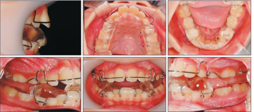
A modified form of Dr. Clark’s Twin-block appliance comprising two separate, upper and lower, removable appliances was used in this study (Figure 1). The bite registration was taken with the incisors in an edge-to-
Figure 1. The Class II Twin-
block appliance used in this
study.
edge position with at least a 5-mm vertical posterior opening. All patients were instructed to wear the appliance 24 hours per day (except when eating). The mean (± SD) total treatment time of these patients was 10 ± 3.9 months.
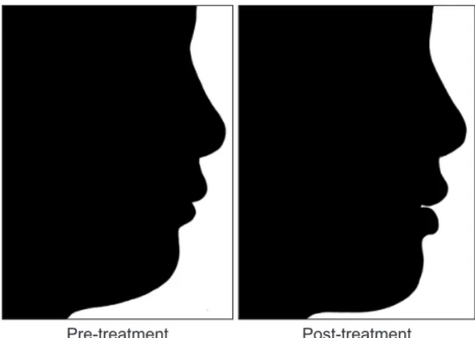
Profile silhouettes were produced from the soft-tissue profile tracings of the pre- and post-treatment lateral cephalograms by the same operator (Figure 2).
13The tracings were arranged with the Frankfort line parallel to the horizontal aspect of the computer monitor and were saved as JPEG images. Using a photo editor program, the profile tracings were filled in black, against a white background, and were standardized for size and range, from the soft-tissue glabella to slightly below the throat point.
For each patient, the silhouette pairs (pre- and post- treatment) were randomly assigned a number (1–45) and were inserted on the same sheet: pre-treatment image on the left side and post-treatment image on the right (Figure 2). The extent of silhouette improvement was evaluated by a panel comprised of three orthodontists, each with an orthodontic experience of more than 10 years. The panel was provided with an explanation of the study and allowed 10 seconds to view each silhouette pair. The panel then recorded the extent of the silhouette improvement following treatment by using an unmarked 100-mm visual analogue scale (VAS) from 0 (no improvement of facial profile) to 100 (facial profile improved close to Class I).
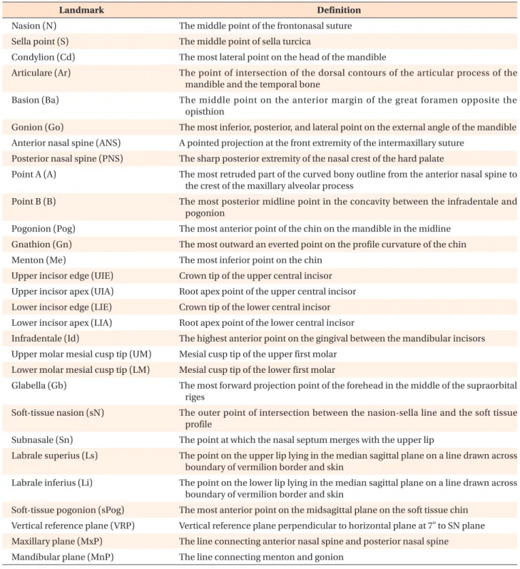
Patients were then divided into two groups based on their VAS scores: a favorable group comprising the upper 30% of VAS scores, and an unfavorable group comprising the lower 30%. Pre- and post-treatment lateral cephalograms of each group were traced and analyzed by one operator. Landmarks used in the study
are presented in Table 1 and Figure 3A, while the linear and angular measurements are shown in Table 2 and Figure 3B–3D.
14Statistical analysis
Cephalometric tracings and analysis were performed by one operator using the software program V ceph
TM6.0 (Osstem Inc., Seoul, Korea). Twenty randomly selected lateral cephalograms were traced 2 weeks after the first measurements were taken. The error of the first and second measurements was compared using Dahlberg’s formula. All statistical analyses were performed using PASW Statistics for Windows, version 18.0 (IBM Co., Armonk, NY, USA). A normal distribution of the variables was confirmed using the Kolmogorov- Smirnov test. Statistical comparison of the pre-treatment measurements between the favorable and unfavorable groups was performed using an independent t-test. A logistic regression test was performed to determine the most highly correlated factor among the pretreatment variables and an intra-class correlation coefficient was used to determine intra-panel reliability; a high correlation coefficient (0.895) was recorded between the orthodontists within the panel.
RESULTS
Analysis using Dahlberg’s formula revealed that the linear measurement error varied between 0.20 mm (lower anterior facial height) and 1.23 mm (condylion to gonion length), and the angular measurement error varied between 0.54
o(sella-nasion line [SN] to pogonion angle) and 1.21
o(upper incisor to maxillary plane angle [MxP]).
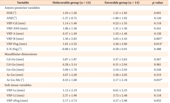
The favorable group was comprised with 14 subjects (mean age, 10.5 ± 1.4 years; 12 boys and 2 girls) and the unfavorable group was comprised with 14 subjects (mean age, 10.4 ± 1.3 years; 12 boys and 2 girls). Table 3 presents the comparative data for pre-treatment cephalometric measurements in both groups. An independent t-test indicated that there were statistically significant pre-treatment cephalometric differences in symphysis inclination, lower incisor to mandibular plane angle (IMPA), lower incisor to occlusal plane angle (L1 to Occ), lower incisor to pogonion distance (L1 to pogonion), ANB, SN to MxP, SN to mandibular plane angle (MnP), and gonial angle between the two groups.
With the exception of L1 to Occ, the favorable group had the lowest cephalometric measurements. There were no statistically significant differences in pre-treatment soft-tissue measurements between the two groups.
Table 4 presents the cephalometric treatment changes;
statistically significant differences were observed in the gonial angle, vertical reference plane (VRP) to B point, and VRP to pogonion between the two groups. Increase Figure 2. An example of a pair of pre-treatment (left)
and post-treatment (right) silhouettes used for evaluation by the panels.
Pre-treatment Post-treatment
in gonial angle, VRP to B point, and VRP to pogonion following treatment were significantly larger in the favorable group compared to the unfavorable group.
Logistic regression analysis was used to determine the predictors of favorable soft-tissue profile outcomes. With one independent variable, L1 to pogonion was the most effective predictor with a coefficient of determination
of 0.473, and with two independent variables, the IMPA and gonial angle were the most strongly related variables, with a coefficient of determination of 0.719.
In combination with L1 to pogonion, the coefficient of determination was 0.751, and this increased to 0.818 when combined with the vertical factor SN to MxP.
Table 1. Description of cephalometric landmarks and planes used in the study
Landmark Definition
Nasion (N) The middle point of the frontonasal suture
Sella point (S) The middle point of sella turcica
Condylion (Cd) The most lateral point on the head of the mandible
Articulare (Ar) The point of intersection of the dorsal contours of the articular process of the mandible and the temporal bone
Basion (Ba) The middle point on the anterior margin of the great foramen opposite the opisthion
Gonion (Go) The most inferior, posterior, and lateral point on the external angle of the mandible Anterior nasal spine (ANS) A pointed projection at the front extremity of the intermaxillary suture
Posterior nasal spine (PNS) The sharp posterior extremity of the nasal crest of the hard palate
Point A (A) The most retruded part of the curved bony outline from the anterior nasal spine to the crest of the maxillary alveolar process
Point B (B) The most posterior midline point in the concavity between the infradentale and pogonion
Pogonion (Pog) The most anterior point of the chin on the mandible in the midline Gnathion (Gn) The most outward an everted point on the profile curvature of the chin
Menton (Me) The most inferior point on the chin
Upper incisor edge (UIE) Crown tip of the upper central incisor Upper incisor apex (UIA) Root apex point of the upper central incisor Lower incisor edge (LIE) Crown tip of the lower central incisor Lower incisor apex (LIA) Root apex point of the lower central incisor
Infradentale (Id) The highest anterior point on the gingival between the mandibular incisors Upper molar mesial cusp tip (UM) Mesial cusp tip of the upper first molar
Lower molar mesial cusp tip (LM) Mesial cusp tip of the lower first molar
Glabella (Gb) The most forward projection point of the forehead in the middle of the supraorbital riges
Soft-tissue nasion (sN) The outer point of intersection between the nasion-sella line and the soft tissue profile
Subnasale (Sn) The point at which the nasal septum merges with the upper lip
Labrale superius (Ls) The point on the upper lip lying in the median sagittal plane on a line drawn across boundary of vermilion border and skin
Labrale inferius (Li) The point on the lower lip lying in the median sagittal plane on a line drawn across boundary of vermilion border and skin
Soft-tissue pogonion (sPog) The most anterior point on the midsagittal plane on the soft tissue chin Vertical reference plane (VRP) Vertical reference plane perpendicular to horizontal plane at 7
oto SN plane Maxillary plane (MxP) The line connecting anterior nasal spine and posterior nasal spine
Mandibular plane (MnP) The line connecting menton and gonion
DISCUSSION
After Class II Twin-block treatment, the extent of soft- tissue profile improvement among patients was variable, despite the fact that treatment resulted in Class I molar relationship and normal overjet and overbite in all the patients. Some patients exhibited an excellent profile
improvement, obtaining almost a Class I orthognathic profile, while some exhibited extremely small profile changes, retaining a retrognathic appearance. Other patients also showed profile improvement however, it was not enough to classify as a straight profile. The objective of this study was to determine factors related to favorable soft-tissue profile outcomes following Class Figure 3. Cephalometric analysis. A, Cephalometric landmarks (and the vertical reference plane) recorded: Nasion (N), sella point (S), condylion (Cd), articulare (Ar), basion (Ba), gonion (Go), anterior nasal spine (ANS), posterior nasal spine (PNS), point A (A), point B (B), pogonion (Pog), gnathion (Gn), menton (Me), upper incisor edge (UIE), upper incisor apex (UIA), lower incisor edge (LIE), lower incisor apex (LIA), infradentale (Id), upper molar mesial cusp tip (UM), lower molar mesial cusp tip (LM), glabella (Gb), soft-tissue nasion (sN), subnasale (Sn), labrale superius (Ls), labrale inferius (Li), soft- tissue pogonion (sPog), and the vertical reference plane perpendicular to the horizontal plane at 7
oto the SN plane (VRP).
B, Cranial base, facial height, and vertical measurements: (1) S-Ar, (2) S-N, (3) Ba-N, (4) N-S-Ar, (5) UAFH, (6) LAFH, (7) UPFH, (8) LPFH, (9), S-Ar-Go, and (10) maxillary plane angle (MxP)-mandibular plane angle (MnP). C, Anteroposterior mandibular dimension measurements: (1) SNA, (2) SNB, (3) VRP-Cd, (4) VRP-ANS, (5) VRP-A, (6) VRP-B, (7) VRP-Pog, (8) B-Pog, (9) S-N-Pog, (10), Cd-Go, (11) Cd-Gn, (12) Go-Gn, (13) Ar-Gn, (14) Ar-Go-Me, and (15) symphysis inclination. D, Dento-alveolar and soft-tissue measurements: (1) U1-MxP, (2) L1-MnP, (3) interincisal angle, (4) UM-MxP, (5) LM-MnP, (6) VRP-UM, (7) VRP-LM, (8) VRP-Gb, (9) VRP-sN, (10) VRP-Sn, (11) VRP-Ls, (12) VRP-Li, and (13) VRP-sPog.
Terms and definitions are listed in Tables 1 and 2.
SN plane Gb
sN N
S
Cd
Ar Ba
PNS
A B
C D
97
ANS
UIA A Sn
Ls LIE
UIE LM
UM Id B LIA
Li
PogsPog MeGn Go
VRP
1 2
3
4 5
6 7
8 9
10
1
2
3
4 5
6
7 8
9
10
VRP 11
12 13 14
15
7 9 8
4
5 2
3 1 10
11 6 7 12
13
VRP VRP