Fluid Retention Associated with Imatinib Treatment in Patients with Gastrointestinal Stromal Tumor: Quantitative Radiologic Assessment and Implications for Management
10
0
0
전체 글
(2) Imatinib Associated Fluid Retention. in periorbital regions, lower extremities and/or body were noted in 74–84% in phase II (3, 4) and phase III trials (5, 6), which was usually mild and manageable. Severe FR resulting in generalized edema (anasarca), ascites, pleural or pericardial effusions was less frequently reported in 2.8–9% of GIST patients (4, 6), which often necessitates dose-reduction or drug-cessation. As the indications and use of imatinib have expanded greatly, the prevalence of adverse effects has also been reported increasingly over the last decade. Nonetheless, the majority of radiology literature regarding GIST patients treated with imatinib has focused on treatment response and recurrence patterns, and the literature about the imaging characteristics of imatinib-associated FR is very limited (7, 8). This becomes important as imaging findings of FR are sometimes misinterpreted as findings of peritoneal metastatic disease. Therefore, we aimed to systematically describe the imaging characteristics of FR in GIST patients treated with imatinib over time and its implications for management.. MATERIALS AND METHODS Patients This study was approved by the Institutional Review Board and the requirement for informed consent was waived. Search from our Research Patient Data Registry, which pools clinical data from hospital billing and electronic medical record systems, revealed 403 patients with GIST who were treated with imatinib from January 2006 through December 2012. The radiology reports of body CT of these patients during the course of imatinib treatment were then queried for terms indicating presence of FR, such as “edema”, “anasarca”, “stranding”, “swelling”, “fluid”, “retention”, “ascites”, or “effusion”, followed by manual confirmation by review of the imaging study by a fellowship-trained oncoradiologist. This search yielded 36 patients who had one or more of the following: subcutaneous edema, anasarca, ascites, pleural or pericardial effusions. During the image review of serial computed tomography (CT) scans of these 36 patients, 21 patients who showed signs of FR at baseline CT before starting imatinib treatment from other potential causes such as post-operative FR (n = 16), renal dysfunction (n = 1), heart failure due to atrial fibrillation (n = 1), and widespread peritoneal metastatic disease with ascites and/or pleural effusion (n = 3) were excluded. Finally, 15 patients (mean age 66.7 ± 18.1 years, 6 men kjronline.org. Korean J Radiol 16(2), Mar/Apr 2015. and 9 women) with GIST, in whom radiologic signs of FR developed during imatinib treatment, were included in this study. Imatinib was orally administered daily. Generally, the standard initial dose was 400 mg/day and high dose of 800 mg/day imatinib was applied to patients with exon 9 mutant GISTs. Dose adjustment was performed during the treatment course according to the clinical need. All included patients underwent contrast-enhanced CT of the chest, abdomen, and pelvis at treatment initiation (baseline CT). The patients were followed up with contrast-enhanced CT every 2–6 months (follow-up CT). CT Acquisition CT scans of the abdomen/pelvis and/or chest were performed by using a 64-row multidetector CT (MDCT) scanner (Aquilion 64; Toshiba America Medical Systems, Tustin, CA, USA). The CT protocols are as follows: 64row MDCT scanner at 0.5 mm collimation, 120 kVp, tube current maximum of 500 mA using dose modulation with noise index of 12.5 Hounsfield units, 0.5 seconds gantry rotation time, and a table speed of 26.5 mm per rotation. One hundred milliliters of iopromide (Ultravist 300; Bayer HealthCare, San Francisco, CA, USA) were injected intravenously at a rate of 2–3 mL/s, with a scan delay of 60 seconds. Oral contrast (Gastrografin, Bracco Diagnostics, Princeton, NJ, USA) was administrated prior to the CT scans. Axial images with 5 mm thickness and coronal images with 4 mm thickness were reconstructed using standard abdominal algorithms. Imaging Analysis and Quantification All CT scans of the included 15 patients performed at baseline and during the course of imatinib treatment were evaluated in consensus by two radiologists (with 13 and 8 years of experience) for the presence of radiologic signs of FR. Imaging findings were analyzed for the presence of four components, including subcutaneous edema, ascites, pleural effusion, and pericardial effusion at baseline and at all available follow-up CT scans. Each of these radiologic signs of FR was graded using a four-point ordinal score scale (none, score 0; mild, score 1; moderate, score 2; severe, score 3), as follows: 1) “Subcutaneous edema” was graded according to the extent of subcutaneous fat stranding and/or the presence of a measurable subcutaneous fluid collection, as mild (less than half of abdominal wall, without fluid collection), 305.
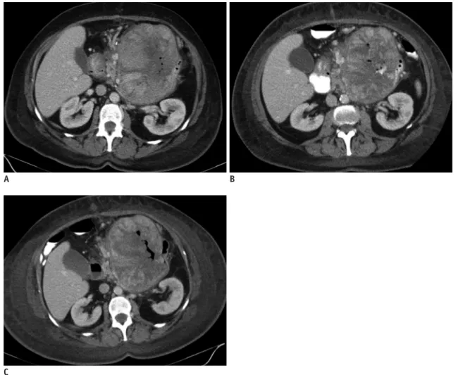
(3) Kim et al.. A. B. C Fig. 1. Grade of subcutaneous edema. In 71-year-old female with gastrointestinal stromal tumor (case 6), subcutaneous edema occurred as mild form involving less than half of abdominal wall at 1st follow-up CT (A) after initiation of imatinib 400 mg, and aggravated to moderate form involving more than half of abdominal wall at 2nd follow-up CT (B) obtained shortly after dose escalation from 400 to 800 mg, followed by severe circumferential subcutaneous edema at 3rd follow-up CT (C).. moderate (more than half of abdominal wall, without fluid collection), or severe (circumferential involvement of fullthickness of abdominal wall, with areas of fluid collection/ dispersion) (Fig. 1). Localized subcutaneous fat stranding along the paraspinal muscles posteriorly was not regarded as FR, because it may be secondary to posturing or dependent changes. 2) “Ascites” was graded as mild (few small collections of fluid confined to the pelvic cavity or dependent areas, with largest dimension ≤ 3 cm), moderate (multiple collections or diffuse dispersion of free fluid, with smallest dimension > 3 cm), and severe (large volume generalized fluid in whole abdominopelvic cavity, with floating bowel) (9). 3) “Pleural effusion” was graded according to the ratio of anteroposterior height of fluid collection to anteroposterior diameter of pleural cavity on axial CT, as mild (≤ 25%), moderate (> 25%, ≤ 50%), and severe (> 50%). In cases of 306. bilateral pleural effusions, the side of the larger effusion was graded (9). 4) “Pericardial effusion” was graded according to the maximal thickness of pericardial fluid on axial or coronal CT, as mild (> 0.5 cm, ≤ 1 cm), moderate (> 1 cm, ≤ 1.5 cm), and severe (> 1.5 cm) (10). Then, the total score of radiologic FR for each patient was calculated by summing up the scores of each of the four radiologic sign of FR. In this study, “maximum FR” refers to a condition when a patient’s total score of radiologic FR is highest during imatinib treatment. Medical Records Review Clinical data regarding patient demographics, the setting of imatinib treatment (neoadjuvant, adjuvant, or metastatic), imatinib dosage, relevant concomitant medications, and clinical signs/symptoms of FR were Korean J Radiol 16(2), Mar/Apr 2015. kjronline.org.
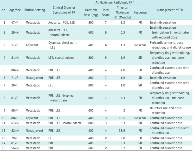
(4) Imatinib Associated Fluid Retention. recorded during review of the medical records for all 15 patients. Laboratory data at baseline and during followup were recorded. The presence of an underlying cardiac comorbidity, such as congestive heart failure, was also reviewed. Note was also made of any medical events at the time of maximum FR, which may have exacerbated the FR (i.e., hypoalbuminemia, renal dysfunction, cardiac disease, and infection). The management of FR were noted, including permanent drug-cessation, temporary withholding of the drug, dose-reduction, and diuretic use. Statistical Analysis Ordinal scoring system to measure the severity of the four radiologic signs of FR was used for quantitative analysis. First, we plotted the scores of the radiologic signs of FR over time during imatinib treatment. The patterns of. radiologic FR were evaluated initially by visual inspection in consensus of two reviewers. Subsequently, two distinct types of FR were categorized by the following quantitative criteria, as described in the results section. Second, the difference in patient demographics, characteristics of radiologic FR and clinical management of FR between two groups of patients with distinct types of FR was evaluated. The unpaired Student t test was used to compare patient’s age, Mann-Whitney U test was used to compare median scores of radiologic FR, and Fisher’s exact test or chisquare test was used to compare the categorical data. The statistical analyses were performed using GraphPad Prism Version 6 (GraphPad Prism Software Inc., La Jolla, CA, USA). All two-sided p values less than 0.05 were considered to indicate a statistically significant difference.. Table 1. Clinical Characteristics and Radiologic Findings of All Patients No. Age/Sex Clinical Setting. Clinical Signs or Symptoms of FR. 1. 47/F. Metastatic. Anasarca, POE, LEE. 2. 29/M. Metastatic. Anasarca, LEE, scrotal edema. 3. 51/F. Adjuvant. Dyspnea, chest pain, LEE. At Maximum Radiologic FR* Time to Management of FR Imatinib Total Maximum Response Dose (mg) Score FR† (Months) 800 7 1.3 PR Imatinib cessation Imatinib cessation 600 5 0.3 PR (reinitiation 6 month later with reduced dose) 400. 6. 1.3. No recur. Pericardiocentesis, dosereduction, and diuretics use Temporary drug withholding, diuretics use, and dosereduction. 4. 81/M. Metastatic. LEE, scrotal edema. 800. 5. 1.9. SD. 5. 88/M. Metastatic. POE, LEE. 400. 4. 4.0. PR. 6. 71/F. Neoadjuvant. POE, LEE. 800. 7. 1.9. SD. 7. 39/F. Metastatic. LEE. 800. 4. 1.0. SD. 8. 81/F. Metastatic. POE, LEE, dyspnea, weight gain. 800. 7. 3.3. PR. 9. 68/F. Metastatic. POE, LEE. 600. 4. 2. PR. 10 11. 80/F 67/M. Adjuvant Metastatic. POE, LEE POE, LEE, scrotal edema. 400 800. 3 3. 10.5 0.3. No recur SD. 12. 82/M. Neoadjuvant. POE, LEE. 400. 4. 23.6. PR. 13 14 15. 79/F 81/F 56/M. Metastatic Metastatic Metastatic. LEE POE POE. 400 400 600. 2 1 2. 3.0 2.3 2.7. PR SD PR. Continued current dose with diuretics use Imatinib cessation Continued current dose with diuretics use Temporary drug withholding, diuretics use, and dosereduction Diuretics use and dosereduction Continued current dose Continued current dose Continued current dose with diuretics use Continued current dose Continued current dose Continued current dose. Note.— *Maximum radiologic FR refers to condition at highest total score of FR, †Time from drug initiation or latest drug escalation. FR = fluid retention, LEE = lower extremity edema, POE = periorbital edema, PR = partial response, SD = stable disease. kjronline.org. Korean J Radiol 16(2), Mar/Apr 2015. 307.
(5) Kim et al.. Fig. 2. Time-course of radiologic signs of fluid retention (FR) of all patients. Vertical axis represents total score and each score of radiologic signs of FR and transverse axis represents time from imatinib initiation. Nine patients (cases 1–9) show acute/progressive FR, while six patients (cases 10–15) show only intermittent/steady FR, with mild persistent FR or occasional episode of mild FR. AS = ascites, PC = pericardial effusion, PL = pleural effusion, SE = subcutaneous edema. 308. Korean J Radiol 16(2), Mar/Apr 2015. kjronline.org.
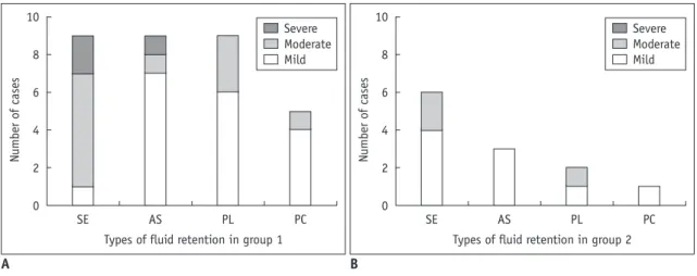
(6) Imatinib Associated Fluid Retention. Table 2. Differences of Characteristics between Two Groups with Distinct Pattern of FR Demographic characteristics Characteristics of FR. Management. Mean age ± SD (range) Sex (male:female) Comorbidity Types of FR Recent dose initiation/change Median maximum total score No management Continued dose + diuretics Dose-reduction + diuretics Temporary drug withholding + dose-reduction Drug-cessation. Group 1 (n = 9) Group 2 (n = 6) 61.7 ± 20.1 (range 29–88) 74.2 ± 10.5 (range 56–82) 3:6 3:3 2 (22%) 2 (33%) Acute/progressive FR with Intermittent/steady FR with improvement after management mild occasional or persistent FR 9 (100%) 2 (33%) 5 (range 4–7) 2.5 (range 1–4) 0 (0%) 4 (67%) 2 (22%) 2 (33%) 2 (22%) 0 (0%) 2 (22%). 0 (0%). 3 (33%). 0 (0%). P 0.201* 0.622† 1.0†. 0.044† 0.002‡. 0.029§. Note.— *Student t test, †Fisher’s exact test, ‡Mann-Whitney test, §Chi-square test. FR = fluid retention, SD = standard deviation. RESULTS. 8. kjronline.org. Korean J Radiol 16(2), Mar/Apr 2015. 6 Score. Radiologic Evaluation and Quantification of FR The clinical and radiologic features of all patients are summarized in Table 1. On the plot of scores of radiologic signs of FR over time during imatinib treatment (Fig. 2), the FR was categorized into two distinct types, acute/ progressive FR and intermittent/steady FR, based on the following quantitative criteria: 1) the slope of the plot between recent drug initiation/dose change to the highest point ≥ 1 score/month, and 2) the highest total score of radiologic FR ≥ 4. Acute/progressive FR was defined with the two criteria were met simultaneously. The first type, acute/progressive FR, is characterized by a relatively acute aggravation of FR and rapid improvement after management, observed in 9 patients (cases 1–9, referred to group 1). The second type, intermittent/steady FR, is characterized by occasional or persistent mild FR, observed in 6 patients (cases 10–15, referred to group 2). Patient demographics, characteristics of radiologic FR, and management of FR in groups 1 and 2 are summarized in Table 2. In group 1, the acute/progressive FR always occurred early after drug initiation/dose escalation (9/9, 100%), with median time to maximum FR from drug initiation/dose escalation of 1.9 months (range 0.3–4.0 months) (Fig. 3). In group 2, an episode of mild FR (total score ≥ 2) occurred at any time during imatinib treatment, including during stable imatinib dosing (n = 4, 67%) and early after imatinib initiation or dose escalation (n = 2, 33%). Acute/progressive FR had higher total score compared to intermittent/steady FR (median score, 5 vs. 2.5, p = 0.002).. Case 1 Case 2 Case 3 Case 4 Case 5 Case 6 Case 7 Case 8 Case 9. 4. 2. 0. 0. 3. 6. 9. 12. Month. Fig. 3. Time-course of acute/progressive fluid retention (FR) in patients of group 1. Vertical axis represents total score of radiologic signs of FR, and transverse axis represents time from imatinib initiation or recent dose escalation. Acute/progressive FR occurs in early period after imatinib initiation or recent dose escalation (9/9, 100%), with median time to maximum FR 1.9 months and median score of maximum FR 5. Thick gray line is representative line of timecourse of exacerbation periods with time to peak 1.9 months and peak height 5.. The proportions of signs of radiologic FR are illustrated in Figure 4. In all patients, the most common imaging manifestation of FR was subcutaneous edema (15/15, 100%), followed by ascites (12/15, 80%), pleural effusion (11/15, 73%), and pericardial effusion (6/15, 40%) at the time of maximum FR. Subcutaneous edema occurred in almost all patients, while ascites, pleural effusion, and pericaridal effusion occur mostly in patients with acute/ progressive FR. These results indicate that subcutaneous edema is the most common and most sensitive radiologic sign of FR. Peritoneal metastases were found in 7 patients (cases 1, 2, 4, 7–9, 11). In these 7 patients, ascites was noted at 309.
(7) Kim et al.. 10. 6 4 2. SE. AS. PL. PC. Types of fluid retention in group 1. Severe Moderate Mild. 8 Number of cases. Number of cases. 8. 0. 10. Severe Moderate Mild. 6 4 2 0. SE. AS. PL. PC. Types of fluid retention in group 2. A B Fig. 4. Proportion of type of radiologic fluid retention of group 1 (A) and group 2 (B). In all types of fluid retention, proportion and severity were higher in group 1 compared to group 2. AS = ascites, PC = pericardial effusion, PL = pleural effusion, SE = subcutaneous edema. A B Fig. 5. 67-year-old male with gastrointestinal stromal tumor presented with new ascites (case 11).. A. Axial contrast-enhanced CT (CECT) shows new small ascites in pelvic cavity (arrow) adjacent to peritoneal metastasis in pelvic cavity (arrowheads), 10 days after starting imatinib treatment. Mild subcutaneous edema is also newly apparent. B. Axial image of CECT shows scrotal edema (curved arrows). Based on scrotal edema, subcutaneous edema, and stability of peritoneal metastasis, new small ascites occurred early after starting imatinib may be manifestation of fluid retention rather than malignant ascites.. the time of maximum FR when the metastatic disease was otherwise stable or improving. In addition, subcutaneous edema and/or pleural or pericardial effusion occurred, as ascites did (Fig. 5), and the ascites resolved or improved after management of FR in all of these 7 patients. These observations may suggest that the ascites was part of imatinib-associated FR rather related to peritoneal metastases. Clinical Features The common clinical signs/symptoms in our series were periorbital edema and lower extremity edema. Less frequently, scrotal edema was present (3/15, 33%). One patient (case 3) had a moderate pericardial effusion which required pericardiocentesis and imatinib dose-reduction. However, echocardiography performed at baseline and at the time of maximum FR showed normal cardiac function, which 310. may mean that FR was not related to the cardiotoxicity in all patients. In our series, the imatinib dose at the time of maximum FR was 400 mg (n = 6, 40%), 600 mg (n = 3, 20%), and 800 mg (n = 6, 40%), and showed moderate correlation with total score of radiologic FR (Pearson correlation coefficient r = 0.526) (Fig. 6A). According to severity of FR and patient’s tolerance, five levels of management were performed as follows: continuing current dose with close monitoring (level 1), continuing the current imatinib dose with diuretic use (level 2), dose-reduction and/or diuretic use (level 3), temporary drug withholding with reinitiating at a lower dose (level 4), and permanent imatinib cessation (level 5). Strong positive correlation (r = 0.879) between the level of management and total score of radiologic FR was observed on scatterplot (Fig. 6B). Aggressive management, drug-cessation/dose-reduction Korean J Radiol 16(2), Mar/Apr 2015. kjronline.org.
(8) Imatinib Associated Fluid Retention. 5. 6. 4. 5. Management level. Total score of radiologic fluid retention. 7. 4 3. 3. 2. 1. 2 r = 0.526 1. r = 0.879 0. 400. 600 Imatinib dose (mg) 1. Management level 2 3 4. 800. 1. 2. 3. 4. 5. 6. 7. 8. Total score of radiologic fluid retention 5. 400. Dose (mg) 600 800. A B Fig. 6. Relationship between total score of radiologic fluid retention (FR) and imatinib dose (A) or management level of FR (B). A. Scatterplot shows moderate positive correlation (r = 0.526) between total score of radiologic FR and imatinib dose. B. Strong positive correlation (r = 0.879) between level of management and total score of radiologic FR was observed on scatterplot.. (level 3–5), was generally required in cases with a score of 5 or higher; while conservative management and continuing current dose and/or diuretics (level 1, 2) were generally required in cases with a score of 4 or lower. Moderate to severe FR requiring aggressive management (level 3–5) occurred more frequently in cases with high imatinib dose 600–800 mg (6/15, 40%) compared with doses of 400 mg (1/15, 7%). Differences in the management of FR between groups 1 and 2 were also observed (Table 2). Aggressive management (level 3–5) was performed in the majority of patients in group 1 (7/9, 78%), while all patients in group 2 were treated with conservative management (level 1, 2).. DISCUSSION Clinical manifestations of imatinib-associated FR have been described generally as mild, but occasionally severe (grade 3–4, according to the Common Terminology Criteria for Adverse Events [CTCAE] of National Cancer Institute) (11) with anasarca, ascites, pleural effusion, and pericardial effusion; and symptomatic FR can occur early in imatinib therapy and diminish over time with management (1, 12). These clinical features of FR are concordant with time-course and radiologic signs of FR in our series, which illustrates the serial changes of FR detailed with quantitative scoring kjronline.org. Korean J Radiol 16(2), Mar/Apr 2015. system. In our study, two distinct types or courses of FR were observed as follows: 1) acute/progressive FR occurred early after drug initiation or dose escalation which improved after clinical management, and 2) intermittent/ steady FR with mild, occasional or persistent FR. Currently, clinical trials and oncologists in routine practice use the CTCAE to grade the severity of adverse events. CTCAE criteria are generally designed to identify acute toxicity of a particular agent, and the evaluation is often based on the severity and level of intervention needed (11). Due to the subjective and qualitative nature of CTCAE criteria, hospitals and individual practitioners may use different criteria based on their own previous experiences and their patients’ expectations (12). In addition to the clinical assessment of FR, radiologic evaluation using a scoring system may be a good complementary method to assess the patient’s condition in an objective and quantitative way. Indeed, strong correlation between the total score of radiologic FR and the management level of FR (r = 0.879) in our study, may indicate that quantitative radiologic evaluation of FR may be helpful to determine the appropriate level of management. The detailed quantitative illustration of the time-course and types of radiologic FR provides several noteworthy points of interest. First of all, the acute/progressive 311.
(9) Kim et al.. FR early after drug initiation or dose escalation, the reversibility of the retention after drug-cessation/dosereduction, and the moderate dose-severity correlation suggest that FR might occur when the pharmacologic effect exceeds the compensation capability of human body, reflecting a degree of intolerance (13, 14). Even during the period of intermittent/steady FR, the drug effect to cause FR is continually balanced by compensation mechanism of human body. These pharmacokinetic/pharmacodynamic characteristics of imatinib are influenced by many factors such as age, sex, body weight, pharmacogenetic characteristics, imatinib dose, interaction with concomitant medications, and comorbidities such as heart disease, renal dysfunction, anemia or hypoalbuminemia (1, 15, 16). Indeed, FR was aggravated in patients on a stable imatinib dose, probably related with predisposing acute medical events in two patients (case 11, 12). In our study, echocardiography performed at baseline and at the time of maximum FR showed normal cardiac contractile function supports the notion that FR in our series is not related to the cardiotoxicity of imatinib. Although a report that imatinib was associated with the development of severe heart failure alarmed the oncology community (17), cardiotoxicity associated with imatinib is nowadays thought to be rare. The estimated incidence of cardiotoxicity evidenced by new onset heart failure or left ventricular dysfunction is very uncommon, 0.2% per year, and similar to the incidence in the general population (2, 18, 19). The fact that subcutaneous edema was the most common sign of FR in all patients while ascites, pleural effusion, and pericardial effusion occurred significantly in acute/ progressive FR only, may suggest the imatinib-specific pharmacological mechanism of action works on the subcutaneous fluid distribution. The mechanism of the FR, however, remains unclear. Even though the most popular theory is that imatinib inhibits the platelet-derived growth factor receptor (PDGFR) which regulates interstitial fluid homeostasis in dermal layer (20, 21), nilotinib, which also inhibits PDGFR in similar way to imatinib, does not cause FR. Dasatinib, which is also an ABL inhibitor like imatinib, specifically causes pleural and pericardial effusion without subcutaneous edema, through its preferential action on the Src kinases within the pleural and pericardial mesothelial cells (22). The mechanism of imatinib-associated FR which predominantly occurs in the form of subcutaneous edema is unknown but may be due to drug-specific mechanism of 312. action. Knowledge about the time-course and types of radiologic FR would be very helpful to interpret CT images of patients treated with imatinib. Any new ascites or pleural effusion should not be mistakenly interpreted as a sign of peritoneal metastases or worsening tumor, especially when tumor elsewhere is stable or improving. The presence of concomitant subcutaneous edema and its interval change should be evaluated, and history of recent dose change or recent acute medical condition is also quite helpful. Furthermore, careful attention of the interval increase in subcutaneous edema and other signs of FR and prompt notification of the treating oncologist will enormously enhance the quality of patient care. For best patient care in the era of molecular targeted therapeutic agents, a multidisciplinary approach with close collaboration between oncologists and radiologists is essential to assess treatment response as well as toxicity of MTTs. Radiologists should be familiar with the clinical and radiological characteristics as well as the overall approach of diagnosis and management of imatinib-associated FR. This study has several limitations. The number of patients was small, partly due to the rarity of symptomatic FR and partly due to the retrospective nature of the study based on search terms. In clinical practice, CT scan at the time of FR may be performed selectively only for clinically severe FR, which may raise the issue of selection bias and underestimation of incidence of the mild FR on imaging. True incidence of the radiologic signs of FR has not been previously examined and could not be evaluated in our study due to its retrospective nature. In summary, regarding the types and time-course of FR, two distinct types were observed, including an acute/ progressive FR usually requiring aggressive management and a intermittent/steady FR with mild fluctuation generally requiring conservative management. Acute/progressive FR generally occurs following treatment initiation or dose-escalation. Regarding the radiologic signs of FR, subcutaneous edema is the most common and earliest finding, while ascites, pleural effusion, and pericaridal effusion occur mostly in acute/progressive FR.. REFERENCES 1. Siddiqui MA, Scott LJ. Imatinib: a review of its use in the management of gastrointestinal stromal tumours. Drugs 2007;67:805-820. Korean J Radiol 16(2), Mar/Apr 2015. kjronline.org.
(10) Imatinib Associated Fluid Retention. 2. Thanopoulou E, Judson I. The safety profile of imatinib in CML and GIST: long-term considerations. Arch Toxicol 2012;86:1-12 3. Demetri GD, von Mehren M, Blanke CD, Van den Abbeele AD, Eisenberg B, Roberts PJ, et al. Efficacy and safety of imatinib mesylate in advanced gastrointestinal stromal tumors. N Engl J Med 2002;347:472-480 4. Verweij J, van Oosterom A, Blay JY, Judson I, Rodenhuis S, van der Graaf W, et al. Imatinib mesylate (STI-571 Glivec, Gleevec) is an active agent for gastrointestinal stromal tumours, but does not yield responses in other soft-tissue sarcomas that are unselected for a molecular target. Results from an EORTC Soft Tissue and Bone Sarcoma Group phase II study. Eur J Cancer 2003;39:2006-2011 5. Verweij J, Casali PG, Zalcberg J, LeCesne A, Reichardt P, Blay JY, et al. Progression-free survival in gastrointestinal stromal tumours with high-dose imatinib: randomised trial. Lancet 2004;364:1127-1134 6. Blanke CD, Rankin C, Demetri GD, Ryan CW, von Mehren M, Benjamin RS, et al. Phase III randomized, intergroup trial assessing imatinib mesylate at two dose levels in patients with unresectable or metastatic gastrointestinal stromal tumors expressing the kit receptor tyrosine kinase: S0033. J Clin Oncol 2008;26:626-632 7. Hong X, Choi H, Loyer EM, Benjamin RS, Trent JC, Charnsangavej C. Gastrointestinal stromal tumor: role of CT in diagnosis and in response evaluation and surveillance after treatment with imatinib. Radiographics 2006;26:481-495 8. Joensuu H, Trent JC, Reichardt P. Practical management of tyrosine kinase inhibitor-associated side effects in GIST. Cancer Treat Rev 2011;37:75-88 9. Kim KW, Choi HJ, Kang S, Park SY, Jung DC, Cho JY, et al. The utility of multi-detector computed tomography in the diagnosis of malignant pleural effusion in the patients with ovarian cancer. Eur J Radiol 2010;75:230-235 10. Figueras J, Juncal A, Carballo J, Cortadellas J, Soler JS. Nature and progression of pericardial effusion in patients with a first myocardial infarction: relationship to age and free wall rupture. Am Heart J 2002;144:251-258 11. Trotti A, Colevas AD, Setser A, Rusch V, Jaques D, Budach V, et al. CTCAE v3.0: development of a comprehensive grading system for the adverse effects of cancer treatment. Semin Radiat Oncol 2003;13:176-181 12. Pinilla-Ibarz J, Cortes J, Mauro MJ. Intolerance to tyrosine. kjronline.org. Korean J Radiol 16(2), Mar/Apr 2015. kinase inhibitors in chronic myeloid leukemia: definitions and clinical implications. Cancer 2011;117:688-697 13. Peng B, Hayes M, Resta D, Racine-Poon A, Druker BJ, Talpaz M, et al. Pharmacokinetics and pharmacodynamics of imatinib in a phase I trial with chronic myeloid leukemia patients. J Clin Oncol 2004;22:935-942 14. Ostro D, Lipton J. Unusual fluid retention with imatinib therapy for chronic myeloid leukemia. Leuk Lymphoma 2007;48:195-196 15. Van Glabbeke M, Verweij J, Casali PG, Simes J, Le Cesne A, Reichardt P, et al. Predicting toxicities for patients with advanced gastrointestinal stromal tumours treated with imatinib: a study of the European Organisation for Research and Treatment of Cancer, the Italian Sarcoma Group, and the Australasian Gastro-Intestinal Trials Group (EORTC-ISG-AGITG). Eur J Cancer 2006;42:2277-2285 16. Marrari A, Wagner AJ, Hornick JL. Predictors of response to targeted therapies for gastrointestinal stromal tumors. Arch Pathol Lab Med 2012;136:483-489 17. Kerkelä R, Grazette L, Yacobi R, Iliescu C, Patten R, Beahm C, et al. Cardiotoxicity of the cancer therapeutic agent imatinib mesylate. Nat Med 2006;12:908-916 18. Verweij J, Casali PG, Kotasek D, Le Cesne A, Reichard P, Judson IR, et al. Imatinib does not induce cardiac left ventricular failure in gastrointestinal stromal tumours patients: analysis of EORTC-ISG-AGITG study 62005. Eur J Cancer 2007;43:974-978 19. Trent JC, Patel SS, Zhang J, Araujo DM, Plana JC, Lenihan DJ, et al. Rare incidence of congestive heart failure in gastrointestinal stromal tumor and other sarcoma patients receiving imatinib mesylate. Cancer 2010;116:184-192 20. Heuchel R, Berg A, Tallquist M, Ahlén K, Reed RK, Rubin K, et al. Platelet-derived growth factor beta receptor regulates interstitial fluid homeostasis through phosphatidylinositol-3’ kinase signaling. Proc Natl Acad Sci U S A 1999;96:1141011415 21. Pietras K, Ostman A, Sjöquist M, Buchdunger E, Reed RK, Heldin CH, et al. Inhibition of platelet-derived growth factor receptors reduces interstitial hypertension and increases transcapillary transport in tumors. Cancer Res 2001;61:29292934 22. Masiello D, Gorospe G 3rd, Yang AS. The occurrence and management of fluid retention associated with TKI therapy in CML, with a focus on dasatinib. J Hematol Oncol 2009;2:46. 313.
(11)
수치

관련 문서
19) Yu Y, Kang J., Clinical studies on treatment of chronic prostatitis with acupuncture
This research was supported by Korea Atomic Energy Research Institute (NTIS-1711139325) and National Research Foundation of Korea (NRF) Grant funded by the Korean
For my study, this being diagnosed with prostate cancer receiving treatment in patients with complementary and alternative therapies for their experience
Glucose level before use of mannitol and peak osmolarity during mannitol treatment were associated with renal failure in univariate analysis.. In logistic regression
Note: N means patients number according to each 4 severity grades in 112 scrub typhus patients without antibiotics treatment
Changes in ABTS, DPPH radical scavenging and total phenolic contents of different soybean milk depending on ultrasonic treatment time with the Lactobacillus sp..
The associations of hepatic steatosis and fibrosis using fatty liver index and BARD score with cardiovascular outcomes and mortality in patients with new‑onset type 2
In the subgroup of patients who reached ASDAS-ID 1 year after TNFi treatment ( n = 327), ASDAS-ID was observed in 66.1% of the subsequent intervals, and only the
