Author contributions: B.L. and D.H.H. performed the conception and design. B.L. performed the carried out the experiments. B.L. and D.H.H. per- formed the acquisition of data. B.L. and D.H.H. performed the analysis and interpretation. B.L. performed the drafting the article. B.L. and D.H.H. per- formed the statistical analysis. I.S. and H.L. performed the study supervision.
This is an Open Access article distributed under the terms of the Creative Commons Attribution Non-Commercial License, which permits unrestricted non-commercial use, distribution, and reproduction in any medium, provided the original work is properly cited.
Copyright © Korean J Physiol Pharmacol, pISSN 1226-4512, eISSN 2093-3827
INTRODUCTION
Post-traumatic stress disorder (PTSD) is a stress-induced psy- chiatric disorder that is associated with marked deficiencies in social functioning [1]. PTSD can develop in response to a trau- matic, life-threatening event [2]. The characteristic signs of PTSD include hyperarousal, insensibility, fear, and nightmares, leading to high rates of comorbidity with psychosocial impairment and anxiety [3,4]. The onset of PTSD often precedes and increases the risk for subsequent development of anxiety disorders, and PTSD enhances the susceptibility to anxiety [5]. Although the neuro- biology of PTSD is not fully understood, preclinical and clinical
studies have implicated altered dopamine (DA) activity in the disorder [6].
It is widely accepted that neuronal system of catecholamine dysfunction in general and DA in particular play an important role in the pathophysiology of PTSD [1]. DAergic neurotrans- mission in the mesolimbic pathway is important for mediating arousal and memory, and altered DA activity may contribute to hyperarousal and re-experiencing symptoms associated with PTSD [2]. Central DA appears to positively affect fear condition- ing [7]. For example, fear extinction is postponed in rats with re- strained prefrontal or hippocampal DA function, indicating that DA is particularly crucial within the cortex [7].
Original Article
Berberine alleviates symptoms of anxiety by enhancing dopamine expression in rats with post-traumatic stress disorder
Bombi Lee 1,2, *, Insop Shim 3 , Hyejung Lee 1 , and Dae-Hyun Hahm 3, *
1
Acupuncture and Meridian Science Research Center, College of Korean Medicine,
2Center for Converging Humanities,
3Department of Physiology, College of Medicine, Kyung Hee University, Seoul 02447, Korea
ARTICLE INFO
Received October 13, 2017 Revised December 22, 2017 Accepted December 28, 2017
*Correspondence Bombi Lee
E-mail: [email protected] Dae-Hyun Hahm E-mail: [email protected] Key Words
Anxiety Berberine Dopamine
Post-traumatic stress disorder Single prolonged stress
ABSTRACT Post-traumatic stress disorder (PTSD) is a trauma-induced psychiatric
disorder characterized by impaired fear extermination, hyperarousal, anxiety, depres-
sion, and amnesic symptoms that may involve the release of monoamines in the fear
circuit. The present study measured several anxiety-related behavioral responses to
examine the effects of berberine (BER) on symptoms of anxiety in rats after single
prolonged stress (SPS) exposure, and to determine if BER reversed the dopamine (DA)
dysfunction. Rats received BER (10, 20, or 30 mg/kg, intraperitoneally, once daily) for
14 days after SPS exposure. BER administration significantly increased the time spent
in the open arms and reduced grooming behavior during the elevated plus maze
test, and increased the time spent in the central zone and the number of central
zone crossings in the open field test. BER restored neurochemical abnormalities and
the SPS-induced decrease in DA tissue levels in the hippocampus and striatum. The
increased DA concentration during BER treatment may partly be attributed to mRNA
expression of tyrosine hydroxylase and the DA transporter in the hippocampus,
while BER exerted no significant effects on vesicular monoamine transporter mRNA
expression in the hippocampus of rats with PTSD. These results suggest that BER had
anxiolytic-like effects on behavioral and biochemical measures associated with anxi-
ety. These findings support a role for reduced anxiety altered DAergic transmission
and reduced anxiety in rats with PTSD. Thus, BER may be a useful agent to treat or al-
leviate psychiatric disorders like those observed in patients with PTSD.
Several rodent models have been used to investigate the fear mechanism of PTSD. The single prolonged stress (SPS) model, which simulates a traumatic stressful event, has been extensively employed because it yields high validity when examining trauma- induced learning or anxiety [8,9]. Rats exposed to SPS show enhanced dysregulation of the hypothalamic-pituitary-adrenal (HPA) axis [10,11], increased anxiety-like behavior on the elevated plus maze (EPM) test, and increased fear conditioning [9]. These responses imitate the clinical symptoms observed in patients with PTSD [12]. Many studies have shown that dysregulation in brain DA circuitry can lead to the pathological state of PTSD and pro- duce anxiety-like symptoms [13].
Fluoxetine (FLX) is an antidepressant known to be effective for treating patients with PTSD [14]. Some studies have shown that FLX increases synaptic plasticity and fear extinction via the sero- tonergic signaling pathway, which alters DA and serotonin levels in the brain [15]. Although selective serotonin reuptake inhibi- tors, including FLX, are efficacious in many patients, they also have deleterious effects that limit their use, including psychiatric symptoms, including loss of weight, sedation, and sexual dysfunc- tion [16]. Therefore, there is an ongoing need for promising PTSD treatments [17]. Consequently, many studies have explored the use of natural medicines that may be safer for long-term therapy [18].
In the present study, we explored the pharmacological activ- ity of berberine (BER, C
20H
18CINO
4), an isoquinoline alkaloid derived from Korean traditional medicinal herbs, such as Ber- beris, Hydrastis canadensis, Rhizoma coptidis, and Cortex phellodendri [19]. BER is widely reported to improve multiple physiological actions and produces a variety of biological effects in the central nervous system (CNS) [20,21]. BER exerts neuro- protective, antioxidant, anti-apoptotic, antitumor, antiviral, and anti-inflammatory properties in various animal models of CNS- related disorders, such as Alzheimer’s disease, Parkinson’s disease (PD), forebrain ischemia, depression, and anxiety [19,22-24]. BER inhibited 6-hydroxydopamine-induced neurotoxicity medicated by the formation of reactive oxygen species in PC12 cells and in an animal model of PD [22]. The pharmacological profile of BER, which includes high-affinity binding to DA receptors, suggests that it may be useful for treating drug addiction, such as cocaine and morphine [25]. Specifically, BER inhibits DA biosynthesis and reduces DA content in PC12 cells [22]. Therefore, BER ap- pears to contribute to many of the therapeutic effects of these preparations, presumably through its interactions with the DA receptor [26]. BER also significantly reduces the total duration of immobility on the forced swimming test and tail-suspension test [19], and ameliorates anxiety-related behavior by activating the serotonergic system in mice [27]. Therefore, the present study hy- pothesized that BER would be protective against the development of anxiety-like behaviors, which represent the core symptoms of PTSD-related abnormalities, in a rat model of PTSD using SPS.
Identifying the neuropathology of the mesolimbic DA system
may help in understanding the functional relationship between DA and PTSD. DA dysfunction may also be relevant to the mech- anisms underlying emotional numbing, a symptom of PTSD.
Although a brief report was published on the antidepressant and anti-stress effects of BER [19,21], questions remain regarding the mechanisms underlying the effect of BER as an alternative therapeutic intervention for treating anxiety-like symptoms fol- lowing SPS exposure in rats. The present study investigated the medicinal effects of BER on anxiety-related behaviors in rats exposed to SPS using the open field test (OFT) and the EPM test, representing the core symptoms of PTSD-related abnormalities.
Moreover, we examined how the behavioral effects were as- sociated with the DAergic system in the brain as an underlying mechanism.
METHODS
Animals and BER administration
Six-week-old adult male Sprague-Dawley rats (Samtako Ani- mal Co., Seoul, Korea), weighing 210-240 g, were used in all experiments. The rats were housed under a 12-h light/dark cycle (lights on at 8:00 am, lights off at 8:00 pm) under a controlled temperature at 23±5°C and relative humidity of 50±10%. All rats were allowed to adapt to these conditions for 7 days after they arrived. All methods and procedures were approved by the Animal Care and Use Committee of Kyung Hee University [KHUASP(SE)-15-115]. All procedures were executed according to the Guide for the Care and Use of Laboratory Animals.
BER (10, 20, and 30 mg/kg, body weight; Sigma-Aldrich Chemical Co., St. Louis, MO, USA) and the positive drug FLX for positive control group (10 mg/kg, fluoxetine hydrochloride;
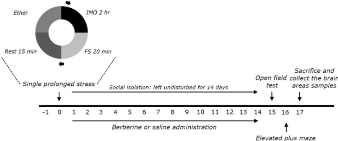
Sigma) were injected by intraperitoneally (i.p.) after exposure to SPS. Six or seven animals per group were used. BER and FLX were dissolved in 0.9% physiological saline before use. The entire experimental schedules of all drug administration and behavioral examinations are shown in Fig. 1.
Single prolonged stress
Rats were subjected to SPS for 14 consecutive days as described
by Patki’s group, with a slight modification [14,28]. Briefly, rats
were held for 2 h in Plexiglas cylinders, then promptly entered a
forced swimming condition for 20 min. The rats were dried and
allowed to recuperate for 15 min, and then were exposed to ether
vapor until they lost consciousness. Following the SPS stressor,
the rats were housed one per cage and left undisturbed for 14 days
to allow PTSD-like symptoms to manifest [14].
Fig. 1. Experimental schedule for developing single prolonged stress (SPS)-induced anxiety-like behaviors, and berberine (BER) treatment in rats. Separate groups of rats (n=6 or 7 animals per group) were used for all experiments.
Elevated plus maze (EPM) test
The EPM test was carried out according to a method described previously [29]. Briefly, the rats were transferred to the EPM, which consisted of a 4-armed wooden platform in the shape of a plus sign. The apparatus was painted with black enamel and was raised 50 cm above the floor. All arms were 50 cm in length, 10 cm in width, and joined in the center to create a 10 cm
2center platform. Two arms facing away from each other were protected, whereas the remaining two arms remained open. At the start of each experiment, the rat could move freely for 5 min. The video footage of these sessions was scored. The ratio of the total time spent in the open arms was used to measure anxiety.
Open field test (OFT)
Before completing the EPM test, the rats were subjected to the OFT. The OFT was carried out according to a method described previously [14]. Each rat was housed individually in a rectangular container (60×60×30 cm) in a dimly lit room. This provided the best contrast for white rats in a dimly lit room equipped with a video camera above the center of the room. Locomotor activi- ties were indicated by the speed and distance of movements and monitored by a computerized video-tracking system using the S- MART program (PanLab Co., Barcelona, Spain). Painted white lines divided the area into 16 squares (15×15 cm each). Each rat could freely explore the arena for 5 min. Locomotion (central zone crossing) and time spent in the central and peripheral zones were observed. The number of rearing events for each rat was also recorded to analyze locomotor activity in the OFT.
DA measurement
Fourteen days after inducing PTSD, DA concentration was assayed in brain tissue using a method described previously [14]. Four rats from each group were deeply anesthetized with
isoflurane (1.2%), and were killed by sacrifice one day after the behavioral testing. The medial prefrontal cortex, hippocampus, striatum, and amygdala were quickly dissected from the rat brains in random order. The DA concentration was assessed by a competitive enzyme-linked immunoassay (ELISA) using a mouse monoclonal DA antibody (DA ELISA Kit; Abcam, Cambridge, MA, USA).
Total RNA isolation and reverse transcription- polymerase chain reaction (RT-PCR)
The levels of tyrosine hydroxylase (TH), DA transporter (DAT), and vesicular monoamine transporter-2 (VMAT-2) mRNA ex- pression were measured by RT-PCR according to a method de- scribed previously [30]. In brief, total RNA was isolated from the hippocampus of each rat using TRIzol reagent according to the manufacturer’s instructions. cDNA was synthesized from 2 μg total RNA using reverse transcriptase (Takara Bio, Otsu, Japan), and then amplified by PCR at 60°C for 30 cycles for TH, at 60°C for 30 cycles for DAT, and at 56°C for 35 cycles for VMAT-2 using Taq DNA polymerase (Takara, Kyoto, Japan) on a thermal cycler (MJ Research, Watertown, MA, USA). Data were normalized against glyceraldehyde 3-phosphate dehydrogenase (GADPH) expression in the corresponding sample.
Statistical analysis
Results are expressed as mean±standard error. Differences within or between normally distributed data were analyzed using an analysis of variance (ANOVA) with SPSS (version 13.0; SPSS, Inc., Chicago, IL, USA) and Tukey’s post hoc tests. A p-value
<0.05 was considered significant.
RESULTS
Effects of BER on anxiety-like behaviors following SPS
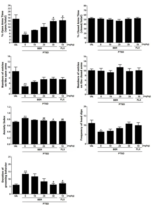
Rats exhibited an obvious anxiety phenotype characterized by decreased open-arm exploration during the EPM test. Statistical analyses of the behavioral results showed that the percentage of time spent in the open arms of the maze significantly differed among the 6 groups [F(5,35)=3.397, p<0.05]. The ANOVA also revealed a significant effect of the number of open arm entries among the six groups [F(5,35)=6.540, p<0.01]. Post-hoc com- parisons indicated that the PTSD groups showed a significantly decrease in both the percentage of time spent and the number of entries in the open arms of the maze than control group (p<0.01).
However, rats in the PTSD+BER30 group showed significant restoration of the time spent in the open arms of the maze com- pared with that of the PTSD group (p<0.05; Fig. 2). Also, rats in the PTSD+BER30 group showed restoration of the number of entries in the open arms of the maze compared with that of the PTSD group, although this result was only marginally signifi- cant. The time and number of enclosed-arm entries did not differ significantly among the six groups [F(5,35)=4.988, p=0.012 and F(5.35)=2.051, p=0.100]. BER administration after SPS elicited anxiogenic or anxiolytic behavior. These findings indicate that the increased time spent in the open arms of the maze in the PTSD+BER30 group was comparable to the exploratory behavior in the PTSD+FLX group. Overall, the anxiety index, calculated based on the number of visits to and time spent in the open and
Fig. 2. Effects of BER administration
on the percentage of time spent in
the open and closed arms, numbers
of entries into the open and closed
arms, anxiety index, unprotected
head dips, and grooming behavior on
the elevated plus maze (EPM) test of
rats exposed to SPS. *p<0.05, **p<0.01
vs. SAL group;
#p<0.05,
##p<0.01 vs. PTSD
group.
closed arms, also differed among the six groups of rats with lower values in the BER-treated rats (p<0.01 or p<0.05). Administering BER significantly decreased the frequency of unprotected head dips compared with that in the PTSD group, although this result was only marginally significant. However, the duration of groom- ing behavior was reversed by 30 mg/kg BER when administered after SPS exposure (p<0.05).
Effects of BER on locomotion and exploratory behavior following SPS
Open-field activity was used to assess exploratory behavior and locomotion among rats exposed to SPS for 14 days (Fig. 3).
Rats exposed to SPS had a significantly decreased amount of time spent in the central zone, with a corresponding increase in time spent in the peripheral zone, compared with the saline (SAL) group (p<0.01). A significant reduction in the number of central zone crossings was observed following the SPS procedure (p<0.01).
Our results indicate that SPS-treated rats developed exploratory activities that were closely associated with the anxiety-like behav- iors observed in the OFT. However, the BER-treated rats (30 mg/
kg) exhibited a significant increase in the number of central zone crossings compared with that in the PTSD group (p<0.05), indi- cating that the anxiety-like behaviors of the PTSD+BER30 group were similar to those of the PTSD+FLX group. In addition, the BER-treated rats (30 mg/kg) exhibited a significant increase in the time spent in the central zone compared with that in the PTSD group (p<0.01), indicating that the anxiety-like behaviors of the PTSD+BER30 group were similar to those of the PTSD+FLX group. A one-way ANOVA was performed, and PTSD-related differences were discovered in locomotor activity (motor func-
tion) and the total number of rearing (exploratory activities) in the OFT (Fig. 3). A significant difference in locomotor activity was observed between saline-treated rats, the PTSD group, and the BER-treated groups [F(5,35)=3.363, p<0.05] and the total times reared [F(5,35)=9.568, p<0.001]. Rats exposed to SPS had significantly decreased moving distance and rearing frequency in the open field compared with the SAL group (p<0.05 and p<0.01).
However, rats in the PTSD+BER30 group showed a significant restoration of the moving distance and rearing frequency in the open field compared with that in the PTSD group (p<0.05 and p<0.01).
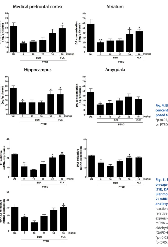
Effects of BER on DA concentration in the hippocampus following SPS
Fig. 4 shows that the brain region levels of DA were signifi- cantly different when the group were compared. Tissue levels of DA were measured in the medial prefrontal cortex, hippocampus, striatum, and amygdala after 2 week later (Day 14). The post-hoc test results indicated a significant decrease in the levels of DA in the hippocampus of the PTSD groups compared with that in the untreated PTSD group (p<0.05). Daily administration of BER sig- nificantly inhibited the SPS-induced decrease in DA concentra- tion in the hippocampus compared with that in the PTSD group (p<0.05). After BER treatment, the levels of DA in the striatum also increased significantly to 256.80% of that in the PTSD group (p<0.05). The DA concentrations in the brain regions of rats re- ceiving 10 mg/kg FLX were similar to those in rats receiving 30 mg/kg BER.
The ELISA showed that SPS exposure for 14 days significantly decreased the DA concentration in the medial prefrontal cortex
Fig. 3. Effects of BER administration on locomotion and exploratory behavior in the open filed test (OFT) of rats exposed to SPS. Change in the number of crossing in the central zone and the time spent in the central and peripheral zones. *p<0.05, **p<0.01 vs. SAL group;
#
p<0.05,
##p<0.01 vs. PTSD group.
of rats by 34.12% compared with rats in the saline-treated group (p<0.01). However, administration of BER inhibited the SPS- induced decrease in DA levels in the medial prefrontal cortex, although this result was only marginally statistically significant.
The ELISA results showed that SPS exposure for 14 days signifi- cantly decreased the DA concentration in the amygdala of rats by 45.03% compared with rats in the saline-treated group (p<0.05).
However, the administration of BER inhibited the SPS-induced
decrease in DA levels in the amygdala; this result was only mar- ginally significant.
Effects of BER on expression of TH, DAT, and VMAT-2 mRNA in the hippocampus following SPS
RC-PCR was conducted to examine the effect of BER on the expression levels of TH, DAT, and VMAT-2 mRNA in the hip-
Fig. 4. Effects of BER on dopamine (DA) concentration in the brains of rats ex- posed to SPS for 14 consecutive days.
*p<0.05, **p<0.01 vs. SAL group;
#p<0.05 vs. PTSD group.
Fig. 5. Effects of BER administration on expression of tyrosine hydroxylase (TH), DA transporter (DAT), and vesic- ular monoamine transporter-2 (VMAT- 2) mRNA in rats during SPS-induced anxiety symptoms. Polymerase chain reaction bands on agarose gel and their relative intensities are indicated. The expression levels of TH, DAT, and VMAT-2 mRNA were normalized to that of glycer- aldehyde 3-phosphate dehydrogenase (GAPDH) mRNA as an internal control.
*p<0.05, **p<0.01 vs. the SAL group,
#