• 검색 결과가 없습니다.

Concurrent treatment with ursolic acid and low-intensity treadmill exercise improves muscle atrophy and related outcomes in rats

N/A
N/A
Protected

Academic year: 2022

Share "Concurrent treatment with ursolic acid and low-intensity treadmill exercise improves muscle atrophy and related outcomes in rats"

Copied!
10
0
0

로드 중.... (전체 텍스트 보기)

전체 글

(1)

Korean J Physiol Pharmacol 2018;22(4):427-436 https://doi.org/10.4196/kjpp.2018.22.4.427

Author contributions: Conception and design of study: S.H.K. and J.C.K.

Acquisition of data, analysis and/or interpretation of data: Y.S.K., E.B.N., B.W.S., S.H.K., and G.D.P. Drafting the manuscript: S.H.K. Revising the manu- script, revising the manuscript critically for important intellectual content:

D.Y.S., G.D.P., and S.H.K.

This is an Open Access article distributed under the terms of the Creative Commons Attribution Non-Commercial License, which permits unrestricted non-commercial use, distribution, and reproduction in any medium, provided the original work is properly cited.

Copyright © Korean J Physiol Pharmacol, pISSN 1226-4512, eISSN 2093-3827

INTRODUCTION

Muscle atrophy, caused by a decrease in physical activity or aging, leads to problems related to an increase in body fat and de- creased muscle strength and cardiovascular endurance, resulting in fatigue [1-4] and deterioration of bone health [5,6], ultimately affecting the activities of daily living and quality of life [7-9].

Thus, efforts to improve muscle atrophy in the increasing elderly population should be explored.

Hypertrophy of the skeletal muscles is required to prevent muscle atrophy [10], and exercising with weights, including resis- tance exercise training, is effective [11]. According to recent stud- ies, even though muscle size and strength can be increased with low-intensity resistance training [12], higher-intensity resistance training (i.e., 80% of one repetition maximum) is more effective [13-15]. However, many adults, including elderly people (more than 70%), are inactive or sedentary, mainly participating only in light physical activities such as stretching or walking [16-18],

Original Article

Concurrent treatment with ursolic acid and low-intensity treadmill exercise improves muscle atrophy and related outcomes in rats

Jae Cheol Kim 1 , Yun Seok Kang 1 , Eun Bi Noh 1 , Baek Woon Seo 1 , Dae Yun Seo 2 , Gi Duck Park 3, *, and Sang Hyun Kim 1, *

1
Department of Sports Science, College of Natural Science, Chonbuk National University, Jeonju 54896,

2

Department of Physiology, Cardiovascular and Meta- bolic Disease Center, Inje University, Busan 47392,

3

Department of Leisure Sport, Kyungpook National University, Sangju 37224, Korea

ARTICLE INFO Received March 2, 2018 Revised May 27, 2018 Accepted June 8, 2018

*Correspondence Sang Hyun Kim E-mail: sh5275@jbnu.ac.kr Gi Duck Park

E-mail: prescript@knu.ac.kr Key Words

Aging Exercise Muscle atrophy Ursolic acid

ABSTRACT The objective of this study was to analyze the concurrent treatment ef- fects of ursolic acid (UA) and low-intensity treadmill exercise and to confirm the ef- fectiveness of UA as an exercise mimetic to safely improve muscle atrophy–related diseases using Sprague-Dawley (SD) rats with skeletal muscle atrophy. Significant muscle atrophy was induced in male SD rats through hind limb immobilization us- ing casting for 10 days. The muscle atrophy–induced SD rats were group into four:

SED, sedentary; UA, daily intraperitoneal UA injection, 5 mg/kg; EX, low-intensity (10- 12 m/min, 0° grade) treadmill exercise; and UEX, daily intraperitoneal UA injection, 5 mg/kg, and low-intensity (10-12 m/min, 0° grade) treadmill exercise. After 8 weeks of treatment, endurance capacity was analyzed using a treadmill, and tissues were extracted for analysis of visceral fat mass, body weight, muscle mass, expression of muscle atrophy- and hypertrophy-related genes, and endurance capacity. Although the effects of body weight gain control, muscle mass increase, and endurance capac- ity improvement were inadequate in the UA group, significant results were confirmed in the UEX group. The UEX group had significantly reduced body weight and visceral fat, significantly improved mass of tibialis anterior and gastrocnemius muscles, and significantly decreased atrophy-related gene expression of MuRF1 and atrogin-1, but did not have significant change in hypertrophy-related gene expression of Akt and mTOR. The endurance capacity was significantly improved in the EX and UEX groups.

These data suggest that concurrent treatment with low-intensity exercise and UA is

effective for atrophy-related physical dysfunctions.

(2)

making them at increased risk of various injuries, such as muscle damage, compared with young adults [19,20]. Therefore, exercis- ing with the lowest intensity possible may be helpful in reduc- ing the risk of injuries. However, as the intensity of the exercise decreases, its effects inevitably decrease. Hence, we hypothesized that if the effects of high-intensity exercise can be generated at a lower intensity using exercise mimetics, health could be safely maintained without the risk of injuries.

Ursolic acid (UA), a pentacyclic triterpenoid compound that is found in herbs and fruits, has various biological activities [21-23].

UA is reported to improve the insulin/insulin-like growth fac- tor 1 (IGF-1) signaling pathway; reduce the expression of muscle ring-finger protein 1 (MuRF1) and atrogin-1, a muscle-specific F-box protein [24]; and induce muscle hypertrophy. It has ef- fects similar to those of resistance training [1,24-26]. In addition, UA is considered to be an effective compound for the treatment of obesity and diseases related to obesity, by decreasing high fat diet–induced obesity and insulin resistance [27]. UA is considered to be an exercise mimetic that is effective for the prevention of, and improvement in, various diseases caused by muscle strength reduction and body fat increase. Although UA intake has a posi- tive effect on muscle atrophy and various disease management by muscle atrophy, it is difficult to improve all fitness factors, includ- ing flexibility, balance, and coordination. Furthermore, it does not have a positive effect on emotional health such as pleasure and happiness, and the effects of many nutrients are somewhat controversial. UA also has cell death–inducing properties [28,29], anti-inflammatory and pro-inflammatory properties [30], and pro-angiogenic and anti-angiogenic properties [31,32]. Therefore, strategies for minimizing the negative effects (such as lowering the UA dose) and maximizing the positive effects are needed.

However, the positive effect of UA may not be fully realized in this case. Thus, we proposed the following hypothesis. A low concentration of UA can help improve several health-related dis- advantages associated with muscle atrophy if it can promote low risk of injury with effective yet low-intensity exercise effects.

This study was aimed at evaluating the use of UA as an exercise mimetic to safely and effectively improve muscle atrophy–related physical dysfunction. We used an animal model of skeletal mus- cle atrophy, one of the major characteristics of aging, and ana- lyzed the concurrent treatment effects of UA and low-intensity treadmill exercise to determine any benefits related to improving muscle atrophy.

METHODS

Animals and experimental design

We used 8-week-old male Sprague-Dawley (SD) rats (n=40), weighing approximately 250 g (Central Lab, Animal Inc., Seoul, Korea). All the SD rats were allowed to acclimate for 1 week.

The rats were randomly allocated into four groups: SED, seden- tary; UA, daily intraperitoneal UA injection, 5 mg/kg; EX, low- intensity (10-12 m/min, 0° grade) treadmill exercise; UEX, daily intraperitoneal UA injection, 5 mg/kg, and low-intensity (10-12 m/min, 0° grade) treadmill exercise. Rats were given ad libitum water and food (carbohydrate, 58.9%; fat, 12.4%; protein, 28.7%;

Purina Corp.MO, USA) during the experimental period. Animals were housed in an environmentally controlled laboratory (tem- perature, 21°C; humidity, 40%-60%) with a 12:12-h dark and light cycle.

The left hind limb of each animal was immobilized using the method employed by Coutinho et al. [33], and muscle atrophy was induced over 10 days. After 10 days, the animals were treated with UA, EX, and UA plus EX for 8 weeks. Body weight was mea- sured once per week during the 8-week period. After 8 weeks of treatment, the rats were anesthetized using sodium pentobarbital (50 mg/kg of body weight). Three rats per group were used for the micro-computed tomography (micro-CT) scan of both hind limbs for 3D structural analysis. Hind limbs were removed, fixed, and preserved using 10% paraformaldehyde. In the remaining rats (7 rats per group), epididymal and retroperitoneal fat 24 h after the last training session was extracted. In addition, tibialis anterior (TA), soleus (SOL), and gastrocnemius (GAS) muscles were removed from both hind limbs, with the contralateral, non- immobilized leg being used as an internal control. The tissues were weighed using an electronic scale (EPG213; Ohaus Co., NJ, USA). The present study received approval from the Institutional Animal Care and Use Committee in Chonbuk National Univer- sity (IACUC approval no. CBNU-2017-0008).

Unilateral immobilization

Rats were anesthetized with pentobarbital sodium (50 mg/kg of body mass; Fort Dodge Animal Health, USA). The left knee joint was stretched, and the ankle joint was fixed by casting in a neutral posture [34,35]. During the immobilization, the rats were monitored for respiration state, discomfort, hind limb edema, and skin color changes. The animals were allowed to move freely, preventing movement of the immobilized leg alone. The muscles from the contralateral non-immobilized leg were used as controls in all experiments.

Ursolic acid injection and exercise program

UA was administered by intraperitoneal injection (5 mg/kg)

once per day, after it was dissolved in distilled water containing

0.1% Tween 80 (Sigma, St. Louis, MO, USA) [36]. For all other

groups, the same amount of vehicle (distilled water containing

0.1% Tween 80) was intraperitoneally injected. Intraperitoneal

administration is a commonly used method in animal studies

and can be a practical alternative in the late phase, but the initial

uptake is lower than that of intravenous and oral routes [37].

(3)

Ursolic acid and low-intensity exercise for muscle atrophy in rats 429 The low-intensity exercise protocol was based on partial revi-

sions to the methods by Kondo et al. [38]. The low-intensity ex- ercise was carried out for 8 weeks using a treadmill (Omnipacer model LC-4, Omnitech, USA). Before the exercise, training for acclimatization on a treadmill was conducted for 3 days, 15 min per day, at a speed of 5-10 m/min on a 0° gradient. For the experi- ment, rats walk or run on the treadmill for 30 min/day 3 days/

week for 8 weeks. The gradient of treadmill was set to 0°, and the speed was set at 10 m/min for the first 3 weeks, 12 m/min for the next 3 weeks, and then 15 m/min (45-55% Vo

2max

) [39] for the last 2 weeks.

Micro-computed tomography and three-dimensional structural analysis

Scanning was carried out using a SkyScan 1076 system (Bruker microCT; Kontich, Belgium) at the following settings: 12.25-μm resolution, 70 kV, 141 μA, and Al 0.5-mm filter. The images were reconstructed using NRecon software (Bruker microCT; Kon- tich). The cross-sectional images of the hind limb were captured using CTAn and CTvox software (Bruker microCT; Kontich).

Quantitative real-time polymerase chain reaction

After total RNA was extracted using TRIzol reagent (Invit- rogen, Carlsbad, CA, USA) from the GAS, it was purified using the DNA-free

TM

Kit (Ambion, CA, USA). After the concentra- tion of purified RNA was measured using an ultraviolet spec- trophotometer (UV-mini 1240, Shimazu Co., Japan), reverse transcription was conducted using 0.5 μg of RNA using the iScript

TM

cDNA Synthesis Kit (Bio-Rad, CA, USA). Real-time polymerase chain reaction analysis was performed with the produced cDNA as a template using the iQ5 Multicolor Real- Time PCR Detection System (Bio-Rad, CA, USA). This was achieved by mixing with MuRF1 (sense: TGTTCTGGTAG- GTCGTTTCCG, antisense: ATGCCGGTCCATGATCACTT), Atrogin-1 (sequence: CCATCAGGAGAAGTGGATCTATGTT, antisense: GCTTCCCCCAAAGTGCAGTA), Akt (sense: GT- GCTGGAGGACAATGACTACGG, antisense: AGCAGCCCT- GAAAGCAAGGA), mTOR (sense: TCCTGAAGAACATGT- GCGAG, antisense: CCAAAGTACAAGCGAGAGGC) and 18S rRNA’s specific primer with the reaction enzyme reagent, 2X SYBR Green Supermix (Bio-Rad, CA, USA). The mRNA expres- sion level was quantified as the ratio to 18S rRNA.

Western blot analysis

Muscle extracts were prepared and Western blotting was per- formed as described previously [40]. Blots were probed with the following antibodies: Akt, P-Akt, mTOR, P-mTOR, MuRF-1, Atrogin-1 (Bio-Rad, CA, USA), and β-actin (Sigma, MO, USA).

The blots were then incubated with the appropriate horseradish-

conjugated secondary antibodies (Jackson Immunoresearch Laboratories, PA, USA). Antibody bound protein was detected by ECL substrate (Bio-Rad, CA, USA). Band intensity was visualized and calculated using a ChemiDoc XRS+ (Bio-Rad, CA, USA).

Endurance capacity

After 8 weeks of treatment, the endurance capacity of each rat was measured using an exercise tolerance protocol on the treadmill. In line with the protocol for a progressive exercise test, treadmill exercise at a speed of 10 m/min was initially carried out at a 15° gradient for 5 min, with the speed increasing at 2 m/min each minute. Any rat that could not run anymore and stayed on the electrical shock bar (on which there was no electric current) was encouraged to continue to run by stimulating the hind limbs with high pressure air. If the rat stayed on the shock bar for more than 3 s despite the air stimulation, the test was suspended [41]

and total running time and distance were calculated. The equa- tion used by Park et al. [42] to calculate total work was used: work (J)=force [body weight (kg)]×vertical distance [sin (15°)×speed (m/

min)×time (min)].

Statistical analysis

The data are presented as mean±standard error (SE). The un- paired t-test was initially conducted for comparison of muscle mass based on pre-immobilization and 10 days after, to deter- mine the muscle atrophy level. A one-way analysis of variance analysis was carried out to verify the differences by each variable between groups. A Bonferroni post hoc test was conducted to de- termine the significance. The significance level was set at p<0.05.

The statistical analyses were performed with SPSS 18.0 (SPSS Inc., Chicago, IL, USA).

RESULTS

Hind limb immobilization induces muscle atrophy

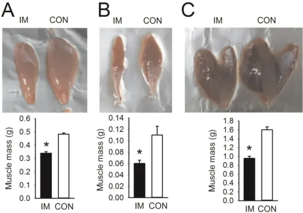
A muscle atrophy model was generated to identify the effects of low-intensity treadmill exercise and UA intake on various physi- cal dysfunctions associated with muscle atrophy. The effect of hind limb immobilization for 10 days was evaluated to determine the extent of muscle atrophy. It was confirmed that the muscle mass in the hind limbs was significantly reduced by immobiliza- tion, as shown in Fig. 1.

Concurrent treatment with low-intensity treadmill exercise and UA reduces body weight and visceral fat

The effects of low-intensity treadmill exercise and UA intake

for 8 weeks on body weight and visceral fat were evaluated (Fig.

(4)

430 Kim JC et al

Fig. 1. Muscle atrophy induced by 10-day hind limb IM. Data are presented as mean±SE. (A) TA. (B) SOL. (C) GAS. TA, tibialis anterior; SOL, soleus;

GAS, gastrocnemius; IM, immobilization; IM, immobilization; CON, contralateral control. *p<0.05 vs. CON by two-sample t-test (n=3 rats).

Figure 2

Fig. 2. Reduction in final body weight and fat mass through UA supplementation with exercise. Rats were given UA and/or performed treadmill

exercise for 8 weeks. Data are presented as mean±SE. (A, B) Initial and final body weights were measured before immobilization and before sacrifice

(n=7 rats per group). (C, D) Weight of epididymal and retroperitoneal fat pads (n=6 rats per group). SED, sedentary; UA, ursolic acid; EX, exercise; UEX,

ursolic acid plus exercise. *p<0.05, one-way analysis of variance.

(5)

Ursolic acid and low-intensity exercise for muscle atrophy in rats 431 2). Although 8 weeks of low-intensity treadmill exercise and UA

treatment had an inadequate effect on body weight, the body weight of the UEX group was significantly lower than the SED and UA groups (p<0.05). Epididymal fat was significantly lower in the EX and UEX groups compared with the SED group (p<0.05), and retroperitoneal fat was significantly lower in the UEX group compared with the SED group (p<0.05).

Concurrent low-intensity treadmill exercise and UA treatment reduces immobilization-induced muscle atrophy

Fig. 3 shows the results as a ratio to the contralateral muscle of the improvement of the muscle mass according to the treatment for 8 weeks. All the muscles analyze tended to improve accord- ing to treatment. Specifically, the UEX group was significantly improved TA and GAS muscles compared to SED group (p<0.05) (Fig. 3 A). The same result was shown in terms of cross-sectional area by micro-CT analysis (Fig. 3B). There was no significant dif- ference between the immobilization-induced muscle and contra- lateral muscle of the UEX group compared with the other groups.

Concurrent low-intensity treadmill exercise and UA treatment reduces atrophic gene expression

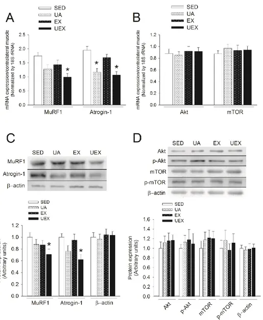
We analyzed muscle atrophy– and hypertrophy-related gene expression to evaluate how the concurrent low-intensity treadmill exercise and UA treatment were effective in improving skeletal muscle mass. As a result, reduction of the mRNA and protein expression of atrophy-related genes was confirmed (Figs. 4A and 4C). Significant differences from the SED group were observed in the UEX group in terms of MuRF1, and in the UA and UEX groups in terms of Atrogin-1 (Fig. 4A). The content of MuRF1 and Atrogin-1 protein was significantly differences between SED group and UEX group. However, mRNA and protein expression of Akt and mTOR, the genes related to muscle hypertrophy, was not different between the groups.

Endurance capacity improves with low-intensity treadmill exercise

As determined by performing an endurance capacity test after 8 weeks of treatment, the exercise time, running distance, and total work in the EX and UEX groups significantly improved Figure 3

Fig. 3. Improvement in atrophied hind limb muscle mass through UA supplementation with treadmill exercise. (A) Skeletal muscle atrophy of the TA, SOL, and GAS induced by IM is expressed in percentage of difference from the contralateral CON leg (non-immobilized). Data are presented as mean±SE. *p<0.05, one-way analysis of variance. (B) Micro-computed tomography (CT) scans of a rat hind limb. Lateral view of a whole limb (top) and mid-tibial transverse micro-CT scans of the same rat hind limb (bottom). TA, tibialis anterior; SOL, soleus; GAS, gastrocnemius; IM, immobilization;

CON, control; SED, sedentary; UA, ursolic acid; EX, exercise; UEX, ursolic acid plus exercise.

(6)

compared with the SED group (Fig. 5). However, UA did not have an additive effect in endurance capacity at all.

DISCUSSION

This study was conducted to evaluate the effects of UA and low- intensity treadmill exercise on the improvement of muscle atro- phy and exercise capacity caused by physical dysfunctions such as aging. Based on these results, we tried to confirm the possibility of UA as an exercise mimetic for the safe and effective improve- ment of physical dysfunctions. Exercise mimetics are defined as biologically active compounds that have an effect on exercise by activating signaling pathways similar to exercise [43]. Interest in

exercise mimetics continuously increased after Narkar et al. [44]

reported that the AMPK-PPARδ pathway not only can improve adaptation according to training, but medicine can be used to target an increase in endurance without performing exercise [44- 47]. However, most relevant research has focused on identifying exercise mimetics for treatment of obesity through endurance capacity improvement or to treat metabolic disorders [44-48].

UA was reported to affect obesity and improve insulin resistance [27] and improve muscle hypertrophy or muscle atrophy [1,24-26]

through increased endurance capacity. This study confirmed that concurrent low-intensity exercise and UA treatment can gener- ate an effect comparable to that of high-intensity exercise and analyzed the possibility of UA as an exercise mimetic. Hence, a muscle atrophy model was made based on casting-induced hind

Fig. 4. Reduction in atrophic gene expression through concurrent low-intensity treadmill exercise and UA treatment. Relative mRNA expres-

sion levels of atrophy- (A) and hypertrophy- (B) related genes and the content of atrophy- (C) and hypertrophy- (D) related protein after 8 weeks of

treatment in the gastrocnemius muscle (n=6 rats per group). Signals were quantified and normalized against the contralateral CON leg (non-immobi-

lized). Data are presented as mean±SE. *p<0.05, one-way analysis of variance. CON, control; SED, sedentary; UA, ursolic acid; EX, exercise; UEX, ursolic

acid plus exercise.

(7)

Ursolic acid and low-intensity exercise for muscle atrophy in rats 433

limb immobilization in SD rats. The hind limb immobilization used in this study for 10 days reduced 30%-45% of the muscle vol- ume and thus confirmed that hind limb immobilization could be used as a model for muscle atrophy (Fig. 1 ). This study analyzed the effects of 8 weeks of treatment using this model.

Muscle atrophy is closely related to physical activity reduction [49], and weight gain as a result of the reduction in physical activ- ity causes various health problems [50]. Therefore, preventative treatment is required. According to the results of the present study, 8 weeks of UA treatment and low-intensity treadmill exer- cise did not significantly contribute to weight gain prevention (Fig.

2B). However, low-intensity treadmill exercise resulted in some visceral fat reduction by reducing (p<0.05) epididymal fat. The retroperitoneal fat weight was decreased 25% but no significantly (p=0.068) changed in EX group. Both epidydimal and retroperi- toneal fat were reduced in the UEX group (Figs. 2C and 2D). We have not determined the reason for the different responses in epididymal fat and retroperitoneal fat in the EX and UEX groups, but we noted a decrease in visceral fat in the EX group. This result indicates that weight gain was effectively prevented with the com- bined treatment. UA reduces interscapular fat by increasing body temperature and energy expenditure with increasing uncoupling protein (UCP) 1 in brown adipose tissue [27]. In addition, it increases energy expenditure by increasing the UCP 3/AMPK- dependent pathway [51] as well as enhancing β-oxidation in skel- etal muscle [51,52]. The effects of UA are similar to the increase of fat oxidation in skeletal muscle [53-55] by activating carnitine palmitoyl transferase (CPT1) by controlling the synthesis of mal- onyl CoA by the fatty acid’s increased oxidation through exercise, namely control of acetyl CoA carboxylase activity by increased AMPK activity. Therefore, it is possible that UA may be an exer- cise mimetic that could promote weight loss, but there are limita- tions in preventing weight gain from muscle atrophy through UA alone. Hence, concurrent low-intensity endurance training and UA intake can be a very effective treatment method.

UA decreases muscle atrophy by decreasing the expression of atrophy-related genes and increases the expression of hypertro- phy-associated genes, inducing muscle hypertrophy [24,26]. It de- creases the expression of MuRF1 and Atrogin-1 and improves the

insulin-like growth factor 1 (IGF-1) signaling pathway for muscle hypertrophy. Resistance training also increases muscle hyper- trophy through increased mTOR signaling action [56,57] and decreases muscle atrophy–related gene expression [9,58]. In this regard, although UA is considered to be an exercise mimetic for muscle atrophy improvement, this study could not confirm the effect of UA alone, which may be similar to the weight loss effect induced through exercise. However, UA administration tended to increase the mass of all muscle compared with the SED group but does not significant (Fig. 3). This may be related to the concentra- tion of UA. A study of 200 mg/kg of UA treatment showed that muscle atrophy was reduced, muscle hypertrophy was stimulated [24], as well as a potential candidate for treatment of pathological conditions associated with muscle atrophy [59]. However, in this study, UA was treated at a concentration of 5 mg/kg. This con- centration improved the soleus muscle mass in the sciatic nerve injury animal model [36], but is much lower than the 200 mg/kg of concentration. Therefore, it would be necessary to carry out a study to compare the different concentrations. The EX group also tend to increase the mass of all muscle compared with the SED group but no significant (Fig. 3). Endurance training is known to improve muscle function, neuromuscular junction, metabolism, and motor-unit adaptability [60]. Moreover, physical activity plays a role in delaying muscle mass loss [60]. Souza et al. [61] report that endurance exercise training prevents heart failure-induced skeletal muscle atrophy. Additionally, Ji and Kang [62] suggest that PGC-1a, which plays an important role in mitochondrial biosynthesis in skeletal muscle, may be contributed to improve the quality of life of elderly people by increasing skeletal muscle health such as suppression of muscle atrophy. Although resistance training is generally associated with muscle hypertrophy, this study confirmed that muscle atrophy can be effectively improved with low-intensity endurance exercise with UA intake (Fig. 3).

These results suggest that the effects of UA and low-intensity ex- ercise are combined.

Atrophy due to aging is mainly caused by a decrease in both number and size of type II fibers [63]. Myosin heavy-chain iso- form of GAS muscle consists of about 6%, 8%, 22%, and 60% for types I, IIa, IIx, and IIb fibers, respectively [64]. Thus, we used Figure 5

Fig. 5. Increased endurance capacity from treadmill exercise, but not ursolic acid, after 8 weeks of treatment. Endurance capacity was mea-

sured using a treadmill after 8 weeks. Data are presented as mean±SE (n=6 rats per group). *p<0.05, one-way analysis of variance.

(8)

GAS muscle to identify signaling pathway to confirm the possi- bility of UA as exercise mimetics to improve physical dysfunction such as aging. UA is effective for Akt/mTORC1 signaling within the skeletal muscle that is associated with muscle hypertrophy [27,65], but some studies reported no effect [66,67]. Furthermore, MuRF1 and Atrogin-1 expression, which are atrophy-related genes, which are atrophy-related genes, are controlled by UA [24,26]. Endurance exercise also prevents muscle atrophy to oc- cur via a decrease in catabolic factors such as atrogin-1, MuRF1, FoxO1, and myostatin [61]. However, as shown in Fig. 4 of this study, UA and low-intensity endurance exercise did not signifi- cantly affect the mechanism associated with atrophy and hyper- trophy. However, the muscle atrophy improvement following UA intake concurrent with low-intensity endurance exercise is effec- tive because the expression of MuRF1 and Atrogin-1 in the UEX group is significantly decreased. It is considered that the effects of anti-catabolic mechanism of UA and low-intensity endurance exercise are additive to each other. Therefore, it will be neces- sary to clarify whether the UA and endurance exercise effects are achieved through different mechanisms or through the same mechanism and whether the effects of each other are additive.

UA increases running distance and exercise time [26,27,68], which not only reduces serum levels of fatigue-related factors such as lactate and lactate dehydrogenase [26], but also increases PGC- 1α and mitochondrial transcription factor A (mtTFA) expression, which plays a key role in mitochondrial biogenesis, mitochondrial enzymes citrate synthase activity, and COX mRNA expression [68], thereby increasing endurance exercise capacity. In the pres- ent study, only the EX and UEX groups showed an increase in exercise time, running distance, and total work compared to the SED group (Fig. 5). Low-intensity endurance exercise alone was found to improve endurance exercise capacity. However, we could not confirm the effect of UA alone on improving endurance ca- pacity. This is also due to the difference in concentration of UA as the result of muscle mass. UA increases serum irisin in human study [25] and increases mitochondrial biogenesis through acti- vation of AMPK-PGC-1α in C2C12 myotubes [68]. Therefore, it may be necessary to evaluate the effect of PGC-1–related mecha- nism in skeletal muscle according to UA concentration.

In conclusion, this study analyzed the effects of concurrent treatment with UA and low-intensity treadmill exercise to im- prove skeletal muscle atrophy using an animal model. The effect of UA alone on muscle atrophy could not be confirmed; however, positive effects could be confirmed for UA combined with low- intensity endurance exercise. Further studies will be needed to verify the differences in concentration of UA and need to ad- ditionally analyze the relevant signaling pathway to confirm a mechanism for use of UA as an exercise mimetic. This study is meaningful in that UA was identified to have a possible use as an exercise mimetic for reduction of body weight or visceral fat mass and inhibition of muscle atrophy.

ACKNOWLEDGEMENTS

This work was supported by the Ministry of Education of the Republic of Korea and the National Research Foundation of Ko- rea (NRF-2016S1A5A8019355), by research funds for newly ap- pointed professors of Chonbuk National University in 2015, and by Kyungpook National University Bokhyeon Research Fund, 2015.

CONFLICTS OF INTEREST

The authors declare no conflicts of interest.

REFERENCES

1. Ebert SM, Dyle MC, Bullard SA, Dierdorff JM, Murry DJ, Fox DK, Bongers KS, Lira VA, Meyerholz DK, Talley JJ, Adams CM. Identifi- cation and small molecule inhibition of an activating transcription factor 4 (ATF4)-dependent pathway to age-related skeletal muscle weakness and atrophy. J Biol Chem. 2015;290:25497-25511.

2. Tanaka H, Desouza CA, Jones PP, Stevenson ET, Davy KP, Seals DR. Greater rate of decline in maximal aerobic capacity with age in physically active vs. sedentary healthy women. J Appl Physiol (1985).

1997;83:1947-1953.

3. Feng HZ, Chen M, Weinstein LS, Jin JP. Improved fatigue resistance in Gsα-deficient and aging mouse skeletal muscles due to adaptive increases in slow fibers. J Appl Physiol (1985). 2011;111:834-843.

4. Wilson JR, Mancini DM, Dunkman WB. Exertional fatigue due to skeletal muscle dysfunction in patients with heart failure. Circula- tion. 1993;87:470-475.

5. Elkin SL, Williams L, Moore M, Hodson ME, Rutherford OM. Rela- tionship of skeletal muscle mass, muscle strength and bone mineral density in adults with cystic fibrosis. Clin Sci (Lond). 2000;99:309- 314.

6. He H, Liu Y, Tian Q, Papasian CJ, Hu T, Deng HW. Relationship of sarcopenia and body composition with osteoporosis. Osteoporos Int. 2016;27:473-482.

7. Kim H, Suzuki T, Saito K, Yoshida H, Kojima N, Kim M, Sudo M, Yamashiro Y, Tokimitsu I. Effects of exercise and tea catechins on muscle mass, strength and walking ability in community-dwelling elderly Japanese sarcopenic women: a randomized controlled trial.

Geriatr Gerontol Int. 2013;13:458-465.

8. Brotto M, Abreu EL. Sarcopenia: pharmacology of today and to- morrow. J Pharmacol Exp Ther. 2012;343:540-546.

9. Gielen S, Sandri M, Kozarez I, Kratzsch J, Teupser D, Thiery J, Erbs S, Mangner N, Lenk K, Hambrecht R, Schuler G, Adams V. Exercise training attenuates MuRF-1 expression in the skeletal muscle of pa- tients with chronic heart failure independent of age: the randomized Leipzig Exercise Intervention in Chronic Heart Failure and Aging catabolism study. Circulation. 2012;125:2716-2727.

10. Bamman MM, Roberts BM, Adams GR. Molecular regulation of exercise-induced muscle fiber hypertrophy. Cold Spring Harb Per- spect Med. 2018;8:a029751. doi: 10.1101/cshperspect.a029751.

11. Lai CC, Tu YK, Wang TG, Huang YT, Chien KL. Effects of resis-

(9)

Ursolic acid and low-intensity exercise for muscle atrophy in rats 435 tance training, endurance training and whole-body vibration on

lean body mass, muscle strength and physical performance in older people: a systematic review and network meta-analysis. Age Ageing.

2018;47:367-373.

12. Watanabe Y, Madarame H, Ogasawara R, Nakazato K, Ishii N. Ef- fect of very low-intensity resistance training with slow movement on muscle size and strength in healthy older adults. Clin Physiol Funct Imaging. 2014;34:463-470.

13. Frontera WR, Meredith CN, O’Reilly KP, Knuttgen HG, Evans WJ.

Strength conditioning in older men: skeletal muscle hypertrophy and improved function. J Appl Physiol (1985). 1988;64:1038-1044.

14. Fiatarone MA, Marks EC, Ryan ND, Meredith CN, Lipsitz LA, Ev- ans WJ. High-intensity strength training in nonagenarians. Effects on skeletal muscle. JAMA. 1990;263:3029-3034.

15. Daly RM. Exercise and nutritional approaches to prevent frail bones, falls and fractures: an update. Climacteric. 2017;20:119-124.

16. Ruuskanen JM, Ruoppila I. Physical activity and psychological well- being among people aged 65 to 84 years. Age Ageing. 1995;24:292- 296.

17. Goodyear LJ. The exercise pill—too good to be true? N Engl J Med.

2008;359:1842-1844.

18. Wen CP, Wai JP, Tsai MK, Yang YC, Cheng TY, Lee MC, Chan HT, Tsao CK, Tsai SP, Wu X. Minimum amount of physical activity for reduced mortality and extended life expectancy: a prospective co- hort study. Lancet. 2011;378:1244-1253.

19. Gorianovas G, Skurvydas A, Streckis V, Brazaitis M, Kamandulis S, McHugh MP. Repeated bout effect was more expressed in young adult males than in elderly males and boys. Biomed Res Int. 2013.

doi: 10.1155/2013/218970.

20. Lavender AP, Nosaka K. Comparison between old and young men for changes in makers of muscle damage following voluntary eccen- tric exercise of the elbow flexors. Appl Physiol Nutr Metab. 2006;

31:218-225.

21. Shishodia S, Majumdar S, Banerjee S, Aggarwal BB. Ursolic acid inhibits nuclear factor-kappaB activation induced by carcinogenic agents through suppression of IkappaBalpha kinase and p65 phos- phorylation: correlation with down-regulation of cyclooxygenase, matrix metalloproteinase 9, and cyclin D1. Cancer Res. 2003;63:

4375-4383.

22. Tsai SJ, Yin MC. Antioxidative and anti-inflammatory protec- tion of oleanolic acid and ursolic acid in PC12 cells. J Food Sci.

2008;73:H174-178.

23. Ullevig SL, Zhao Q, Zamora D, Asmis R. Ursolic acid protects dia- betic mice against monocyte dysfunction and accelerated athero- sclerosis. Atherosclerosis. 2011;219:409-416.

24. Kunkel SD, Suneja M, Ebert SM, Bongers KS, Fox DK, Malmberg SE, Alipour F, Shields RK, Adams CM. mRNA expression signa- tures of human skeletal muscle atrophy identify a natural com- pound that increases muscle mass. Cell Metab. 2011;13:627-638.

25. Bang HS, Seo DY, Chung YM, Oh KM, Park JJ, Arturo F, Jeong SH, Kim N, Han J. Ursolic Acid-induced elevation of serum irisin aug- ments muscle strength during resistance training in men. Korean J Physiol Pharmacol. 2014;18:441-446.

26. Jeong JW, Shim JJ, Choi ID, Kim SH, Ra J, Ku HK, Lee DE, Kim TY, Jeung W, Lee JH, Lee KW, Huh CS, Sim JH, Ahn YT. Apple pomace extract improves endurance in exercise performance by increasing strength and weight of skeletal muscle. J Med Food. 2015;18:1380-

1386.

27. Kunkel SD, Elmore CJ, Bongers KS, Ebert SM, Fox DK, Dyle MC, Bullard SA, Adams CM. Ursolic acid increases skeletal muscle and brown fat and decreases diet-induced obesity, glucose intolerance and fatty liver disease. PLoS One. 2012;7:e39332.

28. Achiwa Y, Hasegawa K, Komiya T, Udagawa Y. Ursolic acid induces Bax-dependent apoptosis through the caspase-3 pathway in endo- metrial cancer SNG-II cells. Oncol Rep. 2005;13:51-57.

29. Choi YH, Baek JH, Yoo MA, Chung HY, Kim ND, Kim KW. Induc- tion of apoptosis by ursolic acid through activation of caspases and down-regulation of c-IAPs in human prostate epithelial cells. Int J Oncol. 2000;17:565-571.

30. Ikeda Y, Murakami A, Ohigashi H. Ursolic acid: an anti- and pro- inflammatory triterpenoid. Mol Nutr Food Res. 2008;52:26-42.

31. Cárdenas C, Quesada AR, Medina MA. Effects of ursolic acid on different steps of the angiogenic process. Biochem Biophys Res Commun. 2004;320:402-408.

32. Sohn KH, Lee HY, Chung HY, Young HS, Yi SY, Kim KW. Anti- angiogenic activity of triterpene acids. Cancer Lett. 1995;94:213-218.

33. Coutinho EL, Gomes AR, Franca CN, Salvini TF. A new model for the immobilization of the rat hind limb. Braz J Med Biol Res. 2002;

35:1329-1332.

34. Vazeille E, Codran A, Claustre A, Averous J, Listrat A, Béchet D, Taillandier D, Dardevet D, Attaix D, Combaret L. The ubiquitin- proteasome and the mitochondria-associated apoptotic pathways are sequentially downregulated during recovery after immobili- zation-induced muscle atrophy. Am J Physiol Endocrinol Metab.

2008;295:E1181-1190.

35. Madaro L, Smeriglio P, Molinaro M, Bouché M. Unilateral immo- bilization: a simple model of limb atrophy in mice. Basic Applied Myology. 2008;18:149-153.

36. Liu B, Liu Y, Yang G, Xu Z, Chen J. Ursolic acid induces neural re- generation after sciatic nerve injury. Neural Regen Res. 2013;8:2510- 2519.

37. Kim C, Kim IH, Kim SI, Kim YS, Kang SH, Moon SH, Kim TS, Kim SK. Comparison of the intraperitoneal, retroorbital and per oral routes for F-18 FDG administration as effective alternatives to intra- venous administration in mouse tumor models using small animal PET/CT studies. Nucl Med Mol Imaging. 2011;45:169-176.

38. Kondo H, Fujino H, Murakami S, Tanaka M, Kanazashi M, Na- gatomo F, Ishihara A, Roy RR. Low-intensity running exercise en- hances the capillary volume and pro-angiogenic factors in the soleus muscle of type 2 diabetic rats. Muscle Nerve. 2015;51:391-399.

39. Bedford TG, Tipton CM, Wilson NC, Oppliger RA, Gisolfi CV.

Maximum oxygen consumption of rats and its changes with vari- ous experimental procedures. J Appl Physiol Respir Environ Exerc Physiol. 1979;47:1278-1283.

40. Kim JC, Park GD, Kim SH. Inhibition of oxidative stress by anti- oxidant supplementation does not limit muscle mitochondrial bio- genesis or endurance capacity in rats. J Nutr Sci Vitaminol (Tokyo).

2017;63:277-283.

41. Copp SW, Davis RT, Poole DC, Musch TI. Reproducibility of endur- ance capacity and VO2peak in male Sprague-Dawley rats. J Appl Physiol (1985). 2009;106:1072-1078.

42. Park JY, Wang PY, Matsumoto T, Sung HJ, Ma W, Choi JW, Ander-

son SA, Leary SC, Balaban RS, Kang JG, Hwang PM. p53 improves

aerobic exercise capacity and augments skeletal muscle mitochon-

(10)

drial DNA content. Circ Res. 2009;105:705-711.

43. Li S, Laher I. Exercise mimetics: running without a road map. Clin Pharmacol Ther. 2017;101:188-190.

44. Narkar VA, Downes M, Yu RT, Embler E, Wang YX, Banayo E, Mihaylova MM, Nelson MC, Zou Y, Juguilon H, Kang H, Shaw RJ, Evans RM. AMPK and PPARdelta agonists are exercise mimetics.

Cell. 2008;134:405-415.

45. Handschin C. Caloric restriction and exercise “mimetics’’: Ready for prime time? Pharmacol Res. 2016;103:158-166.

46. Craig DM, Ashcroft SP, Belew MY, Stocks B, Currell K, Baar K, Philp A. Utilizing small nutrient compounds as enhancers of exer- cise-induced mitochondrial biogenesis. Front Physiol. 2015;6:296.

47. Fan W, Evans RM. Exercise mimetics: impact on health and perfor- mance. Cell Metab. 2017;25:242-247.

48. Stockinger J, Maxwell N, Shapiro D, deCabo R, Valdez G. Caloric restriction mimetics slow aging of neuromuscular synapses and muscle fibers. J Gerontol A Biol Sci Med Sci. 2017;73:21-28.

49. Naseeb MA, Volpe SL. Protein and exercise in the prevention of sar- copenia and aging. Nutr Res. 2017;40:1-20.

50. Spiegelman BM, Flier JS. Obesity and the regulation of energy bal- ance. Cell. 2001;104:531-543.

51. Rao VS, de Melo CL, Queiroz MG, Lemos TL, Menezes DB, Melo TS, Santos FA. Ursolic acid, a pentacyclic triterpene from Sambucus australis, prevents abdominal adiposity in mice fed a high-fat diet. J Med Food. 2011;14:1375-1382.

52. Chu X, He X, Shi Z, Li C, Guo F, Li S, Li Y, Na L, Sun C. Ursolic acid increases energy expenditure through enhancing free fatty acid uptake and β-oxidation via an UCP3/AMPK-dependent pathway in skeletal muscle. Mol Nutr Food Res. 2015;59:1491-1503.

53. Winder WW. Energy-sensing and signaling by AMP-activated pro- tein kinase in skeletal muscle. J Appl Physiol (1985). 2001;91:1017- 1028.

54. Kahn BB, Alquier T, Carling D, Hardie DG. AMP-activated protein kinase: ancient energy gauge provides clues to modern understand- ing of metabolism. Cell Metab. 2005;1:15-25.

55. Minokoshi Y, Kim YB, Peroni OD, Fryer LG, Müller C, Carling D, Kahn BB. Leptin stimulates fatty-acid oxidation by activating AMP- activated protein kinase. Nature. 2002;415:339-343.

56. Ogasawara R, Kobayashi K, Tsutaki A, Lee K, Abe T, Fujita S, Na- kazato K, Ishii N. mTOR signaling response to resistance exercise is altered by chronic resistance training and detraining in skeletal muscle. J Appl Physiol (1985). 2013;114:934-940.

57. Ogasawara R, Fujita S, Hornberger TA, Kitaoka Y, Makanae Y, Na- kazato K, Naokata I. The role of mTOR signalling in the regulation

of skeletal muscle mass in a rodent model of resistance exercise. Sci Rep. 2016;6:31142.

58. Furlanetto R Jr, de Paula Souza A, de Oliveira AA, Nunes PR, Mi- chelin MA, Chica JE, Murta EF, Orsatti FL. Acute resistance exer- cise reduces increased gene expression in muscle atrophy of ovariec- tomised arthritic rats. Prz Menopauzalny. 2016;15:193-201.

59. Bakhtiari N, Hosseinkhani S, Tashakor A, Hemmati R. Ursolic acid ameliorates aging-metabolic phenotype through promoting of skel- etal muscle rejuvenation. Med Hypotheses. 2015;85:1-6.

60. Arthur ST, Cooley ID. The effect of physiological stimuli on sarco- penia; impact of Notch and Wnt signaling on impaired aged skeletal muscle repair. Int J Biol Sci. 2012;8:731-760.

61. Souza RW, Piedade WP, Soares LC, Souza PA, Aguiar AF, Vechetti- Júnior IJ, Campos DH, Fernandes AA4, Okoshi K, Carvalho RF, Cicogna AC, Dal-Pai-Silva M. Aerobic exercise training prevents heart failure-induced skeletal muscle atrophy by anti-catabolic, but not anabolic actions. PLoS One. 2014;9:e110020.

62. Ji LL, Kang C. Role of PGC-1α in sarcopenia: etiology and potential intervention - a mini-review. Gerontology. 2015;61:139-148.

63. Lexell J. Human aging, muscle mass, and fiber type composition. J Gerontol A Biol Sci Med Sci. 1995;50:11-16.

64. Eng CM, Smallwood LH, Rainiero MP, Lahey M, Ward SR, Li- eber RL. Scaling of muscle architecture and fiber types in the rat hindlimb. J Exp Biol. 2008;211:2336-2345.

65. Ogasawara R, Sato K, Higashida K, Nakazato K, Fujita S. Ursolic acid stimulates mTORC1 signaling after resistance exercise in rat skeletal muscle. Am J Physiol Endocrinol Metab. 2013;305:E760-765.

66. Church DD, Schwarz NA, Spillane MB, McKinley-Barnard SK, Andre TL, Ramirez AJ, Willoughby DS. l-Leucine increases skeletal muscle IGF-1 but does not differentially increase Akt/mTORC1 signaling and serum IGF-1 compared to ursolic acid in response to resistance exercise in resistance-trained men. J Am Coll Nutr.

2016;35:627-638.

67. Cho YH, Lee SY, Kim CM, Kim ND, Choe S, Lee CH, Shin JH.

Effect of loquat leaf extract on muscle strength, muscle mass, and muscle function in healthy adults: a randomized, double-blinded, and placebo-controlled trial. Evid Based Complement Alternat Med. 2016. doi: 10.1155/2016/4301621.

68. Chen J, Wong HS, Leong PK, Leung HY, Chan WM, Ko KM. Ur- solic acid induces mitochondrial biogenesis through the activation of AMPK and PGC-1 in C2C12 myotubes: a possible mechanism underlying its beneficial effect on exercise endurance. Food Funct.

2017;8:2425-2436.

수치

Fig. 1. Muscle atrophy induced by 10-day hind limb IM. Data are presented as mean±SE. (A) TA
Fig. 3 shows the results as a ratio to the contralateral muscle of  the improvement of the muscle mass according to the treatment  for 8 weeks
Fig. 4. Reduction in atrophic gene expression through concurrent low-intensity treadmill exercise and UA treatment
Fig. 5. Increased endurance capacity from treadmill exercise, but not ursolic acid, after 8 weeks of treatment

참조

관련 문서

The purpose of this research is to confirm whether voluntary early morning physical exercise effects on the emotional and behavioral development of

The purpose and necessity of this study was to investigate the effect of the 8-week GX complex exercise program on the stress index, blood lipid and APG

12 weeks of combined exercise (body strength exercise and aerobic exercise) was applied to measure health fitness (muscle strength, muscular endurance,

Objective: The purpose of this study was to analyze recent trend in incidence of basal cell carcinoma and squamous cell carcinoma in patients from the Gwangju City

Therefore, as a recovery method during exercise, it is more effective for recovery of exercise fatigue and prevention of body acidification due to high

The purpose of this study was to provide primary material in discussing methods to activate the treatment effects of diabetes and reduce the rate of

The purpose of this study was to investigate how the 8-week aerobic exercise program affects the physical strength and stress of obese women in the

The purpose and necessity of this study was to investigate the effects of lactic acid fatigue and body oxidation through myofascial relaxation exercise using small tools