Received: 2013.6.27. Revised: 2013.10.14. Accepted: 2013.10.18.
Corresponding author: Yoon Ha Kim
Department of Obstetrics and Gynecology, Chonnam National University Medical School, 160 Baekseo-ro, Dong-gu, Gwangju 501-746, Korea
Tel: +82-62-220-6375 Fax: +82-62-227-1637 E-mail: kimyh@jnu.ac.kr
Articles published in Obstet Gynecol Sci are open-access, distributed under the terms of the Creative Commons Attribution Non-Commercial License (http://creativecommons.
org/licenses/by-nc/3.0/) which permits unrestricted non-commercial use, distribution, and reproduction in any medium, provided the original work is properly cited.
Copyright © 2014 Korean Society of Obstetrics and Gynecology
Introduction
Oxidative stress is defined as an imbalance between pro-oxi- dant and antioxidant forces resulting in an overall pro-oxidant insult. Oxygen-derived reactants, collectively termed reactive oxygen species (ROS), are a normal byproduct of cellular me- tabolism; as such, cells also contain a natural antioxidant de- fense system [1,2]. The body contain endogenous antioxidants such as superoxide dismutase, glutathione peroxidase, and bilirubin while total antioxidant capacity is also determined by the exogenous molecules such as vitamin C, vitamin E.
Metabolic activation of molecular oxygen frequently gives rise to ROS; however, when the protective capacity level of
Comparison of oxidative stress markers in umbilical cord blood after vaginal and cesarean delivery
Eun Ji Noh 1 , Yoon Ha Kim 1 , Moon Kyoung Cho 1 , Jong Woon Kim 1 , Jin Wook Kim 1 , Yeung Ja Byun 2 , Tae-Bok Song 1
Department of Obstetrics and Gynecology,
1Chonnam National University Medical School, Gwangju;
2Chonnam National University Research Institute of Medical Sciences, Gwangju, Korea
Objective
The purpose of our study was to investigate the effect of the mode of delivery on the oxidant and antioxidant system in umbilical cord blood.
Methods
We performed gas analysis of umbilical venous blood and umbilical arterial blood immediately after delivery in 38 women; eighteen women had a vaginal delivery while 20 women delivered via cesarean section at over 37 weeks gestation. We examined lipid peroxide concentration by thiobarbituric acid reaction, protein carbonyl content by 2,4-dinitrophenylhydrazine reaction, and total antioxidant capacity by oxygen radical absorbance capacity assay.
Results
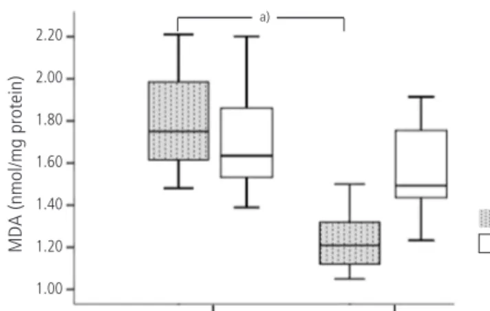
Lipid peroxide levels in umbilical venous blood were significantly higher in patients delivering by planned cesarean section (1.81 ± 0.06 nmol/mg protein) than those with vaginal delivery (1.24 ± 0.05 nmol/mg protein) (P < 0.05).
Antioxidant capacity in umbilical venous blood was significantly higher in patients delivering by planned cesarean section (119.70 ± 0.13 μM/μL) than those with a vaginal delivery (118.70 ± 0.29 μM/μL) (P < 0.05). There was no significant difference in the carbonyl content of umbilical venous blood or in the lipid peroxide, carbonyl content, and total antioxidant capacity of umbilical arterial blood.
Conclusion
Lipid peroxidation levels and antioxidant capacity in umbilical venous blood were higher in patients delivering by planned cesarean section than those with a vaginal delivery. Therefore, we propose that both the mother and neonate are exposed to higher oxidative stress during cesarean section delivery.
Keywords: Cesarean delivery; Oxidative stress marker; Vaginal delivery http://dx.doi.org/10.5468/ogs.2014.57.2.109
pISSN 2287-8572 · eISSN 2287-8580
antioxidants has been exceeded, these products can become toxic to most cellular components, as they stimulate lipid per- oxidation and protein inactivation [3]. The resulting damage is referred to as oxidative stress. Oxidative stress can be as- sessed in vivo using the plasma biomarkers malondialdehyde and protein carbonyl.
In women with normal, uncomplicated pregnancies lipid peroxide and antioxidant levels are elevated as compared to non-pregnant women; this increase is proportional to gesta- tional length [4]. The placenta is a major source of oxidative stress during pregnancy. The placenta is rich in polyunsaturat- ed fatty acids, and it is an abundant source of lipid peroxides which are secreted into the maternal circulation. In normal pregnancy, placental lipid production is believed to be kept under control by placental antioxidant enzymes [5]. When compared to women with normal pregnancies, women with complicating factors such as diabetes mellitus, preeclampsia, and preterm labor experience ever-increasing levels of oxida- tive stress and reduced antioxidant capacity [6].
The increase in oxidative stress during complicated preg- nancy has an effect on neonatal outcome, leading to various short- and long-term problems in neonates such as retinopa- thy, bronchopulmonary dysplasia, intraventricular hemorrhage, and necrotizing enterocolitis, especially in premature babies [7-10]. Pulmonary oxidative stress often occurs in humans during acute lung injury and in acute respiratory distress syn- drome [11]. These increment of oxidative stress markers also reported in perinatal asphyxia and hypoxic ischemic encepha- lopathy in term infants [12].
During parturition, oxidative stress increases more pro- foundly. Increasing energy demand and metabolic activity by the contraction of skeletal muscle during any type of exercise, combined with a rise in using oxygen, is known to result in increased levels of ROS. As labor involves a series of con-
tractions involving both skeletal muscle and uterine smooth muscle, we expect that oxidative stress will increase during vaginal delivery (VD) as compared to a planned cesarean sec- tion (CS).
Despite the well-known adverse effects of oxidative stress on the mother, fetus, and newborn, the effects of the type of delivery on the oxidative stress experienced by both mother and child are still not clear. The purpose of our study is to in- vestigate the effects of the mode of parturition on the oxidant and antioxidant system via umbilical cord blood analysis and on neonatal outcomes.
Materials and methods
Overall, thirty-eight women with pregnancies between 37 and 41 weeks of gestation and their newborns were investigated.
The participants were divided into two groups according to the mode of their labor and delivery: group VD (n=18) and group CS (n=20). Gestational age was determined based on the menstrual history or obstetrical findings.
Planned cesarean delivery is used as a proxy for cesarean delivery on maternal request since these women are planning to undergo a cesarean, although the reason may be breech presentation or previous uterine surgery include CS for some women and maternal desire for others. Mothers who deliv- ered via emergency CS, surgery after prolonged labor, or had gestational problems such as oligohydramnios, eclampsia/
preeclampsia, diabetes mellitus, or preterm labor might have increased levels of oxidative stress due to reasons beyond the mode of delivery and thus were excluded. Further details are summarized in Table 1.
The material used in the study was arterial and venous cord blood. Umbilical cord blood samples were obtained im-
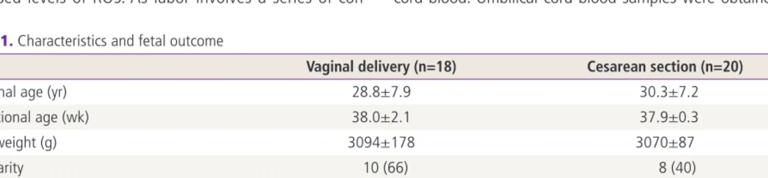
Table 1. Characteristics and fetal outcome
Vaginal delivery (n=18) Cesarean section (n=20)
Maternal age (yr) 28.8±7.9 30.3±7.2
Gestational age (wk) 38.0±2.1 37.9±0.3
Birth weight (g) 3094±178 3070±87
Nulliparity 10 (66) 8 (40)
Apgar score <7
1-min 1 (5.5) 1 (5.0)
5-min 0 (0) 0 (0)
Values are expressed as mean±SD or n (%).
mediately after delivery, before delivery of the placenta, and collected in a tube without anticoagulant. Tubes were centri- fuged at 2,000 ×g for 10 minutes at 4ºC to collect the serum;
samples were stored at -80ºC until analysis.
Lipid perioxidation was measured using the thiobarbituric acid reactionas described by Ohkawa et al. [13]. When thio- barbituric acid is applied to a mixture of acetaldehyde and sucrose, a 532 nm-absorbing chromogen is produced which is indistinguishable from that formed by the malondialdehyde and thiobarbituric acid adduct. The malondialdehyde assay is the most generally used test in the appreciation of the role of oxidative stress in disease. The results are expressed as nmol of malondialdehyde incorporated/mg of protein based on an average absorptivity (electromagnetic wave =1.56×10⁵).
Measurement of protein oxidation was determined by assay for protein carbonyl content using the 2,4-dinitrophenyl hy- drazine reaction. Serum samples were divided into two equal aliquots containing approximately 1.0 mg of protein each.
Both aliquots were precipitated with 10% trichloroacetic acid (w/v, final concentration). One sample was treated with 2N HCl, and the other sample was treated with an equal volume of 0.2% (w/v) dinitrophenylhydrazine (DNPH) in 2 N HCI. Both samples were incubated at 25ºC in 15-mL conical glass cen- trifuge tubes and stirred at 5-minutes intervals. The samples were then re-precipitated with 10% trichloroacetic acid (final concentration) and subsequently extracted with ethanol/ethyl acetate (l:l, v/v) and then re-precipitated at 10% trichloroace- tic acid. The pellets were carefully drained and dissolved in 6M guanidine HCI in 20 mM sodium phosphate buffer, pH 6.5.
Insoluble debris was removed by centrifugation at 6000 ×g at 4ºC. The difference between the spectra of the DNPH-treated sample versus the HCl control was determined and the results are expressed as nmol of DNPH incorporated/mg of protein based on an average absorptivity of 21.0 mM
-1cm
-1for most aliphatic hydrazones [14].
Total antioxidant capacity was measured by oxygen radi- cal absorbance capacity (ORAC) assay kit (Zen-Bio, Research Triangle Park, NC, USA).The ORAC assay was carried out at 37ºC on a spectrofluorometer at an excitation wavelength of 485 nm and an emission wavelength of 528 to 538 nm (cutoff, 530 nm). The procedure was based on the method reported by Cao et al. [15].
All data were presented as mean±standard deviation. The comparison of parameters were performed using an indepen- dent t-test. P<0.05 was considered statistically significant.
Data were analyzed using the IBM SPSS ver. 19.0 (IBM Co., Armonk, NY, USA). At a 2-sided, significance level at 0.05, we calculated a sample size of 15 patients would be necessary to demonstrate the difference between two groups cohort at 95% power. The sample size was increased to 20 patients in planned CS group (G-Power 3.1.5).
Results
As shown in Table 1, the age of the mother, fetal gestational age at delivery, birth weight, and apgar score did not differ among groups (P > 0.05). Lipid peroxide levels in umbilical venous blood were significantly higher in patients delivering by planned CS (1.81±0.06 nmol/mg protein) as compared to those who delivered vaginally (1.24 ± 0.05 nmol/mg
Fig. 1. Lipid peroxide levels in umbilical artery and vein. MDA, malondialdehyde; CS, cesarean section; VD, vaginal delivery.
a)P < 0.05.
Delivery mode
MDA (nmol/mg protein)
CS VD
Vein Artery
a)2.20 2.00 1.80 1.60 1.40 1.20 1.00
Fig. 2. Oxygen radical absorbance capacity (ORAC) levels in umbil- ical artery and vein. CS, cesarean section; VD, vaginal delivery.
a)P < 0.05.
Delivery mode
ORA C ( μM/ μoL)
CS VD
Vein Artery 121.00
120.00
119.00
118.00
117.00
a)