www.ogscience.org 641
Case Report
Obstet Gynecol Sci 2018;61(5):641-644 https://doi.org/10.5468/ogs.2018.61.5.641 pISSN 2287-8572 · eISSN 2287-8580
Introduction
Naturally, uterine fistulas such as utero-vesical or utero-colonic fisula are often seen in the patients with cervical cancer.
However, uterocutaneous fistula which is a pathologic com- munication between adherent uterus and skin surface is a rarely reported condition. The etiologies of uterocutaneous fistula are mostly iatrogenic, such as septic abortion probably after uterine perforation [1], previous uterine surgery includ- ing cesarean section [2], incomplete closure of incision site and incomplete placental removal especially with abnormally invasive placental implantation [3]. Owing to paucity of cases, there is no standard diagnostic and management strategies.
Many diagnostic imaging techniques such as sonography, fistulogram, pelvis magnetic resonance imaging (MRI) and ab- dominal-pelvic computed tomography (CT) are diversely used according to the physicians’ decisions. For management plan, major exploratory surgery including hysterectomy was used previously, but minimally invasive surgery or medical treat- ments are being suggested recently as an option for fertility- preservation. In this case report, we made a diagnosis with
hystero-salpingo contrast sonography and managed with surgical fistula tract excision and conservative medical therapy using gonadotropin-releasing hormone (GnRH) agonist.
Case report
A 30-year-old nulliparous woman was referred to general surgery department in our tertiary medical center for suspi-
Uterocutaneous fistula after pelviscopic myomectomy - successful diagnosis with hystero-salpingo contrast sonography and complete tract resection and medical treatment for fertility preservation in young woman:
a case report
Kyung-Jin Min
*, Jaeeun Lee
*, Seyoung Lee, Sanghoon Lee, Jin-Hwa Hong, Jae-Yun Song, Jae-Kwan Lee, Nak Woo Lee
Department of Obstetrics and Gynecology, Korea University Ansan Hospital, Ansan, Korea
A uterocutaneous fistula is rarely reported clinical condition after uterine procedures. Many diagnostic and management strategies are being suggested. In this case report, uterocutaneous fistula after pelviscopic myomectomy was diagnosed simply with hystero-salpingo contrast sonography and managed by surgical tract excision without hysterectomy and uterine wall dehiscence repair combined with medical treatment using gonadotropin-releasing hormone agonist succeeded to preserve fertility in young woman.
Keywords: Cutaneous fistula; Fertility preservation; Treatment
Articles published in Obstet Gynecol Sci are open-access, distributed under the terms of the Creative Commons Attribution Non-Commercial License (http://creativecommons.
org/licenses/by-nc/3.0/) which permits unrestricted non-commercial use, distribution, and reproduction in any medium, provided the original work is properly cited.
Copyright © 2018 Korean Society of Obstetrics and Gynecology
Received: 2017.07.19. Revised: 2017.09.05. Accepted: 2017.09.21.
Corresponding author: Nak Woo Lee
Department of Obstetrics and Gynecology, Korea University Ansan Hospital, 123 Jeokgeumno, Danwon-gu, Ansan 15355, Korea E-mail: [email protected]
https://orcid.org/0000-0001-9405-7574
*These authors contributed equally to this work as first author.
www.ogscience.org 642
Vol. 61, No. 5, 2018
cious small bowel ileus with possible obstruction 5 days after pelviscopic myomectomy on 10 cm sized myoma in private gynecologic hospital. On 6th day of admission, explorative laparotomy was performed due to suspected mechanical ob- struction on abdominal-pelvic CT and septic condition with persistent fever and aggravated abdominal pain in spite of conservative management such as NPO, hydration and use of antibiotics.
On the surgical exploration, greenish bowel contents were found in whole intra-peritoneal cavity. Three perforation sites of proximal ileum and generally edematous and fragile small intestine from distal jejunum to proximal ileum were visible.
After large amount of saline irrigation, about 1 meter long resection of small intestine and end-to-end anastomosis were performed. During the pelvic exploration, a ruptured uterine fundus was found with exposed endometrium and large he- matoma was visible on the myomectomy site. Dehiscenced myometrium was repaired by interrupted 2 points suture with Vicryl 2-0 (Ethicon, Cincinnati, OH, USA).
Follow-up abdominal-pelvic CT was taken on 8 days after explorative surgery due to bloody discharge from the mid-line abdominal wound with menstrual period. It revealed large he- matoma around uterine fundus and accompanied peritoneal thickening. Percutaneous abdominal drainage was inserted to the hematoma, and bacterial culture from the drainage revealed Citrobacter freundii and Enterococcus faecalis infec- tion. After proper antibiotics treatment, the patient was dis- charged with afebrile condition. After twice a weekly follow- up for 1 month, the bloody discharge was noticed ongoing just during menstrual periods. Biopsy was performed on no improved abdominal wound, and pathologic report suggest- ed endometriosis with acute and chronic inflammation with abscess formation.
Hystero-salpingo contrast sonography was performed on the first visit in our out-patient department, 2 and a half months after initial pelviscopic myomectomy. The result re- vealed endometrial cavity linked to the right side of uterine fundus at which previous myomectomy was performed. Also, contrast spillage to the intra-abdominal cavity and 0.5 cm defect in the lower part of abdominal wound was observed (Fig. 1A). Reoperation was planned for probable myometrial dehiscence repair and simultaneously uterocutaneous fistula tract removal.
On the explorative laparotomy performed 3 months after initial pelviscopic myomectomy, 6 cm low vertical incision
was made including total removal of fistula tract opening.
Fistulous tract connecting abdominal skin to the previously unhealed myomectomy site was completely resected together with surrounding soft tissues. Adhesion of small intestine, sig- moid colon, bladder dome and omentum around the fistula tract were unstucked. Percutaneous abdominal drainage site through the left lower quadrant skin puncture was also com- pletely resected to prevent further fistula formation. Ragged uterine fundus tissue was debrided and dehiscenced uterine wall was repaired layer by layer, Vicryl 3-0 (Ethicon) inter- rupted suture on endometrial layer, Vicryl 2-0 (Ethicon) inter- rupted suture on myometrial layer.
Resected abdominal wall fistula tract specimen revealed acute and chronic inflammation with granulation tissue, ex- tensive fibrosis, and foreign body reaction on a pathologic report. Percutaneous abdominal drainage insertion site tissue Fig. 1. (A) Hysterosalpingo contrast sonography. Each marks mean follow as; Astrix - uterine outline, short arrow - EM cavity with cath- eter ballooning, long arrow - contrast leakage through right side of fundus to the pelvic cavity, and B - bowel. (B) postoperative sono revealing well margined myometrium.
A
B
www.ogscience.org 643 Kyung-Jin Min, et al. Successful treatment of uterocutaneous fistula
revealed chronic inflammation with extensive fibrosis, hemor- rhage and foamy histocytes on a pathologic report.
Transvaginal sonography taken on 4 days after fistulectomy and uterine repair revealed well margined myometrium with- out hematoma and no free fluid collection in pelvic cavity (Fig.
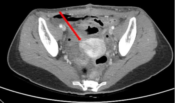
1B). On 6 days after surgery, abdominal skin wound was well healed and stitched out. Also, follow-up abdominal-pelvic CT revealed immediate postoperative state of myomectomy site without any evidence of dehiscence (Fig. 2). The patient was discharged with first 3.75 mg GnRH agonist (Takeda Pharmaceutical Company, Tokyo, Japan) injection. During the 3 months of GnRH agonist after the surgery, there is no evi- dence of recurrent fistula formation until now.
Discussion
Although uterocutaneous fistula is a rare condition, it can be presumed with typical clinical symptoms such as bloody dis- charge during menstrual periods and suspicious clinical histo- ries of uterine procedures.
Many diagnostic methods are attempted so far, and so- nography is the fundamental imaging techniques. However, abdominal fistula hardly detected through transvaginal ap- proaches. After obtaining suggestive image through trans- abdominal sonography, fistulogram can be used by injecting contrast-material via the skin opening [2]. However, narrowed lumen with small cutaneous opening hole cannot be able to visualize the passage of contrast materials [4]. Then, contrast- enhanced MRI or CT would be helpful in planning surgical in- tervention. More precisely, hystero-salpingo sonography with
contrast injection through uterine cervix by vaginal approach can be used instead [4]. In this case, we used hystero-salpingo sonography with contrast material via cervical opening to di- agnose uterocutaneous fistula.
Since 1958 when the first case report about uterocutaneous fistula was published, a complete surgical resection including hysterectomy was the only curative method [5,6] until 2007.
Even medical treatment can be used to relief clinical symp- toms, it did not regarded in achieving complete eradication of the fistula lesion [7]. Therefore even bearing the risk of interval secondary surgery, the aim of the treatment was only complete surgical excision [8].
After 2008, medical treatment solely or combined to surgi- cal treatment for young women who has a strongly desire fer- tility preserving were being reported [3,9]. In these recent re- ports, medical treatment with GnRH agonist might atrophied fistula epithelium and spontaneous closure was achieved [9].
In this way, uterocutaneous fistula can be conservatively treat- ed with timely intervention without major surgery including hysterectomy. In one report, uterocutaneous fistula due to un- derlying peritonitis after septic abortion was completely cured with foreign body removal, irrigation, debridement and use of antibiotics. After 6 months of treatment, normal menstrual cycle was restored [1]. Also minimally invasive laparoscopic or combined laparoscopic and laparotomic surgery after several times of repeated medical treatment reported successful clini- cal outcomes [10].
In this case reports as well, including prolonged use of anti- biotics until eradication of bacteremia, complete resection and debridement of fragile, necrotic, extrauterine endometriotic tissues succeed to repair uterine wall layer by layer without further dehiscence, and complete closure of abdominal wall.
Long term follow-up would be necessary to find whether use of GnRH agonist causing further atrophy of ectopic endo- metrial glands surely prevent recurrence of fistula or develop any sign of treatment failure. Also after the completion of medical therapy, restoration of normalized menstrual cycle and further pregnancy result should be confirmed [11].
In conclusion, hystero-salpingo contrast sonography is in- expensive, convenient method to diagnose uterocutaneous fistula even with narrowed lumen opening. Nowadays, hys- terectomy to completely eradicate fistula lesion is not the only management option any more. Proper conservative manage- ment and adjuvant medical therapy can be used as a fertility- preserving treatment option.
Fig. 2. Postoperative abdominal-pelvic computed tomography; im- mediate postoperative state without evidence of dehiscence. Arrow means sutured uterine wall.
www.ogscience.org 644
Vol. 61, No. 5, 2018
Conflict of interest
No potential conflict of interest relevant to this article was reported.
References
1. Pratibha S, Rajni B. Uterocutaneous fistula following septic abortion: can it heal without major surgical inter- vention? J Obstet Gynaecol 2015;35:651-2.
2. Maddah G, Fattahi AS, Rahnama A, Jamshidi ST. Utero- cutaneous fistula following cesarean section: successful management of a case. Iran J Med Sci 2016;41:157-60.
3. Athanasias P, Krishna A, Karoshi M, Moore J, Chandraha- ran E. Uterocutaneous fistula following classical caesar- ean delivery for placenta percreta with intentional reten- tion of the placenta. J Obstet Gynaecol 2013;33:906-7.
4. Sönmezer M, Sahincioğlu O, Cetinkaya E, Yazici F. Utero- cutaneous fistula after surgical treatment of an incom- plete abortion: methylene blue test to verify the diagno- sis. Arch Gynecol Obstet 2009;279:225-7.
5. Case TC. Uterocutaneous fistula; a case report. Obstet Gynecol 1958.12:233-4.
6. Gupta SK, Shukla VK, Varma DN, Roy SK. Uterocutane- ous fistula. Postgrad Med J 1993;69:822-3.
7. Dragoumis K, Mikos T, Zafrakas M, Assimakopoulos E, Stamatopoulos P, Bontis J. Endometriotic uterocutane- ous fistula after cesarean section. A case report. Gynecol Obstet Invest 2004;57:90-2.
8. Promsonthi P, Herabutya Y. Uterocutaneous fistula in term abdominal pregnancy. Eur J Obstet Gynecol Reprod Biol 2007;132:239-41.
9. Seyhan A, Ata B, Sidal B, Urman B. Medical treatment of uterocutaneous fistula with gonadotropin-releasing hormone agonist administration. Obstet Gynecol 2008;111:526-8.
10. Thubert T, Denoiseux C, Faivre E, Naveau A, Trichot C, Deffieux X. Combined conservative surgical and medical treatment of a uterocutaneous fistula. J Minim Invasive Gynecol 2012;19:244-7.
11. Yadav P, Gupta S, Singh P, Tripathi S. Successful medical management of uterocutaneous fistula. Int J Gynaecol Obstet 2014;124:263-4.