372
ORIGINAL ARTICLEDOI 10.4070 / kcj.2009.39.9.372
Print ISSN 1738-5520 / On-line ISSN 1738-5555 Copyright ⓒ 2009 The Korean Society of Cardiology
The Relationship Between Chronic Atrial Fibrillation and Reduced Pulmonary Function in Cases of Preserved Left Ventricular Systolic Function
Hyunjae Kang, MD, Byung Seok Bae, MD, Jae Hoon Kim, MD, Hee Sang Jang, MD, Bong-Ryeol Lee, MD and Byung-Chun Jung, MD
Department of Cardiology, Fatima General Hospital, Daegu, KoreaABSTRACT
Background and Objectives:
The purpose of this study was to investigate the relationship between chronic atrial fibrillation (AF) and reduced pulmonary function.
Subjects and Methods:Eighty-six chronic AF patients who were enrolled from annual health examination programs were studied using echocardiography and pulmonary function tests (PFT). Echocardiography and PFT matched for age, gender, and year performed were selected by the control group who had normal sinus rhythms. Patients with ejection fractions <50%, valvular heart disease, or ischemic heart disease were excluded.
Results:In the chronic AF patients, the forced expiratory volume at one second (FEV
1), FEV1%, and FEV
1/forced vital capacity (FVC) were significantly reduced, and the right ventricular systolic pressure was significantly increased. Episodes of heart failure were more frequently associated with the chronic AF patients than the controls. In particular, the FEV1% had the most meaningful relationship to chronic AF after an adjustment for cardiovascular risk factors {p=0.003, Exp (B)=0.978, 95% confidence interval (CI):
0.963-0.993}.
Conclusion:Reduced FEV1%, which represents the severity of airway obstruction, was associated with chronic AF, and the greater the pulmonary function impairment, the greater the co-existence with AF and congestive heart failure in those with preserved left ventricular systolic function.
(Korean Circ J 2009;39:372-377)KEY WORDS: Atrial fibrillation; Chronic obstructive lung disease; Pulmonary function tests; Forced expiratory
volumes.
Introduction
Atrial fibrillation (AF) is frequently observed among supraventricular rhythm disorders. The prevalence of AF increases with age.
1-4)Pulmonary function, however, has a tendency to decrease progressively with age. The supra- ventricular and ventricular arrhythmias are also common in chronic obstructive lung disease (COPD).
5-7)The rea- sons are thought to be due to hypoxia, hypercarbia, pul- monary hypertension, and myocardial ischemia, which are easily provoked by this limited ventilatory condi- tion.
8-11)Furthermore, the frequencies of various types of arrhythmias are related to the temporal state of the pulmonary function in these patients. Cardiac arrhyth-
mias have a tendency to become aggravated when venti- latory function deteriorates, and ameliorated when it improves; however, there is still controversy regarding the relationship between AF and reduced pulmonary function.
The purpose of the current study was to determine the relationship between chronic AF and pulmonary function status among subjects with preserved left ven- tricular systolic function (LVSF), and the difference in pulmonary function between the chronic AF group and the controls who presented with sinus rhythm (SR).
Subjects and Methods
Subjects
Eighty-six chronic AF patients were enrolled from an annual health examination program between August 2006 and June 2007, and were studied by echocardiogra- phy and pulmonary function tests (PFT). Echocardio- graphy and PFT matched for age, gender, and year per- formed were selected by the control group who had normal sinus rhythm (SR) (Table 1). The subjects with a
Received: January 21, 2009 Revision Received: April 8, 2009 Accepted: April 24, 2009
Correspondence: Byung-Chun Jung, MD,Department ofCardiology, Fatima General Hospital, 302-1 Sinam-dong, Dong-gu, Daegu 701-600, Korea Tel: 82-53-940-7214, 7459, Fax: 82-53-954-7417
E-mail: augustjbc@yahoo.co.kr
Hyunjae Kang, et al.·
373
left ventricular ejection fraction (LVEF) <50% and with valvular heart disease were excluded from the study.
Also, any patients who had a history of percutaneous co- ronary intervention, coronary artery bypass surgery, or Q-waves on a surface electrocardiogram (ECG) were ex- cluded. Chronic AF was considered when AF was de- tected consecutively >2 ECGs during a 1 month inter- val. Finally, the subject was allocated into either the chronic AF group or the control group, depending on whether the cardiac rhythm was AF or normal SR.
Assessment of risk factors
The clinical data on risk factors for cardiovascular di- sease that were related to AF were obtained from a com- prehensive review of each patient’s medical record. Th- erefore, each patient’s age, gender, and history of syste- mic hypertension, diabetes mellitus, hyperlipidemia, and current smoking were investigated (Table 1). Systemic hypertension was defined by documenting the clinical diagnosis, or with evidence of an elevated systolic blood pressure of 140 mmHg and/or diastolic blood pressure of 90 mmHg, in the absence of any acute medical ill- nesses. Diabetes mellitus was based on the clinical do- cumentation of the condition, and whether the treat- ment consisted of dietary therapy, oral hypoglycemic agents, and/or insulin. Hyperlipidemia was defined by a fasting total cholesterol level of 240 mg/dL and/or a low-density lipoprotein cholesterol level of 160 mg/dL, or by the use of cholesterol-lowering drugs. Current smoking referred to the active use of tobacco products at the time of enrollment in the study. Furthermore, des- pite all the subjects presenting with preserved LVSF, a previous history of heart failure (HF) was also evaluat-
ed by the Framingham heart failure diagnostic criteria
12)because diastolic heart failure is not uncommonly as- sociated with elderly patients and AF with a rapid ven- tricular response is a well-known cause of reversible car- diomyopathy.
Echocardiographic examination
In the echocardiographic study, the conventional two- dimensional grayscale imaging, the pulsed or continuous wave Doppler study, and color Doppler imaging were performed according to previously validated recommen- dations.
13)The semi-quantitative visual estimation and the modified Simpson’s method were used to assess LVSF, and the patients with a LVEF <50% by either of the two methods were excluded. The wall-motion score index was also applied to rule out cases with regional wall motion abnormalities, which were calculated by di- viding the sum of the scores by the number of visualized segments. The left atrial (LA) dimension was measured at the point of end-systole just before the frame that preceded the mitral valve opening from the parasternal long-axis view. The peak systolic pressure of the right ventricle (pRVSPr) was carefully estimated using a previo- usly validated method
13)when tricuspid regurgitation was present. Subjects with insufficient aortic or mitral valves (greater than a mild degree), stenosis of the valves, or structural abnormalities were excluded from the study.
Evaluation of pulmonary function
The forced expiratory volume at one second (FEV
1), forced vital capacity (FVC), mean forced expiratory flow during the middle half of the FVC (FEF
25-75%), and the ratio of the FEV
1-to-FVC (FEV
1/FVC) were measured
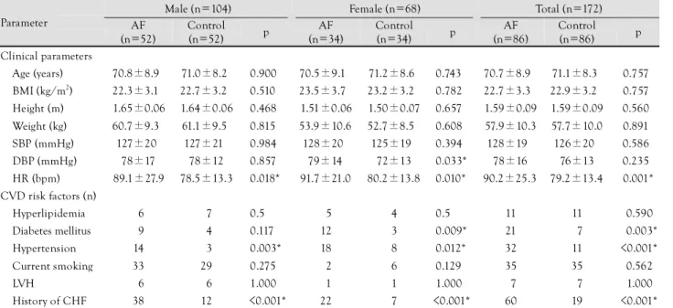
Table 1. Baseline characteristics of the study population
Male (n=104) Female (n=68) Total (n=172)
Parameter AF
(n=52)
Control
(n=52) p AF
(n=34)
Control
(n=34) p AF
(n=86)
Control (n=86) p
Clinical parameters
Age (years) 70.8±8.9 71.0±8.2 <0.900 70.5±9.1 71.2±8.6 <0.743* 70.7±8.9 71.1±8.3 0.757*
BMI (kg/m2) 22.3±3.1 22.7±3.2 <0.510 23.5±3.7 23.2±3.2 <0.782* 22.7±3.3 22.9±3.2 0.757*
Height (m) 1.65±0.06 1.64±0.06 <0.468 1.51±0.06 1.50±0.07 <0.657* 1.59±0.09 1.59±0.09 0.560*
Weight (kg) 60.7±9.3 61.1±9.5 <0.815 53.9±10.6 52.7±8.5 <0.608* 57.9±10.3 57.7±10.0 0.891*
SBP (mmHg) 127±20 127±21 <0.984 128±20 125±19 <0.394* 128±19 126±20 0.586*
DBP (mmHg) 78±17 78±12 <0.857 79±14 72±13 <0.033* 78±16 76±13 0.235*
HR (bpm) 89.1±27.9 78.5±13.3 <0.018* 91.7±21.0 80.2±13.8 <0.010* 90.2±25.3 79.2±13.4 0.001*
CVD risk factors (n)
Hyperlipidemia 06 07 <0.500* 05 4 <0.500* 11 11 0.590*
Diabetes mellitus 09 04 <0.117* 12 3 <0.009* 21 07 0.003*
Hypertension 14 03 <0.003* 18 8 <0.012* 32 11 <0.001*
Current smoking 33 29 <0.275* 02 6 <0.129* 35 35 0.562*
LVH 06 06 <1.000* 01 1 <1.000* 07 07 1.000*
History of CHF 38 12 <0.001* 22 7 <0.001* 60 19 <0.001*
*statistically significant. AF: atrial fibrillation, BMI: body mass index, SBP: systolic blood pressure, DBP: diastolic blood pressure, HR: heart rate, CVD: cardiovascular disease, LVH: left ventricular hypertrophy, CHF: congestive heart failure
374
·Chronic AF and Pulmonary Functionby an electronic spirometer (V
max229 pulmonary func- tion test/cardiopulmonary exercise testing instrument;
Sensomedics, Yorba Linda, CA, USA). Also, the peak expiratory flow (PEF) and the expiratory flow at 25%, 50%, and 75% FVC (FEF
25%, FEF
50%, and FEF
75%, res- pectively) were obtained from the flow-volume curve.
Body plethysmography was applied to the measurement of the residual volume (RV), total lung capacity (TLC), airway resistance (Raw), and lung compliance (Gaw).
The single breath technique with carbon monoxide was adopted for the measurement of the diffusion capacity of the lungs (DLCO). Specially-trained technicians per- formed all of these measurements. Moreover, all of the values, except for the FEV
1/FVC, which was measured with the spirometer, were automatically recalculated as percentile predicted values of the age- and gender-mat- ched normal values using a computer software program.
For example, the FEV1% was obtained from the follow- ing formulae, in which H is the height (cm) and A is the age (year): FEV1%=0.092 H/2.54-0.032 A-1.26 (males);
and FEV1%=0.089 H/2.54-0.025 A-1.932 (females).
Statistical analysis
The data are presented as the mean values ±standard deviations (SDs), unless otherwise stated. Continuous data on the two groups were compared with an indepen- dent sample t-test, and categorical data were compared with a chi-square test. To reveal the relationship of the dichotomous data with the continuous data, logistic re- gression analysis was applied (SPSS, version 13.0; SPSS Inc., Chicago, IL, USA). The statistical differences were considered significant at a p<0.05.
Results
Baseline characteristics
The mean age, body mass index (BMI), and systolic and diastolic blood pressures (BP) of the AF group were similar to the controls. These findings, except for the di- astolic BP in females, were also noted when comparing those parameters between the gender-specific subgroups.
Regardless of gender, the resting heart rate (HR) of the AF group was significantly higher than the controls (p=
0.001). The incidence of hyperlipidemia and current smoking did not differ between the AF and control gro- ups. The AF group, however, had a significantly higher incidence of hypertension for both genders, and diabetes in females. The incidence of previous HF was much hi- gher in the AF than in the control groups, as expected (60% vs. 19%, p<0.001).
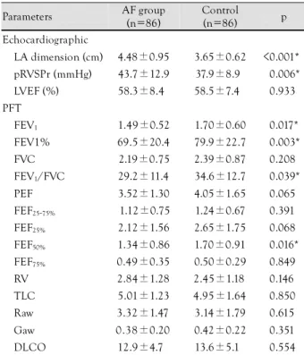
Characteristics of echocardiography and pulmo- nary function tests of the atrial fibrillation and control groups
The LA dimension and pRVSPr of the AF group were
significantly greater than the controls (4.48±0.95 cm vs. 3.65±0.62 cm, p<0.001, and 43.7±12.9 mmHg vs.
37.9±8.9 mmHg, p=0.006, respectively), even though the LVEF of the two groups was similar (Table 2).
Among the parameters of the PFT, the FEV
1, FEV1%, and FEV
1/FVC were significantly lower in the AF group than the control group. The other parameters were not statistically different between the two groups (Table 2).
Therefore, ventilatory dysfunction, rather than SR, ap- peared to be more associated with the AF group. When logistic regression was performed with the FEV
1, FEV1%, and FEV
1/FVC as a function of the cardiac rhythm and with the adjustment of risk factors {hypertension, dia- betes mellitus, and congestive heart failure (CHF)}, FEV 1% had the most meaningful relationship with AF {p
=0.003, Exp (B)=0.978, 95% confidence interval (CI):
0.963-0.993}; FEV
1also exhibited a meaningful relation- ship {p=0.019, Exp (B)=0.520, 95% CI: 0.301-0.897}.
An inverse correlation between FEV1% and the pRVSPr was observed in this study, which was in agreement with the secondary pulmonary hypertension results of the reduced ventilatory function. Furthermore, an increase in the pRVSPr represented a close relationship with
Table 2. Characteristics of echocardiographic examination and pulmonary function tests
Parameters AF group (n=86)
Control
(n=86) p Echocardiographic
LA dimension (cm) 4.48±0.95 3.65±0.62 <0.001*
pRVSPr (mmHg) 43.7±12.9 37.9±8.90 <0.006*
LVEF (%) 58.3±8.4 58.5±7.40 <0.933*
PFT FEV1 1.49±0.52 1.70±0.60 <0.017*
FEV1% 69.5±20.4 79.9±22.7 <0.003*
FVC 2.19±0.75 2.39±0.87 <0.208*
FEV1/FVC 29.2±11.4 34.6±12.7 <0.039*
PEF 3.52±1.30 4.05±1.65 <0.065*
FEF25-75% 1.12±0.75 1.24±0.67 <0.391*
FEF25% 2.12±1.56 2.65±1.75 <0.068*
FEF50% 1.34±0.86 1.70±0.91 <0.016*
FEF75% 0.49±0.35 0.50±0.29 <0.849*
RV 2.84±1.28 2.45±1.18 <0.146*
TLC 5.01±1.23 4.95±1.64 <0.850*
Raw 3.32±1.47 3.14±1.79 <0.615*
Gaw 0.38±0.20 0.42±0.22 <0.351*
DLCO 12.9±4.7 13.6±5.1 <0.554*
AF: atrial fibrillation, LA: left atrium, pRVSPr: peak right ventri- cular systolic pressure, LVEF: left ventricular ejection fraction, PFT:
pulmonary function test, FEV1: forced expiratory volume at on se- cond, FVC: forced vital capacity, FEV1/FVC: ratio of the FEV1 to FVC, PEF: peak expiratory flow, FEF25-75%: mean forced expiratory flow during the middle half of the FVC, FEF25%: expiratory flow at the 25%, FEF50%: expiratory flow at the 50%, FEF75%: expiratory flow at the 75%, RV: residual volume, TLC: total lung capacity, Raw: airway resistance, Gaw: lung compliance, DLCO: diffusion capacity of the lung
Hyunjae Kang, et al.·
375
AF {p=0.009, Exp (B)=1.052, 95% CI: 1.013-1.092}.
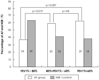
Categorization of the subjects according to the forced expiratory volume at one second range
The FEV1% values were categorized into three sub- groups by applying cutoff levels of 80% and 60% of the predictive value in both groups, which were the levels adopted in many previous studies. In the cases in which the FEV1% was >80% of the predictive value, SR was most frequently observed. AF was most com- monly observed in cases in which the FEV1% was <60%
(Fig. 1). Using the Mantel-Haenszel estimation, the prev- alence of AF in the cases with a FEV1% between 60%
and 80%, and <60% exhibited odds ratios (ORs) of 2.265 (p=0.019) and 2.887 (p=0.007), respectively, when compared to cases with a FEV1% >80%. The prevalence
of AF in the cases with a FEV1% <60% did not differ from the prevalence of AF in the cases with a FEV1%
between 60% and 80% (OR=1.221, p=0.619).
Relationship between congestive heart failure and atrial fibrillation and forced expiratory volume at one second in patients with preserved LVSF
A normal ejection fraction can exist in patients with symptoms of heart failure, and is regarded as an indica- tion of diastolic heart failure. Moreover, AF can induce an enhanced ventricular response, which will result in the aggravation of heart failure. In this study, a history of CHF was observed more frequently in the AF group than in the control group. The FEV1% had a meaningful relationship with CHF {p=0.008, Exp (B)=0.980, 95%
CI: 0.965-0.995}. The mean value of the FEV1% also differed significantly according to whether or not the patient had a history of heart failure (69.6±19.7% vs.
79.0±23.2%, p=0.006). The prevalence in the patients with a FEV1% between 60% and 80% and <60%, had an OR of 3.102 (p=0.003) and 2.910 (p=0.007), res- pectively, compared to a FEV1% >80% (Fig. 2). The pre- valence of CHF in the patients with a FEV1% <60% did not differ from the prevalence of CHF in the patients with a FEV1% between 60% and 80% (OR=0.938, p=0.871).
Discussion
In several studies that used multivariate analyses with correction for age, the major risk factors for AF were hy- pertension, heart failure, diabetes mellitus, and valvular heart disease. Bundle branch block and left ventricular hypertrophy were regarded as potential predictors of AF.
14)However, the results regarding the relationship between AF and pulmonary function have been discordant. The FEV1% was reported to be an important predictive fac- tor for AF in the Copenhagen City Heart Study,
15)which was comprised of a healthy age-stratified cohort (n=
13,430) with a prospective design. Psaty et al.
16)also re- ported that the occurrence of AF was related to reduced pulmonary function in the Cardiovascular Health Study (n=5,201). In contrast, the Renfrew/Paisley study
14)(n=
15,406) did not demonstrate a significant correlation between the FEV1% and AF, whereas it reported the traditional major risk factors to be significant. Similarly, the Framingham study
1)(n=4,731) found no relationship between the FEV
1and AF.
To address these discordant results on the relationship between AF and reduced pulmonary function, the cur- rent study was conducted as a case-control study with consecutive enrollment of patients. In this study, a re- duced FEV1% was significantly associated with chronic AF, and this finding was supported by the results of the preceding two studies. The Renfrew/Paisley study,
14)AF group NSR (control)
Percentage of AF and NSR (%)
90 80 70 60 50 40
30 20 10 0
FEV1%≥80% 80%>FEV1%≥60% FEV1%<60%
p=0.007 p=NS p=0.019
24 43 33 25 29 18
Fig. 1. Histogram showing percentage of atrial fibrillation and normal sinus rhythm groups, stratified into FEV1% categories.
FEV1%: forced expiratory volume at one second, AF: atrial fibrillation, NSR: normal sinus rhythm, NS: not significant.
p=0.007 p=0.003 p=NS
HF (positive) HF (negative)
Percentage of AF and NSR (%)
90 80 70 60 50 40 30 20 10 0
FEV1%≥80% 80%>FEV1%≥60% FEV1%<60%
20 47 33 25 26 21
Fig. 2. Histogram showing relationship of heart failure with atrial fibrillation, stratified into FEV1% categories. FEV1%: forced expiratory volume at one second, AF: atrial fibrillation, NSR: nor- mal sinus rhythm, NS: not significant, HF: heart failure.
376
·Chronic AF and Pulmonary Functionwhich reported the opposite results, observed a small number of new-onset AF cases (n=19) after a short fol- low-up and reported cardiomegaly as the most powerful predictor for AF (OR=14.0) after a long-term follow-up, which means that subjects with a decreased LVSF were enrolled. In the Framingham study,
1)there was a high rate of heart disease. For example, the prevalence of val- vular heart disease was 7% in males and 9% in females;
patients with valvular heart disease were excluded from our study. Therefore, the inclusion of these strong risk factors may have obscured the relationship between the FEV1% and AF in the two aforementioned studies.
The pathophysiologic mechanisms that connect re- duced pulmonary function to chronic AF have not been clearly determined, but there are several suggestive ex- planations. With respect to the first explanation, recent observations have indicated that ectopic beats that initi- ate AF often originate in the pulmonary veins.
17)18)Re- duced ventilatory function could easily trigger ectopic beats by deterioration of the blood gas composition, such as occurs with hypoxia, and pulmonary hypertension results in stress on the right atrium and connecting ves- sels, thus perpetuating AF. In this study, the pulmonary artery pressure (pPAPr) in the AF group was significant- ly higher than the control group. The second explanation can be assumed to be the chronic inflammatory proces- ses that involve the cardiopulmonary system. Anatomi- cally, the pulmonary circulation directly drains into the left atrium. Also, obstructive airway disease is the result of chronic inflammation of the airways of the lungs that consequently manifests in reduced FEV
1or FEV1%.
Recent studies have reported that these processes are as- sociated with an increase in several inflammation-sensi- tive proteins (ISP), such as high sensitivity-C-reactive pro- tein (CRP), fibrinogen, and cytokines, including inter- leukin-6 (IL-6), IL-1, tumor necrosis factor (TNF)-α, and the complement system.
19-22)For example, the level of hs-CRP is known to increase in patients with AF;
hsCRP is synthesized from the liver by stimulation of IL-6, -11, and -12 and is frequently associated with fi- brosis of the atrial tissue and myocarditis on histologic examination. Fibrosis of the atrial tissue is now accept- ed as a perpetuating factor of AF. Therefore, it can be speculated that chronic inflammation is a plausible me- chanism that connects reduced pulmonary function with chronic AF, which was clinically manifested as the relationship between chronic AF and decreased FEV 1% in this study. The third explanation involves the hemodynamic consequences of AF. It is well-recognized that the loss of the atrial contraction decreases cardiac output and causes an increase in backward pressure and congestion of the lungs, especially the small airways.
As a consequence, those hemodynamic alterations not only result in atrial dilation, wall stretching, and elec- trical remodeling of the atrial tissue, but also hinders
the ventilatory function of the lungs.
23)24)In this study, when comparing the parameters for estimating small air- way function, FEF
25-75%, FEF
25%, and FEF
75%, but not FEF
50%, did not differ between the groups. Also, the DLCO exhibited no manifestations compatible with that assumption. Therefore, the results of this study do not sufficiently support the aforementioned assumpt- ion. However, the possibility that these parameters may present meaningful results by an exaggeration of the hemodynamic effects, such as an exercise challenge, can- not be abandoned entirely because these parameters were measured only during the resting state.
In addition, a close relationship existed between the FEV1% and symptomatic episodes of heart failure in the AF group {p=0.011, Exp (B)=0.965, 95% CI: 0.939- 0.002} after adjustment for the risk factors (hypertension and diabetes mellitus), whereas no relationship was ob- served in the control group. AF can cause heart failure through a rapid ventricular response, which results in an elevated end diastolic pressure and backward conges- tion, even in patients with a normal ejection fraction.
Hence, these findings imply that reduced ventilatory function as also attributed to heart failure from a rapid ventricular rate during AF.
Clinical implications and limitations
In clinical practice, β-blockers, which can adversely af- fect ventilation, are commonly administered to alleviate the ventricular rate in AF with preserved LVSF, as are β -agonists, which can aggravate the cardiac rhythm to manage reduced lung function. Prudent use of those dru- gs is likely to be needed because chronic AF co-existing with reduced ventilatory function was not rare in this study.
Similar to other case control studies, the cause-and- effect relationship and the underlying mechanisms were not clearly determined in this study, but the FEV1% was revealed to be a co-morbid factor for chronic AF. Thus, a further study on these issues should be pursued.
In conclusion, this study showed that reduced pul- monary function was related to AF as a co-morbid factor, and was clearly attributed to hemodynamic alterations, such as congestion during chronic AF, even when a nor- mal ejection fraction was present.
REFERENCES
1) Benjamin EJ, Levy D, Vaziri SM, Dágostino RB, Belanger AJ, Wolf PA. Independent risk factors for atrial fibrillation in a popu- lation-based cohort. JAMA 1994;271:840-4.
2) Go AS, Hylek EM, Phillips KA, et al. Prevalence of diagnosed atrial fibrillation in adults: national implications for rhythm ma- nagement and stroke prevention. JAMA 2001;285:2370-5.
3) Tomita F, Kohya T, Sakurai M, et al. Prevalence and clinical cha- racteristics of patients with atrial fibrillation: analysis of 20,000 cases in Japan. Jpn Circ J 2000;64:653-8.
4) Feinberg WM, Blackshear JL, Laupacis A, Kronmal R, Hart RG.
Hyunjae Kang, et al.·
377
Prevalence, age distribution, and gender of patients with atrial fi- brillation: analysis and implication. Arch Intern Med 1995;155:
469-73.
5) Sideris DA, Katsadoros DP, Valianos G, Assioura A. Type of car- diac dysrhythmias in respiratory failure. Am Heart J 1975;89:32-5.
6) McCord J, Borzak S. Multifocal atrial tachycardia. Chest 1998;
113:203-9.
7) Kothari SA, Apiyasawat S, Asad N, Spodick DH. Evidence sup- porting a new rate threshold for multifocal atrial tachycardia.
Clin Cardiol 2005;28:561-3.
8) Khokhar N. Cardiac arrhythmias associated with acute respira- tory failure in chronic obstructive pulmonary disease. Mil Med 1981;146:856-8.
9) Levine PA, Klein MD. Mechanisms of arrhythmias in chronic obstructive lung disease. Geriatrics 1976;31:47-56.
10) Thomas AJ, Valabhji P. Arrhythmia and tachycardia in pulmonary heart disease. Br Heart J 1969;31:491-5.
11) Holford FD, Mithoefer JC. Cardiac arrhythmias in hospitalized patients with chronic obstructive pulmonary disease. Am Rev Re- spir Dis 1973;108:879-85.
12) Ho KK, Anderson KM, Kannel WB, Grossman W, Levy D. Sur- vival after the onset of heart failure in Framingham Heart Study subjects. Circulation 1993;88:107-15.
13) Cheitlin MD, Armstrong WF, Aurigemma GP, et al. ACC/AHA/
ASE 2003 guideline update for the clinical application of echo- cardiography: summary article: a report of the American College of Cardiology/American Heart Association Task Force on Practice Guidelines (ACC/AHA/ASE Committee to Update the 1997 Gui- delines for the Clinical Application of Echocardiography). Cir- culation 2003;108:1146-62.
14) Stewart S, Hart CL, Hole DJ, McMurray JJ. Population prevalence, incidence, and predictors of atrial fibrillation in the Renfrew/Pai-
sley study. Heart 2001;86:516-21.
15) Buch P, Friberg J, Scharling H, Lange P, Prescott E. Reduced lung function and risk of atrial fibrillation in the Copenhagen City Heart Study. Eur Respir J 2003;21:1012-6.
16) Psaty BM, Manolio TA, Kuller LH, et al. Incidence of and risk factors for atrial fibrillation in older adults. Circulation 1997;
96:2455-61.
17) Haissaguerre M, Jais P, Shah DC, et al. Spontaneous initiation of atrial fibrillation by ectopic beats originating in the pulmonary veins. N Engl J Med 1998;339:659-66.
18) Olsson SB. Atrial fibrillation: where do we stand today? J Intern Med 2001;250:19-28.
19) Chung MK, Martin DO, Sprecher D, et al. C-reactive protein ele- vation in patients with atrial arrhythmias: inflammatory mecha- nisms and persistence of atrial fibrillation. Circulation 2001;104:
2886-91.
20) Hwang SJ, Sung KC, Lee YS, et al. Serum C-reactive protein le- vel and its association with atrial fibrillation in Korean adults.
Korean Circ J 2005;35:309-14.
21) Boos CJ, Anderson RA, Lip GY. Is atrial fibrillation an inflam- matory disorder? Eur Heart J 2006;27:136-49.
22) Psychari SN, Apostolou TS, Sinos L, Hamodraka E, Liakos G, Kremastinos DT. Relation of elevated C-reactive protein and interleukin-6 levels to left atrial size and duration of episodes in patients with atrial fibrillation. Am J Cardiol 2005;95:764-7.
23) Wijffels MC, Kirchhof CJ, Dorland R, Allessie MA. Atrial fibril- lation begets atrial fibrillation: a study in awake chronically instrumented goats. Circulation 1995;92:1954-68.
24) Goette A, Honeycutt C, Langberg JJ. Electrical remodeling in at- rial fibrillation: time course and mechanisms. Circulation 1996;
94:2968-74.