Isolation of human mesenchymal stem cells from the skin and their neurogenic differentiation in vitro
Jun-Ho Byun1, Eun-Ju Kang2, Seong-Cheol Park1, Dong-Ho Kang3, Mun-Jeong Choi1, Gyu-Jin Rho2, Bong-Wook Park1
1Department of Oral and Maxillofacial Surgery, School of Medicine and Institute of Health Science, Gyeongsang National University,
2OBS/Theriogenology and Biotechnology, College of Veterinary Medicine, Gyeongsang National University,
3Department of Neurosurgery, School of Medicine, Gyeongsang National University, Jinju, Korea
Abstract(J Korean Assoc Oral Maxillofac Surg 2012;38:343-53)
Objectives: This aim of this study was to effectively isolate mesenchymal stem cells (hSMSCs) from human submandibular skin tissues (termed hSMSCs) and evaluate their characteristics. These hSMSCs were then chemically induced to the neuronal lineage and analyzed for their neurogenic characteristics in vitro.
Materials and Methods: Submandibular skin tissues were harvested from four adult patients and cultured in stem cell media. Isolated hSMSCs were evaluated for their multipotency and other stem cell characteristics. These cells were differentiated into neuronal cells with a chemical induction protocol. During the neuronal induction of hSMSCs, morphological changes and the expression of neuron-specific proteins (by fluorescence-activated cell sorting [FACS]) were evaluated.
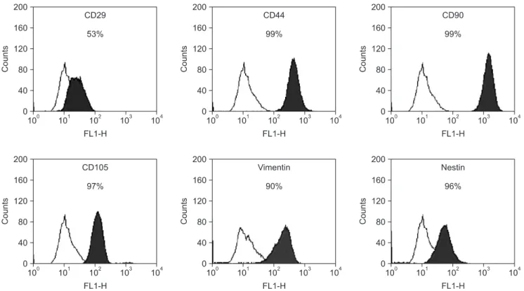
Results: The hSMSCs showed plate-adherence, fibroblast-like growth, expression of the stem-cell transcription factors Oct 4 and Nanog, and positive staining for mesenchymal stem cell (MSC) marker proteins (CD29, CD44, CD90, CD105, and vimentin) and a neural precursor marker (nestin).
Moreover, the hSMSCs in this study were successfully differentiated into multiple mesenchymal lineages, including osteocytes, adipocytes, and chondrocytes. Neuron-like cell morphology and various neural markers were highly visible six hours after the neuronal induction of hSMSCs, but their neuron-like characteristics disappeared over time (24-48 hrs). Interestingly, when the chemical induction medium was changed to Dulbecco's Modified Eagle Medium (DMEM) supplemented with fetal bovine serum (FBS), the differentiated cells returned to their hSMSC morphology, and their cell number increased. These results indicate that chemically induced neuron-like cells should not be considered true nerve cells.
Conclusion: Isolated hSMSCs have MSC characteristics and express a neural precursor marker, suggesting that human skin is a source of stem cells.
However, the in vitro chemical neuronal induction of hSMSC does not produce long-lasting nerve cells and more studies are required before their use in nerve-tissue transplants.
Key words: Skin, Mesenchymal stem cell, In vitro neuronal differentiation
[paper submitted 2012. 8. 10 / revised 2012. 11. 15 / accepted 2012. 11. 22]
neural stem cells have been transplanted into nerve defect sites to improve peripheral and central nerve functions1-3. Among them, mesenchymal stem cells (MSCs) have been the focus in improving nerve regeneration because of their capability to provide multi-lineage differentiations and self- renewal potential4. Bone marrow-derived MSCs (BMSCs) can trans-differentiate in vitro into Schwann cell-like cells, which produce remarkable in vivo nerve regeneration when transplanted into a peripheral nerve defect4-7. Note, however, that bone marrow aspiration sometimes requires invasive procedures, possibly inducing a range of complications in patients such as pain, hemorrhage, and fear. Therefore, more accessible tissues such as skin or fat are investigated
I. Introduction
Recently, many researchers have tried to regenerate nerve tissue with tissue engineering techniques. Multipotent or pluripotent stem cells, cultured Schwann cells, and isolated
Bong-Wook Park
Department of Oral and Maxillofacial Surgery, School of Medicine and Institute of Health Science, Gyeongsang National University, 79, Gangnam-ro, Jinju 660-702, Korea
TEL: +82-55-750-8263 FAX: +82-55-761-7024 E-mail: parkbw@gnu.ac.kr
*This research was supported by the Basic Research Program through the National Research Foundation of Korea (NRF) funded by the Ministry of This is an open-access article distributed under the terms of the Creative Commons
Attribution Non-Commercial License (http://creativecommons.org/licenses/by-nc/3.0/), which permits unrestricted non-commercial use, distribution, and reproduction in any medium, provided the original work is properly cited.
CC
to evaluate the efficiency of their neural differentiation, hSMSCs were differentiated into neural cells under the chemically neural induction protocol of MSCs16,17, and various neurogenic and angiogenic proteins were evaluated by immunocytochemistry (ICC).
II. Materials and Methods
1. Isolation and culture of hSMSCs
Human facial skin samples were obtained from four patients (2 males and 2 females; 24-45 years old, average of 33.3 years) who had undergone head and neck surgery via the submandibular approach.(Fig. 1. A) All experiments were authorized by the Gyeongsang National University Hospital Ethics Committee, and the patients gave their informed consent to tissue donation. Fresh human skin samples were transported to the laboratory, and hSMSCs were isolated as previous protocol15. Briefly, all hairs and subcutaneous fat tissues were removed, and the samples were then cut into 1-3 mm2 explants containing the epidermis and dermis. Skin explants were attached to the culture plates, and 2 mL of Dulbecco’s Modified Eagle Medium (DMEM)/F12 (1 : 1) (Invitrogen, Carlsbad, CA, USA) supplemented with 10%
fetal bovine serum (FBS; Invitrogen), 10 ng/mL epidermal as alternative sources of adult stem cells for the tissue
engineering technique nowadays.
Recently, skin has been considered a potential adult stem cell source. It is highly accessible, and enough auto logous tissue could be easily obtained with minimal donor site complications. Moreover, skin is an abundant pluri potent, multipotent cell source with immune privilege and potential for self-replication8-10. Several researchers have demonstrated that there are several different types of stem cells - such as skin-derived precursors (SKPs), skin-derived mesenchymal stem cells (SMSCs), and epidermal stem cells -- in the dermis and epidermis of skins10-14. In the previous study, we isolated porcine skin-derived cells from the ear skin of miniature pigs and showed the multipotency and MSC characteristics15. The cells were isolated from the epidermis and dermis in serum-containing medium, and they proliferated adherently on the culture plate and expressed MSC-marker proteins. In this study, human SMSCs (hSMSCs) from submandibular skin were isolated and cultured, and in vitro differentiation into mesenchymal cells such as osteocytes, adipocytes, and chondrocytes was evaluated under specific induction media. In addition, isolated hSMSCs were characterized by evaluating the expression of various cell surface markers (CD29, CD44, CD90, CD105, vimentin, and nestin) and transcription factors (Oct 4, Nanog, and Sox 2). Finally,
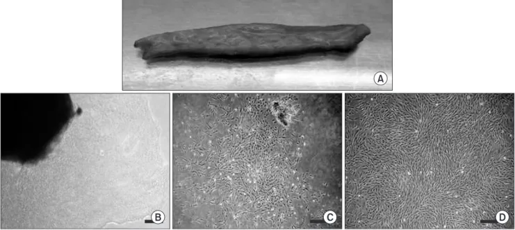
Fig. 1. Isolation and primary culture of human skin-derived cells with serum-containing adherent cell culture method (scale bar=100 μm).
A. Harvested submandibular skin tissue. B-D. Culturing human skin-derived cells (hSDCs) on the 3rd (B), 7th (C), and 14th (D) day can be observed during primary culture (P0). B. Irregular and heterogeneous hSDCs isolated from a skin fragment (black shadow) in the primary culture plates. C. After 7 days of P0, proliferating irregularly shaped hSDCs were detected in the plates. D. After about 2 weeks of P0, plate-adherent, fibroblast-like homogeneous cells were detected in the culture plates.
Jun-Ho Byun et al: Isolation of human mesenchymal stem cells from the skin and their neurogenic differentiation in vitro. J Korean Assoc Oral Maxillofac Surg 2012
anti-human CD29 [1 : 100, BD Pharmingen], mouse anti- human vimentin [1 : 100, Sigma-Aldrich], and mouse anti- human nestin [1 : 100, BD Pharmingen]) for 45 min at 37oC, followed by labeling with the FITC-conjugated secondary goat anti-mouse antibody (1 : 100, BD Pharmingen) for an hour.
3. In vitro neuronal differentiation
Neuronal differentiation of hSMSCs was performed using a modified chemical neural induction protocol for BMSCs differentiation16,17. Briefly, when hSMSCs at passage 3 reached 70% confluence, the cells were transferred to a neuronal preinduction medium containing DMEM (Invitrogen) with 20% FBS (Invitrogen) and 10 ng/mL bFGF (Sigma- Aldrich) for 24 hr. The cells were washed with PBS and cultured in neuronal induction medium consisting of DMEM supplemented with 2% dimethylsulfoxide (DMSO), 200 μM butylated hydroxyanisole (BHA), 25 mM KCl, 2 mM Valproic acid, 10 μM Forskolin, 1 μM Hydrocortisol, 5 μg/mL Insulin, and 2 mM L-glutamine without FBS for up to 48 hr. The cells were fixed for ICC at 0 hr, 6 hr, 24 hr, and 48 hr of induction. All supplemented chemicals in the neural induction medium were manufactured by Sigma-Aldrich Company.
To compare the morphological changes of the neuronally differentiated cells, the chemically inductive medium of the control cells was changed to DMEM supplemented with 20% FBS after 24 hr of chemical induction, and their morphological changes were observed after an additional 24 hr.
growth factor (EGF; Sigma-Aldrich, St. Louis, MO, USA), 10 ng/mL basic fibroblast growth factor (bFGF; Sigma- Aldrich), 100 U/mL penicillin (Sigma-Aldrich), and 100 μg/
mL streptomycin (Sigma-Aldrich) were added. The culture plates were incubated at 37oC in a humidified atmosphere containing 5% CO2 in air for 3 or 5 days. After removing the remaining skin fragments, the attached cells were expanded in vitro, with the culture medium changed twice a week.
Once confluent, the cells were dissociated using 0.25% (w/v) trypsin-ethylenediaminetetraacetic acid (EDTA; Invitrogen) solution and pelleted at 500 × g for 5 min. The cells were then re-grown and incubated until passage 3.
2. Cell surface and intracellular markers analysis
The cell surface and intracelluar markers of hSMSCs at passage 3 were analyzed using a flow cytometer (BD FACSCalibur; Becton Dickinson and Company, Franklin Lakes, NJ, USA) in triplicate. Briefly, cells that reached 90% confluence were harvested using 0.25% EDTA and washed twice in Dulbecco’s phosphate buffered saline (DPBS; Invitrogen) supplemented with 10% FBS. The cells for detecting CD44, CD90, and CD105 were labeled directly with fluorescein isothiocyanate (FITC)-conjugated CD markers (rat anti-mouse CD44 [1 : 100, BD Pharmingen;
BD Biosciences, Franklin Lake, NJ, USA], mouse anti- human CD90 [1 : 100, BD Pharmingen], and goat anti- mouse CD105 [1 : 100, BD Pharmingen]). The cells were fixed in 3.7% formaldehyde for an hour to analyze the levels of CD29, vimentin, and nestin. After washing with DPBS, the samples were labeled with primary antibodies (mouse
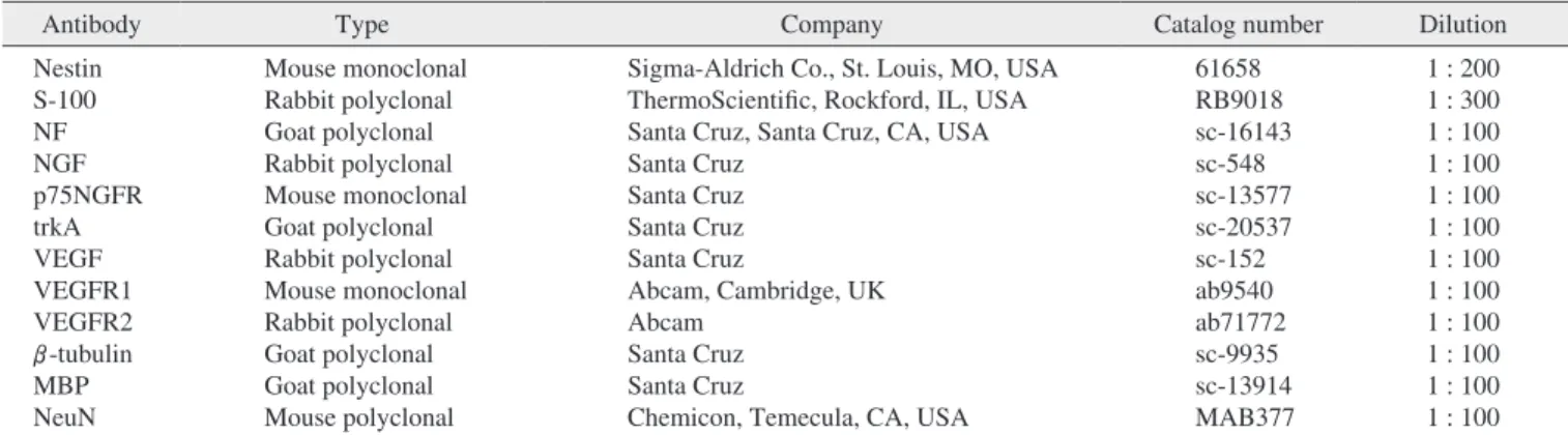
Table 1. Primary antibodies for the immunocytochemical study of chemically neural induced cells
Antibody Type Company Catalog number Dilution
Nestin S-100 NF NGF p75NGFR trkA VEGF VEGFR1 VEGFR2 β-tubulin MBP NeuN
Mouse monoclonal Rabbit polyclonal Goat polyclonal Rabbit polyclonal Mouse monoclonal Goat polyclonal Rabbit polyclonal Mouse monoclonal Rabbit polyclonal Goat polyclonal Goat polyclonal Mouse polyclonal
Sigma-Aldrich Co., St. Louis, MO, USA ThermoScientific, Rockford, IL, USA Santa Cruz, Santa Cruz, CA, USA Santa Cruz
Santa Cruz Santa Cruz Santa Cruz
Abcam, Cambridge, UK Abcam
Santa Cruz Santa Cruz
Chemicon, Temecula, CA, USA
61658 RB9018 sc-16143 sc-548 sc-13577 sc-20537 sc-152 ab9540 ab71772 sc-9935 sc-13914 MAB377
1 : 200 1 : 300 1 : 100 1 : 100 1 : 100 1 : 100 1 : 100 1 : 100 1 : 100 1 : 100 1 : 100 1 : 100 (NF: neurofilament, NGF: nerve growth factor, trkA: tyrosine kinase receptor A, VEGF: endothelial cell growth factor, MBP: myelin basic protein, NeuN: neural-specific nuclear protein)
Jun-Ho Byun et al: Isolation of human mesenchymal stem cells from the skin and their neurogenic differentiation in vitro. J Korean Assoc Oral Maxillofac Surg 2012
5. Reverse transcription-polymerase chain reaction (RT-PCR) analysis
The hSMSCs at passage 3 were evaluated by RT-PCR for the expression of transcription factors Oct 4, Sox 2, and Nanog. Table 2 lists the RT-PCR primers for the markers used in this study. The total RNA was extracted from the cultured cells using an RNeasy Mini Kit (Qiagen, Valencia, CA, USA). cDNA synthesis was performed for 30 min at 55oC using an Omniscript Reverse Transcription Kit (Qiagen) with oligo-dT primers. The cDNAs produced were used as template for PCR amplification. PCR was performed using Maxime PCR Premix (iNtRON Biotechnology, Seongnam, Korea) under the following conditions: pre-denaturation at 94oC for 3 min, followed by 34 cycles of denaturation at 94oC for 45 s, annealing at 60oC or 58oC for 30 s, elongation at 72oC for 45 s, and final extension at 72oC for 10 min using a Thermocycler (PTC-200; GMI, Anoka, MN, USA).
6. Statistical analysis
All values of the counted and calculated cell numbers and intensities of immunostainings of the in vitro neural differentiated cells were statistically analyzed by the Manova test, and independent grouping variables were compared using Bonferroni and SPSS software version 18.0 (SPSS Inc., Chicago, IL, USA). Data were expressed as mean±SD.
Differences were considered to be significant when P<0.05.
4. Immunocytochemical analysis of hSMSCs and in vitro neural induced cells
When the hSMSCs reached passage 3, they were rinsed with PBS and fixed in 4% neutral buffered formaldehyde for 30 min at room temperature. ICC for the transcription factors (Oct 4, Nanog, and Sox 2) was conducted. A 1 : 200 dilution of primary goat polyclonal anti-human Oct 3/4 (sc-8628;
Santa Cruz, Santa Cruz, CA, USA), a 1 : 200 dilution of primary goat polyclonal anti-human Nanog (sc-30331, Santa Cruz), and a 1 : 200 dilution of primary rabbit polyclonal anti-human Sox 2 (sc-20088, Santa Cruz) were used to detect the expression of transcription factors.
In the neural induction medium, the differentiated cells were fixed at 0 hr (immediately after preinduction), 6 hr, 24 hr, and 48 hr after neural induction. Table 1 lists the primary antibodies used for the evaluation of neural differentiation.
Dilutions (1 : 100) of FITC-conjugated donkey anti-goat polyclonal IgG (Jackson ImmunoResearch Laboratories Inc., West Grove, PA, USA), FITC-conjugated donkey anti-rabbit polyclonal IgG (711-095-152, Jackson ImmunoResearch Laboratories Inc.), and FITC-conjugated goat anti-mouse polyclonal IgG (115-096-003, Jackson ImmunoResearch Laboratories Inc.) were used as secondary antibodies.
Densitometric analyses of each immunostaining were performed using analySIS TS software (Olympus Soft Imaging Solution, Münster, Germany). For the evaluation of one antibody’s expression, at least three slides were immunostained and statistically analyzed at each time point.
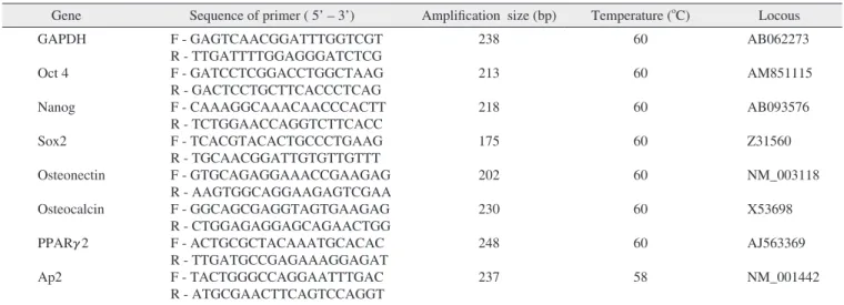
Table 2. RT-PCR primers used for evaluating transcription factors and osteogenic and adipogenic differentiations
Gene Sequence of primer ( 5’ – 3’) Amplification size (bp) Temperature (oC) Locous GAPDH
Oct 4 Nanog Sox2 Osteonectin Osteocalcin PPARγ2 Ap2
F - GAGTCAACGGATTTGGTCGT R - TTGATTTTGGAGGGATCTCG F - GATCCTCGGACCTGGCTAAG R - GACTCCTGCTTCACCCTCAG F - CAAAGGCAAACAACCCACTT R - TCTGGAACCAGGTCTTCACC F - TCACGTACACTGCCCTGAAG R - TGCAACGGATTGTGTTGTTT F - GTGCAGAGGAAACCGAAGAG R - AAGTGGCAGGAAGAGTCGAA F - GGCAGCGAGGTAGTGAAGAG R - CTGGAGAGGAGCAGAACTGG F - ACTGCGCTACAAATGCACAC R - TTGATGCCGAGAAAGGAGAT F - TACTGGGCCAGGAATTTGAC R - ATGCGAACTTCAGTCCAGGT
238 213 218 175 202 230 248 237
60 60 60 60 60 60 60 58
AB062273 AM851115 AB093576 Z31560 NM_003118 X53698 AJ563369 NM_001442 (RT-PCR: reverse transcription-polymerase chain reaction, GAPDH: glyceraldehyde 3-phosphate dehydrogenase)
Jun-Ho Byun et al: Isolation of human mesenchymal stem cells from the skin and their neurogenic differentiation in vitro. J Korean Assoc Oral Maxillofac Surg 2012
Sox 2 was hardly observed by ICC or RT-PCR.(Figs. 2. A, 2.
B) The hSMSCs at passage 3 were positive for typical MSC markers (CD29, CD44, CD90, CD105, and vimentin) based on FACS analysis. In addition, the neural precursor marker, nestin, was detected in the hSDCs.(Fig. 3) The results above demonstrate that human skin-derived cells in this study were multipotent MSCs exhibiting the characteristics of a neural precursor. Moreover, the cultured hSMSCs in this study were successfully differentiated into mesenchymal lineage cells, osteocytes, adipocytes, and chondrocytes in specific induction media.(Fig. 4. A) These differentiated cells from hSMSCs also showed specific osteogenic and adipogenic marker proteins by RT-PCR.(Fig. 4. B)
3. In vitro neural induction of hSMSCs
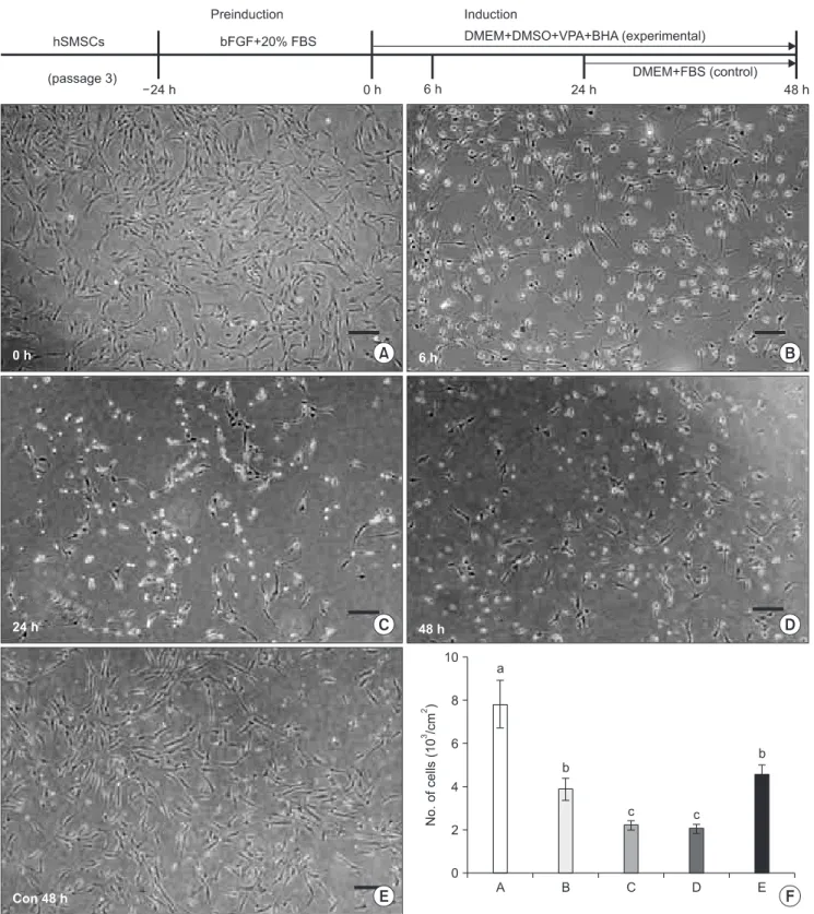
Specific morphological change from hSMSCs was not observed after neural preinduction.(Fig. 5. A) When hSMSCs were neuronally differentiated using various chemical compounds under FBS-deprived conditions, most strongly resembled neurons and included the retraction of cell body and process elaboration, and they were observed after 6 hr of induction.(Fig. 5. B) As the neuronal induction time reached 24 hrs and 48 hrs, however, the differentiated neuron- like cells decreased in number, and their cell morphology
III. Results
1. Cell isolation and culture
Both floated sphere-forming cells and plate-adherent, fibroblast-like cells were co-detected in the culture medium from the first day of primary culture. After 3 days of primary culture, most cells had shown plate-adherent growth in the gelatin-coated plates. Initially, the attached skin-derived cells showed heterogeneously irregular shapes, and partial colony formations were observed. Note, however, that homogeneously shaped and plate-adherent fibroblast-like cells were mainly detected at the end - about 2 weeks later - of the primary culture.(Figs. 1. B-D) These homogeneously shaped, plate-adherent fibroblast-like cells were allowed to proliferate, reaching passage 2 or 3.
2. Expression of transcription factors determined by RT-PCR and cell surface markers measured by FACS analysis
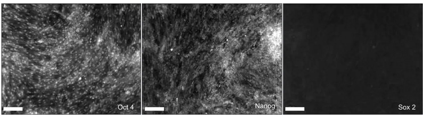
After human skin-derived cells were cultured to passage 3, the expression of transcription factors such as Oct 4, Nanog, and Sox 2 was evaluated by ICC and RT-PCR. Oct 4 and Nanog were highly visible in cultured adult hSMSCs, whereas
Fig. 2. Expression of early transcription factors Oct 4, Nanog, and Sox 2 by immunocytochemistry (A: scale bar=100 μm) and reverse transcription-polymerase chain reaction (B) in human skin-derived cells (hSDCs) at passage 3. Positive expression of Oct 4 and Nanog, even though Sox 2 was hardly expressed, indicates that the hSDCs in this study are multipotential primitive cells. (GAPDH: glyceraldehyde 3-phosphate dehydrogenase)
Jun-Ho Byun et al: Isolation of human mesenchymal stem cells from the skin and their neurogenic differentiation in vitro. J Korean Assoc Oral Maxillofac Surg 2012
Fig. 3. Fluorescence-activated cell sorting analysis of cultured human skin-derived cells. Skin-derived cells at passage 3 were positive for specific mesenchymal stem cell markers (CD29, CD44, CD90, CD105, and vimentin) and neural precursor cell marker (nestin). Open histograms represent staining with negative control, with the black histograms depicting the fluorescence intensity of each of the cell surface antibodies.
Jun-Ho Byun et al: Isolation of human mesenchymal stem cells from the skin and their neurogenic differentiation in vitro. J Korean Assoc Oral Maxillofac Surg 2012
Fig. 4. Mesenchymal-lineage differentiations of human skin-derived mesenchymal stem cells (hSMSCs) into ostocytes (a, b), adiopcytes (c), and chondrocytes (d) for 4 weeks (A: scale bar=100 μm). A. In vitro differentiated cells showed positive staining in the specific staining methods. (a, b) Calcium deposits were observed on the cell surface by von Kossa (a) and Alizalin red (b) staining. (c) Lipid droplets were noted in the cytoplasm of cells by Oil red O staining. (d) Proteoglycans were confirmed on the cell surface using Alcian blue. B. Reverse transcription-polymerase chain reaction results for in vitro differentiated osteocytes and adipocytes from hSMSCs. (a) ON and OC were detected in osteogenic differentiated cells. (b) PPARγ2 and aP2 were expressed in adipogenic differentiated cells. (OC: osteocalcin, ON:
osteonectin, GAPDH: glyceraldehyde 3-phosphate dehydrogenase, aP2: adipocyte protein 2)
Jun-Ho Byun et al: Isolation of human mesenchymal stem cells from the skin and their neurogenic differentiation in vitro. J Korean Assoc Oral Maxillofac Surg 2012
Fig. 5. The upper graph illustrates the schematic in vitro neural induction protocol used in this study. Cultured hSMSCs at passage 3 were preinduced for 24 hrs. The experimental group was neurally induced by a chemical protocol for 48 hr. In the control group, 24 hr after neural induction, the inductive medium was changed to DMEM supplemented with 20% FBS, and morphologic changes were then observed after an additional 24 hrs of media change. A-E. The microphotographs show the morphologic changes of hSMSCs after chemical neural induction (scale bar=100 μm). A. Immediately after neuronal preinduction (0 hr). There are no remarkable morphological changes compared to the original hSMSCs. B. Six hours (6 hr) after neural induction, the neuron-like cells exhibit peak activity. C, D. After the passage of neural induction time (24 and 48 hrs post-neural induction), the neuron-like cells decreased in number, and their shape deteriorated. E. In the control cells, neural differentiated cells returned to the original hSMSC morphology, and cell number increased 24 hrs after media change as DMEM with 20% FBS. F. The number of cells decreased with the passage of neural induction time, but the number increased after the inductive medium was changed. (hSMSCs: human skin-derived mesenchymal stem cells, bFGF: basic fibroblast growth factor, FBS: fetal bovine serum, DMEM: Dulbecco’s Modified Eagle Medium, DMSO: dimethylsulfoxide, VPA: valporic acid, BHA: butylated hydroxyanisole)
Jun-Ho Byun et al: Isolation of human mesenchymal stem cells from the skin and their neurogenic differentiation in vitro. J Korean Assoc Oral Maxillofac Surg 2012
of these proteins was then observed after 24 hr and 48 hr of neurogenic induction.(Fig. 6)
IV. Discussion
Among the skin-derived stem cells, SKPs - believed to be endogenous embryonic neural crest-derived precursor cells that persist into adulthood - have been studied most widely.
SKPs originated with the dermis of the skin and formed floating spheres in the serum-free culturing conditions supplemented with various growth factors - including FGF-2, EGF, and B27 - after enzymatic digestion and cell dissociation of the skin. Moreover, they have distinct characteristics from MSCs, and their full potential is normally restricted by the local environment, but such can be revealed under culture conditions18-21. These results suggest that SKPs have many characteristics that would be beneficial in nervous system regeneration22,23. In the previous study, however, deteriorated (Figs. 5. C, 5. D) In the control cells, the
induction medium was changed to DMEM with 20% FBS after 24 hrs of neural induction, and the neural differentiated cells returned to the original shape of hSMSCs.(Fig. 5. E) In addition, the number of cells increased 24 hrs after medium change.(Fig. 5. F)
In the immunocytochemical studies of differentiated neuron- like cells, neuron- and angiogenesis-related proteins were highly expressed, peaking 6 hrs after neural induction. Note, however, that the expression intensity of all proteins decreased as induction time reached 24 hrs and 48 hrs. Inter estingly, the neural precursor marker, nestin, was substantially expressed in the pre-induced cells (0 hr). In addition, the enhanced co-expression of the nerve growth factor (NGF) and its two receptors (p75NGFR and trkA) as well as the vascular endothelial growth factor (VEGF) and its two receptors (VEGFR1 and VEGFR2) was detected at the early stages of neurogenic induction (6 hrs). The decreased expression
Fig. 6. A. Immunocytochemical studies for various neuronal and angiogenic marker proteins after the in vitro chemical neural induction of hSMSCs (scale bar=100 μm). Most marker proteins were highly visible 6 hrs after neural induction. Nestin was expressed in the 0 hr specimen (before nerve induction), which is similar to the result of FACS analysis. NGF and VEGF were highly visible with their receptors (p75NGFR, trkA, VEGFR1, and VEGFR2) during neuronal diffe- rentiation. B. Immunocytochemical intensities for specific proteins. The expression of most proteins, except NeuN, peaked 6 hrs after neural induction, and then decreased over time (24 hrs and 48 hrs after induction).
Data represent the mean±SE of four independent experiments. A star (*) indicates a significant difference from the control (P<0.05). (S-100: S-100 protein, NF: neurofilament, MBP:
myelin basic protein, NeuN: neural- specific nuclear protein, NGF: nerve growth factor, p75NGFR: p75 nerve growth factor receptor, trkA: tyrosine kinase receptor A, VEGF: vascular endothelial cell growth factor, VEGFR:
vascular endothelial cell growth factor receptor)
Jun-Ho Byun et al: Isolation of human mesenchymal stem cells from the skin and their neurogenic differ- entiation in vitro. J Korean Assoc Oral Maxillofac Surg 2012
produces toxic and stressful culture conditions. Therefore, these conditions contributed to the rapid disruption of the actin cytoskeleton and shape change to a form resembling nerve cells34,35. These nerve-like cells were not truly differentiated nerve cells, and they did not exhibit electrical conductions.
Moreover, the chemical induction led to cell apoptosis and death33,36. In this study, we observed rapid neuron-like morphological changes of hSMSCs after chemical induction.
After 6 hrs of induction, the highest neuron-like cells morphology and peak expression of neural and angiogenic- related proteins were detected. With the passage of induction time, however, the morphology of neural inducted cells deteriorated, and the cell number and expression of nerve- related proteins decreased. Interestingly, when the induction medium was changed to DMEM with FBS, the neural differentiated cells returned to their original shape, and the cell number also increased. These results indicate that the chemically induced neuron-like cells of this study were not regarded as true nerve cells. When stem cells differentiate into target cells, it is difficult for them to return to stem cells especially in a short time period37. Therefore, the chemical induction protocol wherein the induction media contained BME, DMSO, and BHA was not a suitable choice for the neural differentiation method of hSMSCs, even though neuron-like morphology and various nerve-related proteins were observed shortly after induction.
In previous reports, SKPs, considered one of the neural crest originated-stem cells, were differentiated into nerve cells and maintained by supplementation with various growth factors and neurotrophins such as B27, NGF, brain-derived neurotrophic factor (BDNF), neuro-3 (NT-3), and FBS8,18,19. This is similar to previously reported neural differentiation methods of BMSCs involving various growth factors and neurotrophins25. Although further studies are needed to determine the most optimal method for in vitro neural differentiation from MSCs, the use of these growth factors and neurotrophins can be considered a substitute method for the in vitro neural induction of SMSCs.
Several researchers have demonstrated the enhanced nerve regeneration after the in vivo transplantation of differentiated Schwann cells or neurons from BMSCs4,5,38. Nonetheless, undifferentiated MSCs are well-known to have potential for differentiation into target cells after in vivo transplantation.
Moreover, undifferentiated BMSCs expressed various neurotrophic factors after in vivo transplantation into the nerve defect site4. Since previous studies have not suggested an obvious in vitro neural differentiation method we isolated and cultured a different type of porcine skin-
derived stem cells under culture con dition different from that of SKPs as well as different serum-containing and adherent cell culturing method and observed the distinct MSC characteristics in these porcine skin-derived cells15. Similarly, in this study, human skin-derived cells were isolated from the submandibular skin segments, and multipotent MSC characteristics were detected, i.e., expression of transcription factors (Oct 4 and Nanog), detection of MSC markers (CD29, CD44, CD90, CD105, and vimentin), and potential of in vitro osteogenic, adipogenic, and chondrogenic differentiations.
Interestingly, unlike the porcine SMSCs of a previous study15, we could not observe Sox 2 expression in the hSMSCs of this study. This is consistent with the result of other research, i.e., high visibility of Oct 4 and Nanog but negative expression of Sox 2 in human BMSCs24. In addition, they observed strongly expressed nestin, a neural precursor maker, in all kinds of MSCs originating in the bone marrow, dermis, and adipose tissues, similar to this study. Actually, this neural precursor marker was usually seen in SKPs, regarded as neural crest-originated stem cell in skin19. Taken together, the isolated and cultivated human skin-derived cells in this study are considered multipotent, hSMSCs demonstrating the characteristics of a neural precursor.
The in vitro neural differentiation methods of BMSCs have been investigated by several researchers. First, various molecules involved in neural development, such as growth factors, neurotrophins, cytokines, and retinoic acid, were used25,26. Second, neural differentiation was obtained by increasing the intracellular cyclic adenosine monophosphate27. Third, specific chemical compounds such as β-mercaptoethanol (BME), DMSO, and BHA were used in the serum-free culturing medium for neural induction16,17. Most recently, the neural differentiation of BMSCs was also observed when cultured in inflammatory astrocyte-containing medium28. In these various in vitro neural induction protocols, chemical protocols have been widely used and studied because it was a simple approach that rapidly yielded neuron-like cell differentiation from BMSCs29-31. Morphological changes into nerve-like cells and expression of neuron-specific makers have been observed within a few hours of chemical neural induction. In addition, more than 70% of differentiated neuron-like cells from BMSCs were observed after chemical neural induction, which was extremely higher concentration compared with the other induction method32,33.
In recent studies, however, the use of chemical compounds in serum-free conditions for the neural induction of BMSCs
masison CA, Hopping SB, et al. Characterization and isolation of stem cell-enriched human hair follicle bulge cells. J Clin Invest 2006;116:249-60.
14. Riekstina U, Muceniece R, Cakstina I, Muiznieks I, Ancans J.
Characterization of human skin-derived mesenchymal stem cell proliferation rate in different growth conditions. Cytotechnology 2008;58:153-62.
15. Kang EJ, Byun JH, Choi YJ, Maeng GH, Lee SL, Kang DH, et al. In vitro and in vivo osteogenesis of porcine skin-derived mesenchymal stem cell-like cells with a demineralized bone and fibrin glue scaffold. Tissue Eng Part A 2010;16:815-27.
16. Woodbury D, Schwarz EJ, Prockop DJ, Black IB. Adult rat and human bone marrow stromal cells differentiate into neurons. J Neurosci Res 2000;61:364-70.
17. Woodbury D, Reynolds K, Black IB. Adult bone marrow stromal stem cells express germline, ectodermal, endodermal, and mesodermal genes prior to neurogenesis. J Neurosci Res 2002;69:908-17.
18. Fernandes KJ, Mckenzie IA, Mill P, Smith KM, Akhavan M, Barnabé-heider F, et al. A dermal niche for multipotent adult skin- derived precursor cells. Nat Cell Biol 2004;6:1082-93.
19. Fernandes KJ, Kobayashi NR, Gallagher CJ, Barnabé-heider F, Aumont A, Kaplan DR, et al. Analysis of the neurogenic potential of multipotent skin-derived precursors. Exp Neurol 2006;201:32- 48.
20. Hunt DP, Morris PN, Sterling J, Anderson JA, Joannides A, Jahoda C, et al. A highly enriched niche of precursor cells with neuronal and glial potential within the hair follicle dermal papilla of adult skin. Stem Cells 2008;26:163-72.
21. Zhao M, Isom SC, Lin H, Hao Y, Zhang Y, Zhao J, et al. Tracing the stemness of porcine skin-derived progenitors (pSKP) back to specific marker gene expression. Cloning Stem Cells 2009;11:111- 22.
22. Shih DT, Lee DC, Chen SC, Tsai RY, Huang CT, Tsai CC, et al.
Isolation and characterization of neurogenic mesenchymal stem cells in human scalp tissue. Stem Cells 2005;23:1012-20.
23. Marchesi C, Pluderi M, Colleoni F, Belicchi M, Meregalli M, Farini A, et al. Skin-derived stem cells transplanted into resorbable guides provide functional nerve regeneration after sciatic nerve resection. Glia 2007;55:425-38.
24. Riekstina U, Cakstina I, Parfejevs V, Hoogduijn M, Jankovskis G, Muiznieks I, et al. Embryonic stem cell marker expression pattern in human mesenchymal stem cells derived from bone marrow, adipose tissue, heart and dermis. Stem Cell Rev 2009;5:378-86.
25. Sanchez-ramos J, Song S, Cardozo-pelaez F, Hazzi C, Stedeford T, Willing A, et al. Adult bone marrow stromal cells differentiate into neural cells in vitro. Exp Neurol 2000;164:247-56.
26. Kim BJ, Seo JH, Bubien JK, Oh YS. Differentiation of adult bone marrow stem cells into neuroprogenitor cells in vitro. Neuroreport 2002;13:1185-8.
27. Deng W, Obrocka M, Fischer I, Prockop DJ. In vitro differentiation of human marrow stromal cells into early progenitors of neural cells by conditions that increase intracellular cyclic AMP. Biochem Biophys Res Commun 2001;282:148-52.
28. Wang FW, Jia DY, Du ZH, Fu J, Zhao SD, Liu SM, et al. Roles of activated astrocytes in bone marrow stromal cell proliferation and differentiation. Neuroscience 2009;160:319-29.
29. Bossolasco P, Cova L, Calzarossa C, Rimoldi SG, Borsotti C, Deliliers GL, et al. Neuro-glial differentiation of human bone marrow stem cells in vitro. Exp Neurol 2005;193:312-25.
30. Lei Z, Yongda L, Jun M, Yingyu S, Shaoju Z, Xinwen Z, et al.
Culture and neural differentiation of rat bone marrow mesenchymal stem cells in vitro. Cell Biol Int 2007;31:916-23.
31. Yamaguchi S, Kuroda S, Kobayashi H, Shichinohe H, Yano S, Hida K, et al. The effects of neuronal induction on gene expression profile in bone marrow stromal cells (BMSC)--a preliminary study using microarray analysis. Brain Res 2006;1087:15-27.
from hSMSCs, undifferentiated autologous SMSCs could be suggested as reasonable transplant material for the regeneration of nerve defects.
V. Conclusion
Isolated hSMSCs from adult skin showed MSC characte- ristics possessing multipotency and neural precursor marker, suggesting that human skin may be used as available adult stem cell source. Nonetheless, whether the in vitro neurogenic differentiated cells from hSMSCs under the chemical neural induction protocol produce actual nerve cells was doubtful, even though neuron-like cell morphology and various nerve-related proteins were detected in these chemically induced cells. Therefore, we suggest that the undifferentiated autologous hSMSCs - instead of chemically induced neuron- like cells - be considered for use as transplant material for in vivo nerve regeneration in the future.
References
1. Mosahebi A, Woodward B, Wiberg M, Martin R, Terenghi G.
Retroviral labeling of Schwann cells: in vitro characterization and in vivo transplantation to improve peripheral nerve regeneration.
Glia 2001;34:8-17.
2. Murakami T, Fujimoto Y, Yasunaga Y, Ishida O, Tanaka N, Ikuta Y, et al. Transplanted neuronal progenitor cells in a peripheral nerve gap promote nerve repair. Brain Res 2003;974:17-24.
3. Heine W, Conant K, Griffin JW, Höke A. Transplanted neural stem cells promote axonal regeneration through chronically denervated peripheral nerves. Exp Neurol 2004;189:231-40.
4. Tohill M, Mantovani C, Wiberg M, Terenghi G. Rat bone marrow mesenchymal stem cells express glial markers and stimulate nerve regeneration. Neurosci Lett 2004;362:200-3.
5. Dezawa M. Systematic neuronal and muscle induction systems in bone marrow stromal cells: the potential for tissue reconstruction in neurodegenerative and muscle degenerative diseases. Med Mol Morphol 2008;41:14-9.
6. Cuevas P, Carceller F, Garcia-gómez I, Yan M, Dujovny M. Bone marrow stromal cell implantation for peripheral nerve repair.
Neurol Res 2004;26:230-2.
7. Hou SY, Zhang HY, Quan DP, Liu XL, Zhu JK. Tissue-engineered peripheral nerve grafting by differentiated bone marrow stromal cells. Neuroscience 2006;140:101-10.
8. Toma JG, Akhavan M, Fernandes KJ, Barnabé-heider F, Sadikot A, Kaplan DR, et al. Isolation of multipotent adult stem cells from the dermis of mammalian skin. Nat Cell Biol 2001;3:778-84.
9. Toma JG, Mckenzie IA, Bagli D, Miller FD. Isolation and characterization of multipotent skin-derived precursors from human skin. Stem Cells 2005;23:727-37.
10. Shi C, Zhu Y, Su Y, Cheng T. Stem cells and their applications in skin-cell therapy. Trends Biotechnol 2006;24:48-52.
11. Dyce PW, Zhu H, Craig J, Li J. Stem cells with multilineage potential derived from porcine skin. Biochem Biophys Res Commun 2004;316:651-8.
12. Blanpain C, Fuchs E. Epidermal stem cells of the skin. Annu Rev Cell Dev Biol 2006;22:339-73.
13. Ohyama M, Terunuma A, Tock CL, Radonovich MF, Pise-
rapid morphological changes and mimics neuronal phenotype. J Neurosci Res 2004;77:192-204.
36. Rismanchi N, Floyd CL, Berman RF, Lyeth BG. Cell death and long-term maintenance of neuron-like state after differentiation of rat bone marrow stromal cells: a comparison of protocols. Brain Res 2003;991:46-55.
37. Rho GJ, Kumar BM, Balasubramanian SS. Porcine mesenchymal stem cells--current technological status and future perspective.
Front Biosci 2009;14:3942-61.
38. Shimizu S, Kitada M, Ishikawa H, Itokazu Y, Wakao S, Dezawa M.
Peripheral nerve regeneration by the in vitro differentiated-human bone marrow stromal cells with Schwann cell property. Biochem Biophys Res Commun 2007;359:915-20.
32. Muñoz-elías G, Woodbury D, Black IB. Marrow stromal cells, mitosis, and neuronal differentiation: stem cell and precursor functions. Stem Cells 2003;21:437-48.
33. Barnabé GF, Schwindt TT, Calcagnotto ME, Motta FL, Martinez GJ, De OA, et al. Chemically-induced RAT mesenchymal stem cells adopt molecular properties of neuronal-like cells but do not have basic neuronal functional properties. PLoS One 2009;4:e5222.
34. Lu P, Blesch A, Tuszynski MH. Induction of bone marrow stromal cells to neurons: differentiation, transdifferentiation, or artifact? J Neurosci Res 2004;77:174-91.
35. Neuhuber B, Gallo G, Howard L, Kostura L, Mackay A, Fischer I. Reevaluation of in vitro differentiation protocols for bone marrow stromal cells: disruption of actin cytoskeleton induces