Delayed Rupture of Flexor Pollicis Longus after Volar Plating for a Distal Radius Fracture
Chul-Hyun Cho, MD, Kyung-Jae Lee, MD, Kwang-Soon Song, MD, Ki-Cheor Bae, MD
Department of Orthopaedic Surgery, Dongsan Medical Center, Keimyung University School of Medicine, Daegu, Korea
Case Report
Clinics in Orthopedic Surgery 2012;4:325-328 • http://dx.doi.org/10.4055/cios.2012.4.4.325Received December 14, 2009; Accepted February 9, 2010 Correspondence to: Ki-Cheor Bae, MD
Department of Orthopaedic Surgery, Dongsan Medical Center, Keimyung University School of Medicine, 56 Dalseong-ro, Jung-gu, Daegu 700-712, Korea
Tel: +82-53-250-7729, Fax: +82-53-250-7205 E-mail: [email protected]
Open reduction and volar plate fixation for distal radius fractures is becoming very popular, but several reports have documented complications associated with volar plating.1) Extensor tendon rupture after volar plating is a relatively common complication,2,3) but flexor tendon rup- tures have rarely been reported. Most reported instances of a flexor tendon rupture have involved improper plate placement, increased prominence of the distal edge of the plate because of collapse of the fracture site, use of custom- made plates, current steroid use by the patient, or a history of tendon injury.4-9) We report a case of delayed rupture of the flexor pollicis longus (FPL) tendon 40 months after volar plating with a 3.5-mm T-locking compression plate (T-LCP, Synthes, Paoli, PA, USA).
Although extensor tendon rupture often occurs after volar plating for a distal radius fracture, a flexor tendon rupture is extremely rare. Most reported instances of flexor tendon ruptures after volar plating have involved improper placement of the plate, in- creased prominence of the distal edge of the plate because of collapse of the fracture site, use of custom-made plates, current steroid use by the patient, or a history of tendon injury. We report a case of delayed rupture of the flexor pollicis longus tendon 40 months after volar plating with a 3.5-mm T-locking compression plate for which the distal edge was located at the transverse ridge level of the distal radius. If symptoms such as tendon irritation occur in this situation, surgeons should consider removing the plate as soon as possible after bony union is achieved.
Keywords: Flexor pollicis longus, Distal radius fracture, Volar plating, Delayed rupture
CASE REPORT
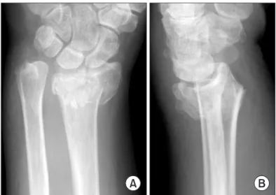
A 63-year-old right-handed woman sustained a right in- tra-articular comminuted distal radius fracture with dorsal displacement (Fig. 1), a right femoral shaft fracture, and a hemothorax in a motor vehicle accident. Open reduction and internal fixation was performed using 3.5-mm T-LCP for the distal radius fracture at 12 days after the trauma,
Copyright © 2012 by The Korean Orthopaedic Association
This is an Open Access article distributed under the terms of the Creative Commons Attribution Non-Commercial License (http://creativecommons.org/licenses/by-nc/3.0) which permits unrestricted non-commercial use, distribution, and reproduction in any medium, provided the original work is properly cited.
Clinics in Orthopedic Surgery • pISSN 2005-291X eISSN 2005-4408
Fig. 1. Initial anteroposterior (A) and lateral (B) plain radiographs show the intra-articular comminuted distal radius fracture with dorsal displacement.
326
Cho et al. Delayed Rupture of Flexor Pollicis Longus after Volar Plating for Distal Radius Fracture Clinics in Orthopedic Surgery • Vol. 4, No. 4, 2012 • www.ecios.org
and the plate was covered by the pronator quadratus after fixation. Immediate postoperative plain radiographs re- vealed acceptable restoration of radial inclination, radial length, and volar tilting, except an approximate 2 mm step-off of the articular surface. The plate was placed tight- ly to the distal radius without any gap, and even though the distal edge of the plate was very close to the transverse ridge of the distal radius, it did not reach the distal por- tion of the transverse ridge. Plain radiographs at 8 months after surgery revealed bony union without further collapse of the distal fragment compared to that of the immedi- ate postoperative radiographs, and range of motion of the right wrist was normal. Accordingly, we recommended plate removal, but the patient refused, stating that she was experiencing no specific discomfort.
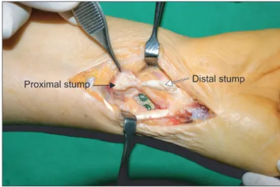
She visited our hospital at 40 months after surgery and reported that she had a sudden thumb flexion limita- tion during washing 10 days ago. She had no associated pain and no warning symptoms such as tendon irrita- tion. A physical examination revealed that she could not actively flex the right interphalangeal joint of her thumb, although the range of motion of her other joints and sen- sory findings were normal. Radiographic findings revealed no definite interval change (Fig. 2). Ultrasonographic findings showed that the FPL tendon had ruptured, and the proximal stump was found at the radiocarpal joint. We performed a surgical exploration and found that the distal edge of the plate was exposed even though the pronator quadratus was nearly completely covering the plate. The FPL tendon had ruptured over the distal edge and was frayed (Fig. 3). We performed complete debridement of the granulated and frayed tissue in the ruptured margin, removed the plate, and performed a primary repair of
the tendon using the modified Kessler method. The pa- tient wore a thumb spica cast for 3 weeks and then began passive-motion exercises. The patient had no movement restriction of the right thumb at 6 months after surgery.
DISCUSSION
When a plate is fixed on the dorsal side, it causes various complications such as extensor tendon irritation or rupture and metal breakage due to the difficulty of manufactur- ing a plate to fit the irregular bone contour.2,3) In contrast, volar plating significantly reduces these complications, but reports have been published on the complications as- sociated with volar plating, including complex regional pain syndrome, carpal tunnel syndrome, tendon injury, or infection.1,6,7) Most reports of complications related to volar plating have considered extensor tendon damage; in particular, damage to the extensor pollicis longus. Flexor tendon injuries after volar plating are extremely rare.
Orbay and Touhami10) emphasized the importance of the transverse ridge or watershed line of the distal radi- us to prevent a flexor tendon injury. Plates placed distal to the transverse ridge have the potential to impinge on the traversing flexor tendons. Bell et al.4) reported their experi- ence with four patients who had FPL ruptures after volar plating and noted that all patients were taking steroids.
They concluded that steroid use is an indication for early removal of the device. Additionally, they recommended that volar plates be removed early if they are placed on the distal part of the distal radius transverse ridge or if the distal edge of the plate becomes prominent because of col- lapse of the fracture site, as these conditions could result in irritation to the tendon. Cross and Schmidt5) reported their experience with two patients who had FPL ruptures after Fig. 2. Anteroposterior (A) and lateral (B) plain radiographs obtained at
40 months after surgery show no interval change in comparison with radiographs obtained immediately after surgery.
Fig. 3. Intraoperative findings show a total rupture of the flexor pollicis longus tendon with granulation and fraying of tissue in the ruptured margin.
327
Cho et al. Delayed Rupture of Flexor Pollicis Longus after Volar Plating for Distal Radius Fracture Clinics in Orthopedic Surgery • Vol. 4, No. 4, 2012 • www.ecios.org
their fractures were repaired by volar plates manufactured by two different companies. They also emphasized the role of placing the plate in relation to the transverse ridge for a tendon rupture due to friction. They recommended that volar plates be removed early and that patients be advised of the possibility of FPL damage if simple radiographs revealed that the plates were located on the distal part of the transverse ridge. Duncan and Weiland6) reported their experience with a patient whose FPL ruptured 9 months after surgery and noted that the distal part of plate adhered tightly to the bone without a gap. They emphasized that the plate edge must be covered with the pronator quadra- tus to prevent irritation to the tendon (Table 1).
In our case, there was no long-term use of steroids and the Synthes 3.5-mm T-LCP used was widely available and not custom-made. Additionally, no history of injury before surgery or of thumb abnormality after surgery were noted. Follow-up plain radiographs revealed bony union without further collapse of the distal fragment, as compared with radiographs obtained immediately after surgery. However, even though the plate was not violat- ing the distal part of the transverse ridge, it was at the transverse ridge level. We found that the plate shaft was covered by the pronator quadratus, but that the distal edge was exposed. Additionally, the FPL tendon ruptured right at the point where it met the exposed distal edge. We sug- gest that the rupture was due to friction from the exposed distal edge of the plate at the transverse ridge, because the pronator quadratus does not attach to the distal part of the
transverse ridge. Therefore, we believe that it is very im- portant for the distal edge of a plate to be completely cov- ered by the pronator quadratus to prevent a flexor tendon injury.
As reported by some authors, when the FPL rup- tures because of friction against the plate over a long pe- riod of time, primary repair is impossible in most cases because of a hypertrophied tenosynovium and a tendon defect with fibrous tissue and retraction; thus, a tendon graft or tendon transfer is necessary. However, although the rupture in our patient occurred 40 months after the initial surgery, findings from the exploratory surgery re- vealed little retraction; thus, a primary end-to-end repair was possible even after removing some fibrous tissues and a frayed tendon. It is unclear why such unusual circum- stances occurred.
We have found that when the plate is located near the transverse ridge after surgery, it is necessary to make thorough and careful observations to prevent flexor ten- don damage. If symptoms such as tendon irritation occur in this situation, surgeons should consider removing the plate as soon as possible after bony union is achieved.
CONFLICT OF INTEREST
No potential conflict of interest relevant to this article was reported.
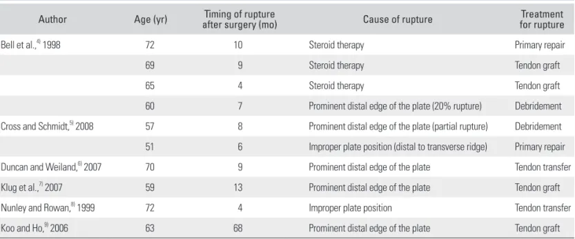
Table 1. Cases of Rupture of Flexor Pollicis Longus after Volar Plating for Distal Radius Fracture
Author Age (yr) Timing of rupture
after surgery (mo) Cause of rupture Treatment
for rupture
Bell et al.,4) 1998 72 10 Steroid therapy Primary repair
69 9 Steroid therapy Tendon graft
65 4 Steroid therapy Tendon graft
60 7 Prominent distal edge of the plate (20% rupture) Debridement
Cross and Schmidt,5) 2008 57 8 Prominent distal edge of the plate (partial rupture) Debridement 51 6 Improper plate position (distal to transverse ridge) Primary repair
Duncan and Weiland,6) 2007 70 9 Prominent distal edge of the plate Tendon transfer
Klug et al.,7) 2007 59 13 Prominent distal edge of the plate Tendon graft
Nunley and Rowan,8) 1999 72 4 Improper plate position Tendon transfer
Koo and Ho,9) 2006 63 68 Prominent distal edge of the plate Tendon graft
328
Cho et al. Delayed Rupture of Flexor Pollicis Longus after Volar Plating for Distal Radius Fracture Clinics in Orthopedic Surgery • Vol. 4, No. 4, 2012 • www.ecios.org
1. Arora R, Lutz M, Hennerbichler A, Krappinger D, Espen D, Gabl M. Complications following internal fixation of unstable distal radius fracture with a palmar locking-plate. J Orthop Trauma. 2007;21(5):316-22.
2. Louis DS. Prospective multi-center trial of a plate for dor- sal fixation of distal radius fractures. J Hand Surg Am.
1998;23(2):353-4.
3. Schnur DP, Chang B. Extensor tendon rupture after internal fixation of a distal radius fracture using a dorsally placed AO/ASIF titanium pi plate: Arbeitsgemeinschaft fur Osteo- synthesefragen/Association for the Study of Internal Fixa- tion. Ann Plast Surg. 2000;44(5):564-6.
4. Bell JS, Wollstein R, Citron ND. Rupture of flexor pollicis longus tendon: a complication of volar plating of the distal radius. J Bone Joint Surg Br. 1998;80(2):225-6.
5. Cross AW, Schmidt CC. Flexor tendon injuries following locked volar plating of distal radius fractures. J Hand Surg Am. 2008;33(2):164-7.
6. Duncan SF, Weiland AJ. Delayed rupture of the flexor pollicis longus tendon after routine volar placement of a T-plate on the distal radius. Am J Orthop (Belle Mead NJ).
2007;36(12):669-70.
7. Klug RA, Press CM, Gonzalez MH. Rupture of the flexor pollicis longus tendon after volar fixed-angle plating of a distal radius fracture: a case report. J Hand Surg Am.
2007;32(7):984-8.
8. Nunley JA, Rowan PR. Delayed rupture of the flexor pollicis longus tendon after inappropriate placement of the pi plate on the volar surface of the distal radius. J Hand Surg Am.
1999;24(6):1279-80.
9. Koo SC, Ho ST. Delayed rupture of flexor pollicis longus tendon after volar plating of the distal radius. Hand Surg.
2006;11(1-2):67-70.
10. Orbay JL, Touhami A. Current concepts in volar fixed-angle fixation of unstable distal radius fractures. Clin Orthop Relat Res. 2006;445:58-67.