Ameloblastoma is an uncommon, slow-growing, and locally invasive benign tumor that constitutes 11% to 18%
of odontogenic tumors that arise in both jaws.1-5It has several different histological and clinico-radiologic types with a high rate of recurrence despite being classified as a benign tumor.6 Most ameloblastomas occur between age 30 and 60 years, peaking around age 35 and show no sex predilection.7,8 Approximately 80% of ameloblastomas occur in the mandible, usually in the vicinity of the molars and ramus.3,7-11In one report, symptomatic ameloblasto- mas accounting for 35% of 60 patients were discovered incidentally on radiographic studies, and ameloblastoma usually appears as unilocular or typical multilocular cystic radiolucencies.2
Desmoplastic ameloblastoma is rare, accounting for approximately 4% to 13% of ameloblastomas.12-16 The main histological types of ameloblastoma are the follicu- lar and plexiform patterns. The desmoplastic ameloblas- toma is one of the other less common histological vari- ants.17The desmoplastic variant of ameloblastoma usually appears in the anterior and premolar regions as a mixed radiolucent and radiopaque lesion, sometimes resembling a benign fibro-osseous lesion.12
Ameloblastoma is a locally aggressive tumor that may cause recurrence and in rare cases, malignant transforma- tion, with repeated postsurgical reccurences.18,19Recur- rence after incomplete removal is a feature of ameloblas- tomas and it is, therefore, recommended that desmoplastic ameloblastomas be treated by complete surgical resec- tion.18,20An autogenous bone graft for subsequent recon- struction after resection prevents the site from becoming affected by a recurrence of the neoplasm.21,22
This report presents a case of a patient who was initially diagnosed with a developmental cyst resembling unicystic
A repeatedly recurrent desmoplastic ameloblastoma after removal and allobone graft:
Radiographic features compared with histological changes
Jae-Duk Kim1, Hyun-Seon Jang2, Yo-Seob Seo1, Jin-Soo Kim1,*
1Department of Oral and Maxillofacial Radiology, School of Dentistry, Chosun University, Gwangju, Korea
2Department of Oral Pathology, School of Dentistry, Chosun University, Gwangju, Korea
ABSTRACT
A 40-year-old man suffered from a repeatedly recurrent desmoplastic ameloblastoma in the right maxillary anterior and premolar regions. During the first visit, the patient was provisionally histopathologically diagnosed with a developmental cyst, and it was confirmed to be unicystic ameloblastoma and resected. Four years later, the lesion recurred, and was diagnosed as a desmoplastic type of ameloblastoma and removed again. Then, 5 years after the second surgery, the lesion recurred again, and was diagnosed as a type containing a follicular pattern, recurrent ameloblastoma. A panoramic radiograph showed a multilocular and mixed radiolucent/radiopaque expansile lesion at the first visit, a unilocular cystic lesion confined to the premolar area at the second visit, and a small soap bubble appearance in the molar area in the final visit. Cone-beam computed tomographic images of the final recurrence of the tumor revealed multiple small cyst-like structures in the right maxillary anterior and posterior regions. (Imaging Sci Dent 2013; 43: 201-7)
KEY WORDS: Ameloblastoma; Recurrence; Cone-Beam Computed Tomography; Radiography, Panoramic
*This study was supported by research funds from Chosun University Dental Hospi- tal, 2013.
Received March 30, 2013; Revised April 15, 2013; Accepted April 27, 2013
*Correspondence to : Prof. Jin-Soo Kim
Department of Oral and Maxillofacial Radiology, College of Dentistry, Chosun University, 421 Susuk-dong, Dong-gu, Gwangju, Korea
Tel) 82-62-220-3880, Fax) 82-62-227-0270, E-mail) [email protected]
Copyright ⓒ 2013 by Korean Academy of Oral and Maxillofacial Radiology
This is an Open Access article distributed under the terms of the Creative Commons Attribution Non-Commercial License (http://creativecommons.org/licenses/by-nc/3.0) which permits unrestricted non-commercial use, distribution, and reproduction in any medium, provided the original work is properly cited.
Imaging Science in Dentistry∙pISSN 2233-7822 eISSN 2233-7830
ameloblastoma in the anterior and the right premolar max- illary regions and secondly with desmoplastic ameloblas- toma at recurrence after enucleation and bone grafting, which repeatedly recurred. The lesion was successfully controlled with the third resection.
Case Report
A 40-year-old man visited the Chosun University Dental Hospital with a tender swelling on his anterior palatal area.
The overlying mucosa appeared normal and all the teeth in the affected area were caries free and reacted positively to both thermal and electrical stimuli. His medical history was non-contributory.
A panoramic radiograph revealed a multilocular and mixed radiolucent/radiopaque expansile lesion extending
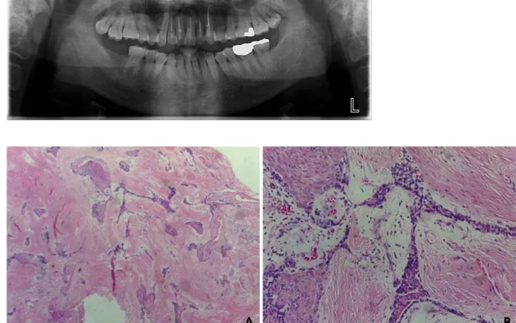
from the right second premolar to the left canine of the maxilla, presenting a soap bubble appearance in the mid- dle third of the lesion, and ill-defined radiopacity in the left third of the lesion (Fig. 1). The lesion showed an often curved and scalloped border extending to the middle por- tion of the roots of the upper anterior teeth. The lesion was diagnosed provisionally and radiographically as amelo- blastoma with histological evidence of a developmental cyst showing a cystic structure lined by epithelium with incisional biopsy (Fig. 2A). After endodontic treatment of multiple upper anterior teeth, he underwent enucleation of the lesion and an allobone graft by an oral surgeon. Histo- pathologically, the enucleated mass showed unicystic ame- loblastoma with peripheral palisading of hyperchromatic epithelium and loose fibrous stroma (Fig. 2B). The fistula formation process near the bone graft area was persistent.
Fig. 1.Panoramic radiograph reveals a multilocular and mixed radiolu- cent/radiopaque expansile lesion extending from the right second pre- molar to the left canine of the max- illa, showing a soap bubble appear- ance in the middle third of the lesion, and an ill-defined radiopacity in the left third of the lesion.
Fig. 2. A. The incisional biopsy shows a cystic structure lined by epithelium (H&E stain, 200×). B. The histopathologic examination after the first surgery on the tumor shows peripheral palisading of hyperchromatic epithelium and loose fibrous stroma (H&E stain, 200×).
A B
After being treated repeatedly with antibiotics and dressing for about 5 months, he finally underwent removal of the allobone.
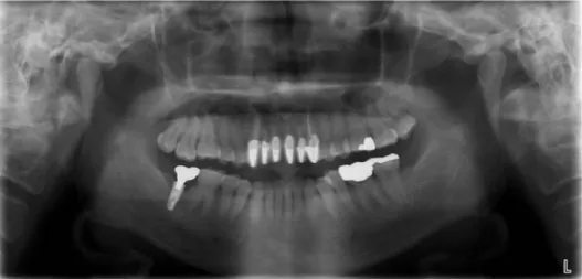
Four years after the first surgery, a recurrent tumor was found in the same area. A panoramic radiograph showed a unilocular cystic radiolucency in the area corresponding to the right part of the primary lesion (Fig. 3), and re-enu- cleation of only the cystic lesion and Tutoplast grafting were performed. Histopathologically, the enucleated mass had changed from a developmental cyst to the desmoplas- tic type of ameloblastoma that contained small islands and thin cords of ameloblastic epithelium within a dense fib- rous connective tissue stroma (Fig. 4).
The patient returned with the symptom of mild pain at the site of the operation 5 years after the second surgery.
A panoramic radiograph revealed a soap bubble appear-
ance in the periapical area of the right first and second molars (Fig. 5). On the axial cone-beam computed tomo- graphic (CBCT) images, multiple small cyst-like structures were found adjacent to the maxillary sinus and in the ante- rior area, which were separated by normal bone. The lesion at the molar area was well demarcated from the maxillary antrum (Fig. 6). Sagittal CBCT images showed small locu- lations in the molar and anterior area, respectively, and they were separated by normal bone (Fig. 7). The tumor showed a relatively defined border, and it was adjacent to the maxillary sinus (Fig. 7A).
Finally, the tumor histology became a follicular pattern next to the desmoplastic type that contained small cystic tumor islands and thin cords of ameloblastic epithelium within connective tissue stroma (Fig. 8).
Fig. 3.A panoramic radiograph re- veals a unilocular radiolucent cystic area confined to the premolar region of the right maxilla, 4 years after the first surgery.
Fig. 4. A. Histopathologic examination 4 years after the first surgery shows the desmoplastic type of ameloblastoma (H&E stain, 40×). B.
The histopathologic features contain small islands and thin cords of ameloblastic epithelium within a dense fibrous connective tissue stroma (H&E stain, 200×).
A B
Discussion
In 1984, Eversole et al17first described the “desmoplas- tic ameloblastoma.” Histologically, a desmoplastic amelo- blastoma consists of an abundant and excessive stromal collagenization or desmoplasia in which irregularly shaped islands of odontogenic epithelium are scattered.12Wald- ron and El-Mofty12 introduced the term “hybrid lesions”
to describe the conditions in which desmoplastic amelo- blastoma was located adjacent to follicular or plexiform ameloblastoma. Wakoh et al23presented a case of a pati- ent demonstrating follicular-type ameloblastoma with desmoplasia, in whom radiological findings suggested the coexistence of a fibro-osseous lesion with a solitary cystic lesion and proposed it to be hybrid follicular/desmoplastic ameloblastoma.
Histopathologically, the original biopsy of our case
showed features that were consistent with those of a devel- opmental cyst suspected to be unicystic ameloblastoma.
In the second histological study, the features of the enu- cleated mass had changed from a developmental cyst to a desmoplastic type of ameloblastoma. Finally, the tumor histology became a small cystic and follicular pattern next to the desmoplastic type of ameloblastoma. However, in the desmoplastic type of this case, a histologically fibro- osseous lesion-like pattern and hybrid pattern were not recognizable and radiologically, the relationship to the mixed lesion was also unclear.
Analyzing the radiographical features in order of histo- logical changes, this tumor initially showed a multilocular radiolucent lesion with soap-bubble appearance in the right maxilla and concomitant radiopaque area in the left anterior region of the maxilla on the panoramic radiograph with the histopathologic features of a developmental cyst next to a unicystic ameloblastoma.
At the second visit, only panoramic radiography reveal- ed a unilocular cystic radiolucency confined to the right maxillary premolar region; however, the biopsy result of the enucleated mass was desmoplastic ameloblastoma.
Although Philipsen et al,24 in their report of 2 cases of desmoplastic ameloblastoma, remarked that the presence of osteoplasia in desmoplastic ameloblastoma might cor- respond to the radiologic appearance of mixed radiolu- cency and radiopacity in some of the desmoplastic amelo- blastomas, thereby presenting radiographic features of a fibro-osseous lesion. However, Sun et al18 reported that desmoplastic ameloblastomas radiologically presented as radiolucent (44.4%) and mixed radiolucent/radiopaque (55.6%) in the cases they reviewed. Effiom and Odukoya25 also reported that multilocular radiolucency accounted for 82.4% of cases in a desmoplastic series and the remaining
Fig. 6. An axial view of the CBCT image shows multiple small cyst-like structures in the right anterior and posterior regions. They are separated by normal bone. The lesion in the molar region is well demarcated, and separated from the maxillary antrum.
Fig. 5.Five years after the second surgery. The panoramic radiograph reveals a soap bubble appearance in the periapical area of the right first and second molars of the maxilla.
17.6% presented with a mixed radiolucent and radiopaque radiographic appearance, thereby mimicking a fibro-osse- ous lesion. Kawai et al26reported that desmoplastic amelo- blastoma appeared either as a diffuse, poorly delineated, mottled radiolucent/radiopaque lesion similar to a benign fibro-osseous lesion or as a lesion with a honeycomb or soap-bubble appearance with indistinct borders. However, the relationship to the mixed lesion was not discovered in this case.
At the final visit, a panoramic radiograph revealed a small area of soap bubble appearance in the periapical area of the right maxillary first and second molars. An axial CBCT image showed that multiple small cyst-like structures adjacent to the maxillary sinus and in the ante- rior area, and they were separated by normal bone (Fig.
8). The sagittal CBCT images showed small loculations in the molar and anterior areas, respectively, and they were separated by normal bone (Fig. 7). The histological result was the desmoplastic type containing a small cystic and follicular pattern as recurrent ameloblastoma. The radio- graphic features of our final recurrent ameloblastoma showed agreement with what another author mentioned, that the recurrent tumor has a characteristic appearance of multiple small cyst-like structures with very coarse scle- rotic cortical margins sometimes separated by normal bone.1
Regarding the behavior of desmoplastic ameloblastomas, Philipsen et al24suggested that desmoplastic ameloblasto- mas in the maxilla would be more aggressive than those in the mandible. Mendenhall et al7 also suggested that
Fig. 7.Sagittal CT images (A; molar area B; premolar area C: anterior area) show small loculations in the molar and anterior region of the maxilla, respectively, separated by normal bone. The tumor shows a relatively defined border, and it is adjacent to the maxillary sinus.
Fig. 8. A. The histopathologic examination 5 years after the second surgery reveals small cystic tumor islands and thin cords of ameloblas- tic epithelium within connective tissue stroma (H&E stain, 40×). B. A follicular pattern next to the desmoplastic type of ameloblastoma (H&E stain, 200×).
A B
A B C
maxillary ameloblastomas tended to have a higher local recurrence rate because the thinness of the cortical bone was a less effective barrier to tumor invasion compared with the mandible. In this report, the tumor developed between the anterior and the right premolar regions of the maxilla.
Concerning the recurrence, it was mentioned in the WHO classification of odontogenic tumors that desmoplastic ameloblastoma possibly has a lower recurrence rate than other ameloblastomas.27In contrast, Keszler et al28report- ed that desmoplastic ameloblastoma showed a higher re- currence rate (21.4%) than the other type of ameloblasto- mas (10.1%). Desmoplastic ameloblastomas have the po- tential for recurrence because they fail to produce a cap- sule.14,29Sun et al18 analyzed 115 cases of desmoplastic ameloblastoma from 35 published papers and reported that whereas enucleation provided a recurrence rate of 21.1%, resection reduced this rate remarkably to 3.1%.
The average period until recurrence was 36.9 months.
When applying conservative techniques such as enuclea- tion, this recurrence originates from small fragments of the tumor left in situ and jeopardizes possible good results from immediate reconstruction of the surgical defect.30,31 In this report, the patient’s tumor recurred after conserva- tive treatment and an immediate autobone graft.
At the first visit, our patient showed the histological fea- ture of unicystic ameloblastoma. Actually, the surgeon thought of the tumor as a dentigerous cyst by clinical examination. Unicystic ameloblastoma may form from the epithelial lining of a dentigerous cyst and show radio- graphical similarities with dentigerous cysts.1 Phillipsen and Reichart32found that 66 cases among 152 cases of unicystic ameloblastoma were initially diagnosed as denti- gerous cysts from the literature and that unicystic amelo- blastoma radiographically might appear not only as a uni- locular, but indeed also as a multilocular bone defect.
References
1. White SC, Pharoah MJ. Oral radiology: principles and inter- pretation. 6th ed. St. Louis: Mosby Elsevier; 2009. p. 373-5.
2. Becelli R, Carboni A, Cerulli G, Perugini M, Iannetti G. Man- dibular ameloblastoma: analysis of surgical treatment carried out in 60 patients between 1977 and 1998. J Craniofac Surg 2002; 13: 395-400.
3. Hertog D, Schulten EA, Leemans CR, Winters HA, Van der Waal I. Management of recurrent ameloblastoma of the jaws:
a 40-year single institution experience. Oral Oncol 2011; 47:
145-6.
4. Rapidis AD, Andressakis DD, Stavrianos SD, Faratzis G, Arnogiannaki-Liappi N, Lagogiannis GA, et al. Ameloblasto-
mas of the jaws: clinico-pathological review of 11 patients. Eur J Surg Oncol 2004; 30: 998-1002.
5. Siar CH, Lau SH, Ng KH. Ameloblastoma of the jaws: a retro- spective analysis of 340 cases in a Malaysian population. J Oral Maxillofac Surg 2012; 70: 608-15.
6. Dissanayake RK, Jayasooriya PR, Siriwardena DJ, Tilakaratne WM. Review of metastasizing (malignant) ameloblastoma (METAM): pattern of metastasis and treatment. Oral Surg Oral Med Oral Pathol Oral Radiol Endod 2011; 111: 734-41.
7. Mendenhall WM, Werning JW, Fernandes R, Malyapa RS, Mendenhall NP. Ameloblastoma. Am J Clin Oncol 2007; 30:
645-8.
8 Ladeinde AL, Ogunlewe MO, Bamgbose BO. Ameloblastoma:
analysis of 207 cases in a Nigerian teaching hospital. Quintes- sence Int 2006; 37: 69-74.
9. Zemann W, Feichtinger M, Kowatsch E, Kärcher H. Exten- sive ameloblastoma of the jaws: surgical management and immediate reconstruction using microvascular flaps. Oral Surg Oral Med Oral Pathol Oral Radiol Endod 2007; 103: 190-6.
10. Reichart PA, Philipsen HP, Sonner S. Ameloblastoma: biolo- gical profile of 3677 cases. Eur J Cancer B Oral Oncol 1995;
31B: 86-99.
11. Kim SG, Jang HS. Ameloblastoma: a clinical, radiographic, and histopathologic analysis of 71 cases. Oral Surg Oral Med Oral Pathol Oral Radiol Endod 2001; 91: 649-53.
12. Waldron CA, El-mofty SK. A histopathologic study of 116 ameloblastomas with special reference to the desmoplastic variant. Oral Surg Oral Med Oral Pathol 1987; 63: 441-51.
13. Kaffe I, Buchner A, Taicher S. Radiologic features of desmo- plastic variant of ameloblastoma. Oral Surg Oral Med Oral Pathol 1993; 76: 525-9.
14. Ng KH, Siar CH. Desmoplastic variant of ameloblastomas in Malaysians. Br J Oral Maxillofac Surg 1993; 31: 299-303.
15. Takata T, Miyauchi M, Ogawa I, Zhao M, Kudo Y, Sato S, et al. So-called ‘hybrid’ lesion of desmoplastic and convention-al ameloblastoma: report of a case and review of the literature.
Pathol Int 1999; 49: 1014-8.
16. Philipsen HP, Reichart PA, Takata T. Desmoplastic ameloblas- toma (including “hybrid” lesion of ameloblastoma). Biolo- gical profile based on 100 cases from the literature and own files. Oral Oncol 2001; 37: 455-60.
17. Eversole LR, Leider AS, Strub D. Radiographic characteristics of cystogenic ameloblastoma. Oral Surg Oral Med Oral Pathol 1984; 57: 572-7.
18. Sun ZJ, Wu YR, Cheng N, Zwahlen RA, Zhao YF. Desmo- plastic ameloblastoma - A review. Oral Oncol 2009; 45: 752-9.
19. Huang CM, Chen JY, Chen CH, Huang CJ. Radiotherapy for a repeatedly recurrent ameloblastoma with malignant transfor- mation. Head Neck (in press).
20. Thompson IO, van Rensburg LJ, Phillips VM. Desmoplastic ameloblastoma: correlative histopathology, radiology and CT- MR imaging. J Oral Pathol Med 1996; 25: 405-10.
21. Bianchi S, Tarello F, Polastri F, Valente G. Ameloblastoma of the mandible involving an autogenous bone graft. J Oral Maxillofac Surg 1998; 56: 1187-91.
22. Stea G. Recurrence of an ameloblastoma in an autogenous iliac bone graft. J Oral Maxillofac Surg 1985; 43: 374-7.
23. Wakoh M, Harada T, Inoue T. Follicular/desmoplastic hybrid
ameloblastoma with radiographic features of concomitant fibro-osseous and solitary cystic lesions. Oral Surg Oral Med Oral Pathol Oral Radiol Endod 2002; 94: 774-80.
24. Philipsen HP, Ormiston IW, Reichart PA. The desmo- and osteoplastic ameloblastoma. Histologic variant or clinicopatho- logic entity? Case reports. Int J Oral Maxillofac Surg 1992;
21: 352-7.
25. Effiom OA, Odukoya O. Desmoplastic ameloblastoma: analy- sis of 17 Nigerian cases. Oral Surg Oral Med Oral Pathol Oral Radiol Endod 2011; 111: e27-31.
26. Kawai T, Kishino M, Hiranuma H, Sasai T, Ishida T. A unique case of desmoplastic ameloblastoma of the mandible: report of a case and brief review of the English language literature.
Oral Surg Oral Med Oral Pathol Oral Radiol Endod 1999; 87:
258-63.
27. Gardner DG, Heikinheimo K, Shear M, Philipsen HP, Cole- man H. Ameloblastomas. In: Barnes L, Eveson JW, Reichart
P, Sidransky D. World Health Organization classification of tumors: pathology and genetics of head and neck tumors. 3rd ed. Lyon: IARC Press; 2005. p.296-300.
28. Keszler A, Paparella ML, Dominguez FV. Desmoplastic and non-desmoplastic ameloblastoma: a comparative clinicopatho- logical analysis. Oral Dis 1996; 2: 228-31.
29. Ashman SG, Corio RL, Eisele DW, Murphy MT. Desmoplas- tic ameloblastoma. A case report and literature review. Oral Surg Oral Med Oral Pathol 1993; 75: 479-82.
30. Zachariades N. Recurrences of ameloblastoma in bone grafts.
Report of 4 cases. Int J Oral Maxillofac Surg 1988; 17: 316-8.
31. Gold L, Upton GW, Marx RE. Standardized surgical termino- logy for the excision of lesions in bone: an argumentum for accuracy in reporting. J Oral Maxillofac Surg 1991; 49: 1214- 7.
32. Philipsen HP, Reichart PA. Unicystic ameloblastoma. A review of 193 cases from the literature. Oral Oncol 1998; 34: 317-25.