Introduction
Vertical root fracture is the most severe form of longi- tudinal tooth fracture,1which can cause an inflammatory process, leading to bone resorption and the formation of granulation tissue.2Clinical and radiographic signs of ver- tical root fractures are variable and nonspecific; they may be similar to those of periodontal lesions and endodontic
failures.3-5 Nowadays, in the diagnosis of a vertical root fracture, the patient’s history and symptoms, such as pain, swelling, the existence of deep isolated periodontal pockets, and the accompanying periapical and lateral radiolucencies associated with the root, provide valuable information.6A radiographic diagnosis of vertical root fractures is difficult due to the various fracture patterns and the fractures are not found on the radiographs in many cases.7Unfortunate- ly, only one-third of these fractures can be diagnosed on conventional radiographs.8-10 Conventional radiography can show only two dimensions of the three-dimensional (3D) anatomical structures.11Due to the limitations of intraoral radiographs, 3D imaging systems such as con-
Assessment of vertical root fracture using cone-beam computed tomography
Ehsan Moudi1, Sina Haghanifar1, Zahrasadat Madani2, Abdolhamid Alhavaz3, Ali Bijani4, Mohammad Bagheri1,*
1Oral and Maxillofacial Radiology Department, Dental Material Research Center, Dental School, Babol University of Medical Sciences, Babol, Iran
2Endodontics Department, Dental Material Research Center, Dental School, Babol University of Medical Sciences, Babol, Iran
3Prosthodontics Department, Dental Material Research Center, Dental School, Babol University of Medical Sciences, Babol, Iran
4Social Determinants of Health Research Center, Babol University of Medical Sciences, Babol, Iran
ABSTRACT
Purpose: The aim of this study was to investigate the accuracy of cone-beam computed tomography (CBCT) in the diagnosis of vertical root fractures in a tooth with gutta-percha and prefabricated posts.
Materials and Methods: This study selected 96 extracted molar and premolar teeth of the mandible. These teeth were divided into six groups as follows: Groups A, B, and C consisted of teeth with vertical root fractures, and groups D, E, and F had teeth without vertical root fractures; groups A and D had teeth with gutta-percha and prefabricated posts; groups B and E had teeth with gutta-percha but without prefabricated posts, and groups C and F had teeth without gutta-percha or prefabricated posts. Then, the CBCT scans were obtained and examined by three oral and maxillofacial radiologists in order to determine the presence of vertical root fractures. The data were analyzed using IBM SPSS 20.0 (IBM Corp., Armonk, NY, USA).
Results: The kappa coefficient was 0.875±0.049. Groups A and D showed a sensitivity of 81% and a specificity of 100%; groups E and B, a sensitivity of 94% and a specificity of 100%; and groups C and F, a sensitivity of 88% and a specificity of 100%.
Conclusion: The CBCT scans revealed a high accuracy in the diagnosis of vertical root fractures; the accuracy did not decrease in the presence of gutta-percha. The presence of prefabricated posts also had little effect on the accuracy of the system, which was, of course, not statistically significant. (Imaging Sci Dent 2014; 44 : 37-41)
KEY WORDS: Cone-Beam Computed Tomography; Gutta-Percha; Tooth Fractures
Received May 19, 2013; Revised June 17, 2013; Accepted June 28, 2013
*Correspondence to : Dr. Mohammad Bagheri
Oral and Maxillofacial Radiology Department, Dental School, Babol University of Medical Sciences, Ganj Afrooz Ave., Babol, Mazandaran Province, Iran
Tel) 98-918-610-7767, Fax) 98-851-221-2261, E-mail) drbagheri110@gmail.com
Copyright ⓒ 2014 by Korean Academy of Oral and Maxillofacial Radiology
This is an Open Access article distributed under the terms of the Creative Commons Attribution Non-Commercial License (http://creativecommons.org/licenses/by-nc/3.0) which permits unrestricted non-commercial use, distribution, and reproduction in any medium, provided the original work is properly cited.
Imaging Science in Dentistry∙pISSN 2233-7822 eISSN 2233-7830
ventional computed tomography (CT), cone-beam com- puted tomography (CBCT), and multi-detector computed tomography have been introduced. In recent years, various studies have been performed to investigate the accuracy of each of these systems. According to various studies, CT systems are highly accurate in diagnosing root frac- tures.3-10,12-15 However, despite the many capabilities of CT, the main disadvantages of its dental applications are the high radiation dose in comparison with conventional radiography, the existence of artifacts, and the relatively low spatial resolution.13These limitations led to the devel- opment of CBCT, which is considered a better alternative.3
The aims of this study were to investigate the accuracy of CBCT in detecting vertical root fractures and to assess the influence of root canal filling and intra-canal prefabri- cated posts on the visibility of a root fracture.
Materials and Methods
Ninety-six human mandibular premolars and molars without fracture, periapical pathology, root resorption, or any other anomaly were collected. The teeth had not under- gone any restorative or root canal treatment. The teeth were inspected under a stereomicroscope (Zeiss Stemi SV6, Zeiss, Göttingen, Germany) to confirm the absence of vertical root fractures. Access cavity preparation and root canal treatment were done with a Pro Taper Rotary System (Dentsply Maillefer, Tulsa, OK, USA) up to size F3.
In order to create root fractures in half of the teeth (24 canals of premolars and 24 distal canals of the molars), the teeth were mounted in acrylic resin after covering the root surfaces with a thin layer of modeling wax to simulate soft tissue. Then, a screw driver-tape wedge was placed inside the canals, and fractures were created in the intend- ed canals by applying gentle pressure on the hammer.
Therefore, in 48 canals (half of the teeth), fractures were created, and well-fitting gutta-percha (Pro Taper F3) was placed in the root canal in 32 teeth (16 canals of premolars and 16 distal canals of molars); further, gold-plated screw posts (Nordin, Montreux, Switzerland) having the sizes of
#4 and #5 were placed in the other 16 canals (8 canals of premolars and 8 distal canals of the molars). The same pro- cedures were performed for the canals without fracture, and therefore, the teeth were divided into the six groups (8 premolars and 8 molars in each group) listed in Table 1.
The teeth were again evaluated using a stereo micro- scope to ensure the absence of vertical root fractures in the teeth without fracture. Then, the teeth were coded, and
a wax rim was made from 3 to 4 layers of wax designed on the acrylic base in the shape of the mandible.
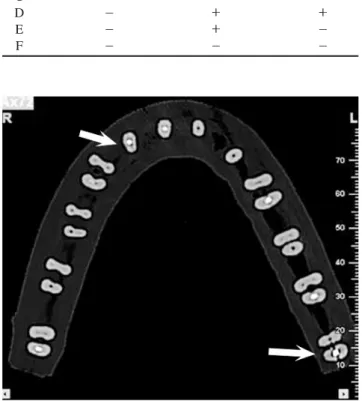
CBCT scans were obtained using the denture scan mode of a Newtom 5G system (QR s.r.l., Verona, Italy) set at 110 kV. Axial and multi-planar reformation (MRP) images were evaluated by using NNT viewer software version 3.0 (QR s.r.l., Verona, Italy). In this study, the slice thick- ness was 0.3 mm and the interval space was 0.3 mm. All of the obtained scans were examined by three oral and max- illofacial radiologists in the axial (Fig. 1) and MPR planes (Fig. 2) in a low-light room by using a Flatron 18.5-inch monitor (LG, Seoul, Korea). The observers were free to choose the magnification degree. All of the teeth were examined for the presence or absence of a vertical root fracture.
The data were analyzed by using IBM SPSS 20.0 statis- tical software (IBM Corp., Armonk, IL, USA). The sensi- tivity, specificity, positive predictive value (PPV), negative predictive value (NPV), and likelihood ratio (LR++, LR-)
Assessment of vertical root fracture using cone-beam computed tomography
Table 1.The characteristics of the groups A, B, C, D, E, and F in this study
Group Vertical root
Gutta-percha Prefabricated
fracture posts
A ++ ++ ++
B ++ ++ -
C ++ - -
D - ++ ++
E - ++ -
F - - -
Fig. 1.An axial CBCT image shows the molar and premolar teeth with vertical root fracture.
with 95% confidence interval were calculated based on an evaluation of at least two observers for each tooth. The kappa coefficient was used to assess the agreement among all the observers, and a p value of 0.05 was considered significant.
Results
Our results showed that the specificity in the diagnosis of a vertical root fracture using CBCT was 100% in all groups. Further, the sensitivity of vertical root fractures in the teeth with gutta-percha and without prefabricated posts was a little higher than in those without gutta-percha and prefabricated posts even though there was no statistical significance. The sensitivity in the diagnosis of vertical root fractures in the groups having teeth with gutta-percha and prefabricated posts was lower than in the other groups.
The PPV was 100% in all of the groups, and the NPV in the groups containing teeth with gutta-percha and without prefabricated posts was higher than in the other groups;
further, NPV in those without prefabricated posts and gutta- percha teeth was higher than in the groups containing teeth with gutta-percha and prefabricated posts.
In the case of the first observer, the kappa coefficient was 0.875±0.049 and the p value was ⁄0.001; in the case of the second observer, the kappa coefficient was 0.896±
0.045 and the p value was ⁄0.001; and in the case of the third observer, the kappa coefficient was 0.771±0.064 and the p value was ⁄0.001. The sensitivity, specificity, PPV, NPV, and LRs of each observer are listed in Table 2.
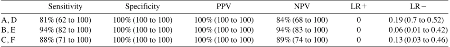
The kappa coefficient was 0.813±0.101 for groups A and D, 0.938±0.061 for groups B and E, and 0.875±
0.085 for groups C and F. The sensitivity, specificity, PPV, NPV, and LRs of each group are listed in Table 3.
A
B
C
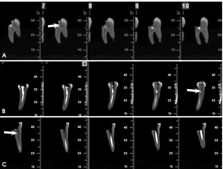
Fig. 2.Cross sectional images of molar and premolar teeth with vertical root fracture. A. A molar tooth with vertical root fracture. B. A premolar tooth with prefabricated post and gutta-percha has vertical root fracture. C. A premolar tooth with gutta-percha shows vertical root fracture.
Discussion
In this study, the presence of gutta-percha did not decr- ease the accuracy of CBCT scans in the diagnosis of a vertical root fracture. This result was consistent with that of the studies conducted by Hassan et al3and Melo et al,7 but inconsistent with the result of the study conducted by Khedmat et al.5Interestingly, in this study, the sensitivity of CBCT scans in the teeth with gutta-percha and without prefabricated posts was a little higher than in the teeth without gutta-percha and prefabricated posts. In the study conducted by Melo et al,7the sensitivity of CBCT in the teeth with gutta-percha having a voxel size of 0.2 mm was higher than in those without gutta-percha. This study was in accordance with our results.
According to the results of this study, the prefabricated posts reduced the accuracy of the CBCT scans. The reduc- tion of the sensitivity and the negative predictive value in teeth with gutta-percha and prefabricated posts could be attributed to the artifacts caused by the posts, which con- cealed the fracture lines in some teeth of this group. The specificity in the diagnosis of a vertical root fracture was the same in the group containing teeth with gutta-percha and prefabricated posts as that in the other groups, and this result did not agree with that of Melo et al.7In their study, the specificity decreased in the teeth with the posts.
We believed that a possible reason for the same specificity and positive predictive value was the absence of the dark strip artifacts that could stimulate the fracture patterns in the presence of prefabricated posts and gutta-percha.
Further, the general overall specificity obtained in this
study was higher than that obtained in the studies con- ducted by Melo et al. Finally, the overall sensitivity of CBCT in the diagnosis of vertical root fractures was 88%
and the specificity was 100%. The value of agreement of all the observers (kappa coefficient) for the evaluation of each tooth by the observers was 0.875±0.046; this showed a high level of agreement (k==0.7). In addition, the kappa coefficient for each observer was higher than 0.7 which represented the high agreement of each observer. To the best of our knowledge, in few studies on the effects of pre- fabricated posts, the posts were examined to determine the accuracy of CBCT in diagnosing vertical root fractures.16 It is suggested that in future research, a smaller voxel size and field of view should be used to examine the diagnostic accuracy of CBCT in vertical root fractures.
In conclusion, CBCT scans showed a high accuracy in the diagnosis of vertical root fractures. Moreover, the pre- sence of gutta-percha and prefabricated posts had little effect on the accuracy of this system.
References
1. Tamse A, Fuss Z, Lustig J, Ganor Y, Kaffe I. Radiographic features of vertically fractured, endodontically treated maxil- lary premolars. Oral Surg Oral Med Oral Pathol Oral Radiol Endod 1999; 88: 348-52.
2. Tamse A. Vertical root fractures in endodontically treated teeth:
diagnostic signs and clinical management. Endod Topics 2006;
13: 84-94.
3. Hassan B, Metska ME, Ozok AR, van der Stelt P, Wesselink PR. Detection of vertical root fractures in endodontically treated teeth by a cone beam computed tomography scan. J Endod 2009; 35: 719-22.
Assessment of vertical root fracture using cone-beam computed tomography
Table 3.Sensitivity, specificity, positive predictive value (PPV), negative predictive value (NPV), negative likelihood ratio (LR-), positive likelihood ratio (LR++) with (C1 95%) for each group
Sensitivity Specificity PPV NPV LR++ LR-
A, D 81% (62 to 100) 100% (100 to 100) 100% (100 to 100) 84% (68 to 100) 0 0.19 (0.7 to 0.52) B, E 94% (82 to 100) 100% (100 to 100) 100% (100 to 100) 94% (83 to 100) 0 0.06 (0.01 to 0.42) C, F 88% (71 to 100) 100% (100 to 100) 100% (100 to 100) 89% (74 to 100) 0 0.13 (0.03 to 0.46) A-F: See Table 1.
Table 2.Sensitivity, specificity, positive predictive value (PPV), negative predictive value (NPV), negative likelihood ratio (LR-), positive likelihood ratio (LR++) with (C1 95%) for each of observer
Sensitivity Specificity PPV NPV LR++ LR-
Observer1 88% (78 to 97) 100% (100 to 100) 100% (100 to 100) 89% (81 to 97) 0 0.13 (0.06 to 0.26) Observer2 90% (81 to 98) 100% (100 to 100) 100% (100 to 100) 91% (93 to 98) 19.50 (4.99 to 76.25) 0.10 (0.05 to 0.24) Observer3 81% (70 to 92) 96% (90 to 100) 95% (89 to 100) 84% (74 to 93) 19.50 (4.99 to 76.25) 0.20 (0.11 to 0.33) Observer* 88% (78 to 97) 100% (100 to 100) 100% (100 to 100) 89% (81 to 97) 0 0.13 (0.06 to 0.26)
*diagnosis of at least two observers in each tooth
4. Mora MA, Mol A, Tyndall DA, Rivera EM. In vitro assess- ment of local computed tomography for the detection of lon- gitudinal tooth fractures. Oral Surg Oral Med Oral Pathol Oral Radiol Endod 2007; 103: 825-9.
5. Khedmat S, Rouhi N, Drage N, Shokouhinejad N, Nekoofar MH. Evaluation of three imaging techniques for the detection of vertical root fractures in the absence and presence of gutta- percha root fillings. Int Endod J 2012; 45: 1004-9.
6. Edlund M, Nair MK, Nair UP. Detection of vertical root frac- tures by using cone-beam computed tomography: a clinical study. J Endod 2011; 37: 768-72.
7. Melo SL, Bortoluzzi EA, Abreu M, Corrêa LR, Corrêa M.
Diagnostic ability of a cone-beam computed tomography scan to assess longitudinal root fractures in prosthetically treated teeth. J Endod 2010; 36: 1879-82.
8. Rud J, Omnell KA. Root fractures due to corrosion. Diagnostic aspects. Scand J Dent Res 1970; 78: 397-403.
9. Youssefzadeh S, Gahleitner A, Dorffner R, Bernhart T, Kain- berger FM. Dental vertical root fractures: value of CT in detec- tion. Radiology 1999; 210: 545-9.
10. Kamburo˘glu K, Murat S, Yüksel SP. Cebeci AR, Horasan S.
Detection of vertical root fracture using cone-beam computeriz- ed tomography: an in vitro assessment. Oral Surg Oral Med Oral Pathol Oral Radiol Endod 2010; 109: e74-81.
11. Scarfe WC, Levin MD, Gane D, Farman AG. Use of cone beam computed tomography in endodontics. Int J Dent 2009; 2009:
634567.
12. Kamburo˘glu K, Ilker Cebeci AR, Gröndahl HG. Effectiveness of limited cone-beam computed tomography in the detection of horizontal root fracture. Dent Traumatol 2009; 25: 256-61.
13. Bernardes RA, de Moraes IG, Húngaro Duarte MA, Azevedo BC, de Azevedo JR, Bramante CM. Use of cone-beam volu- metric tomography in the diagnosis of root fractures. Oral Surg Oral Med Oral Pathol Oral Radiol Endod 2009; 108: 270-7.
14. Bornstein MM, Wölner-Hanssen AB, Sendi P, von Arx T.
Comparison of intraoral radiography and limited cone beam computed tomography for the assessment of root-fractured permanent teeth. Dent Traumatol 2009; 25: 571-7.
15. Ozer S. Detection of vertical root fractures of different thick- nesses in endodontically enlarged teeth by cone beam comput- ed tomography versus digital radiography. J Endod 2010; 36:
1245-9.
16. Taramsari M, Kajan ZD, Bashirzadeh P, Salamat F. Compari- son of high-resolution and standard zoom imaging modes in cone beam computed tomography for detection of longitudinal root fracture: An in vitro study. Imaging Sci Dent 2013; 43:
171-7.