Received on August 21, 2013. Revised on September 2, 2013. Accepted on September 11, 2013.
CC This is an open access article distributed under the terms of the Creative Commons Attribution Non-Commercial License (http://creativecommons.org/licenses/by-nc/3.0) which permits unrestricted non-commercial use, distribu- tion, and reproduction in any medium, provided the original work is properly cited.
*Corresponding Author. Jongseon Choe, Department of Microbiology and Immunology, Kangwon National University School of Medicine, 192-1, Hyoja-dong, Chuncheon, Korea. Tel: 82-33-250-8862; Fax: 82-33-244-3286; E-mail:
jchoe@kangwon. ac.kr
#These authors contributed equally to this work.
Keywords: Syntenin, Follicular dendritic cell, FAK
Abbreviations: ECM, extracellular matrices; FA, focal adhesion; FAK, focal adhesion kinase; FDC, follicular dendritic cell;
PDZ, postsynaptic density protein
Syntenin Is Expressed in Human Follicular Dendritic Cells and Involved in the Activation of Focal Adhesion Kinase
Whajung Cho1#, Hyeyoung Kim1#, Jeong-Hyung Lee2, Seung Hee Hong3 and Jongseon Choe1*
1Department of Microbiology and Immunology, Kangwon National University School of Medicine, Chuncheon 200-701, 2Department of Biochemistry, College of Natural Sciences, Kangwon National University, Chuncheon 200-701, 3Department of Food Nutritional Sciences, Hanbuk University, Dongducheon 483-777, Korea
Syntenin is an adaptor molecule containing 2 PDZ domains which mediate molecular interactions with diverse integral or cytoplasmic proteins. Most of the results on the biological function of syntenin were obtained from studies with malig- nant cells, necessitating exploration into the role of syntenin in normal cells. To understand its role in normal cells, we in- vestigated expression and function of syntenin in human lym- phoid tissue and cells in situ and in vitro. Syntenin expression was denser in the germinal center than in the extrafollicular area. Inside the germinal center, syntenin expression was ob- vious in follicular dendritic cells (FDCs). Flow cytometric analysis with isolated cells confirmed a weak expression of syntenin in T and B cells and a strong expression in FDCs. In FDC-like cells, HK cells, most syntenin proteins were found in the cytoplasm compared to weak expression in the nucleus. To study the function of syntenin in FDC, we exam- ined its role in the focal adhesion of HK cells by depleting syntenin by siRNA technology. Knockdown of syntenin mark- edly impaired focal adhesion kinase phosphorylation in HK cells. These results suggest that syntenin may play an im- portant role in normal physiology as well as in cancer patho- logy.
[Immune Network 2013;13(5):199-204]
INTRODUCTION
Syntenin was originally identified as a melanoma differ- entiation-associated gene-9 (mda-9) and as a syndecan-bind- ing protein (1). It has 2 postsynaptic density protein (PDZ) domains which mediate molecular interactions with diverse integral or cytoplasmic proteins in addition to syndecans (2).
For example, syntenin interacts with CD6 (3), CD63 (4), and TRAF6 (5). It is a scaffolding protein implicated in a variety of cellular processes. In particular, the role of syntenin in can- cer metastasis has been extensively studied. Elevated levels of syntenin expression were observed in cancer cells com- pared to controls (6). Melanoma cells express syntenin, the levels of which correlate with metastatic progression (7).
Syntenin is reported to promote metastasis in human melano- ma cells by activating c-Src (8). Syntenin is also suggested as a new regulator of endocytosis (4). Therefore, syntenin ap- pears to have various functions in a cell type-dependent manner.
Since most previous studies of syntenin have been con- ducted with cancer cells, this study focuses on its expression and function in normal tissues and cells. In this study, we examined the expression levels of syntenin in lymphoid tissue and immune cells and further investigated its potential role
in focal adhesion. Our results indicated that human follicular dendritic cells (FDCs) exhibited a strong expression of synte- nin while lymphocytes revealed a weak expression of synte- nin both in situ and in vitro. Knockdown of syntenin im- paired focal adhesion kinase (FAK) activation in FDC-like cells, suggesting that syntenin may play an important role in normal FDCs.
MATERIALS AND METHODS
Preparation of lymphocytes and HK cells
Human B, T, and HK cells were prepared from human tonsils obtained from children undergoing tonsillectomy at Asan Medical Center (Seoul, Korea). This study was approved by the Institutional Review Board of Asan Medical Center (Approval number, 2012-0636; approval date, August 31, 2012). Tonsillar B cells were prepared as previously de- scribed (9). Briefly, tonsillar mononuclear cells were sub- jected to 2 sessions of T cell depletion by rosetting with SRBC; the resulting cells contained more than 98% CD20+ cells as analyzed by FACScan (Becton Dickinson, Sunnyvale, CA, USA). T cells were isolated from the rosette-forming cell pellets after lysing SRBC; the resulting cells contained more than 95% CD3+ cells. HK cells were primary cells obtained from human tonsils and were used until they displayed de- generate features in culture. They were prepared as described by Kim et al. (10) and maintained in RPMI-1640 (Irvine Scientific, Santa Ana, CA, USA) containing 10% fetal calf serum (Hyclone, Logan, UT, USA), 2 mM L-glutamine (Invitrogen, Carlsbad, CA, USA), 100 U/ml penicillin G (Sigma-Aldrich, St. Louis, MO, USA), and 100 μg/ml strepto- mycin (Invitrogen). The purity and phenotype of typical HK cells are presented elsewhere (11).
Immunohistochemistry and confocal microscopy Cryostat sections of human tonsils were fixed in cold acetone for 10 min. The sections were rehydrated in PBS and blocked for 10 min with Protein block (Dako Korea Ltd, Seoul, Korea). The slides were double-stained first with anti-syntenin antibody (IgG1; Santa Cruz Biotechnology, Paso Robles, CA) at 4oC for 12 h. After wash, goat anti-mouse IgG1-FITC (Jackson Immunoresearch, West Grove, PA, USA) was added, followed by incubation with PE-labeled anti-CD19, anti-CD3 antibodies (Becton Dickinson), or unconjugated CNA.42 (IgM; Abcam, Cambridge, UK). CNA.42 was visualized with goat anti-mouse IgM-PE. The coverslips were mounted onto
slides using Fluorescent-mounting media (Dako). The relative positional distribution of the 2 fluorochromes was visualized and scanned using a confocal laser microscope (Fluoview FV300, Olympus, Tokyo, Japan). HK cells cultured in cham- ber slides were fixed with 4% formaldehyde for 10 min and incubated with anti-syntenin antibody for 1 h at room temper- ature. After wash, the slides were incubated with FITC-con- jugated goat anti-mouse IgG1 and DAPI for nuclear staining.
Preparation of cytoplasmic and nuclear fractions of HK cells
Confluent HK cells were harvested by scraping, followed by the addition of cold PBS for centrifugation at 4oC, 500 g for 1 min. After discarding the supernatants, 100μl of a mixture containing 10 mM HEPES (pH 7.9), 10 mM KCl, 0.1 mM EDTA, 0.1 mM EGTA, 0.5 mM PMSF, 0.5 mM leupeptin, and 10% NP-40 was added before centrifugation at 4oC, 17000 g for 5 min. The supernatants were taken as the cytoplasmic fraction. The nuclear fraction of pellets were lysed in PRO- PREP protein extraction solution (iNTRON, Seongnam, Korea).
Immunoblotting
Immunoblotting was carried out as previously described (12).
Used antibodies were against syntenin, FAK, phosphorylated FAK (Cell Signaling Technology, Danvers, MA, USA), β-actin (Sigma-Aldrich), HRP-conjugated anti-mouse IgG (Jackson Immunoresearch), and HRP-conjugated anti-rabbit IgG (KOMA Biotech, Seoul, Korea). The membranes were incubated with SuperSignal West Pico Chemiluminescent Substrate (PIERCE, Rockford, IL, USA) and exposed to X-ray films. Densitometry was carried out on the blots by using the LabWorks image acquisition and analysis software (UVP, Upland, CA, USA).
siRNA transfection
HK cells were cultured to 50∼60% confluence in 100-mm plates. For each plate, 40 nM of each siRNA (Ambion Inc, Austin, TX, USA) and 24μl of LipofectamineTM (Invitrogen) were separately diluted in 400μl of serum-free medium with- out antibiotics, mixed together, and incubated at room tem- perature for 45 min. The sequences of syntenin siRNA du- plexes used; control (Neg- siRNA#2, sequence not disclosed by Ambion); sense (5'-GCACCAAGCAUUAUGAAAATT-3'), antisense (5'-UUUUCAUAAUGCUUGGUGCCA-3'). The plates were then washed with serum-free medium, added with 5 ml serum-free medium, and then with the diluted solutions. The plates were incubated at 37oC for 8 h, followed by the addi-
Figure 1. Distribution of syntenin molecules in human tonsil tissue.
Cryosections of a normal tonsil were subjected to immunohisto- chemical analyses. Sections were dual stained with anti-syntenin and anti-CD3 (T cell) (A), anti-CD19 (B cell) (B), or CNA.42 (FDC) (C) anti- bodies. Colocalization of syntenin (green) with other molecules (red) was visualized as yellow in merged panels of confocal microscopic analyses. The results were reprodu- ced in three independent experi- ments.
Figure 2. Syntenin expression levels in lymphocytes and HK cells. T and B cells were freshly isolated from tonsils. HK cells were prepared as described in MATERIALS AND METHODS. (A) Immunoblotting analysis of syntenin expression in lymphocytes and HK cells. β-actin was used to demonstrate equal loading of lysates. (B) After mem- brane permeabilization, the syn- tenin expression levels were de- termined by a flow cytometer. Gray histograms were obtained by using isotype-matched control antibody.
Representative results of three re- producible experiments.
tion of a growth medium containing 10% serum. After 48 h of additional incubation, cells were used in the experiments.
The degree of gene-silencing was assayed by immunoblott- ing.
Flow cytometry
Purified B, T, and HK cells were stained with anti-syntenin antibody and then with goat anti-mouse IgG-FITC after mem- brane permeabilization with saponin. Flow cytometric analy- sis was carried out on a FACSCalibur with CELLQuest soft- ware (Becton Dickinson).
RESULTS
Syntenin is expressed in human follicular dendritic cells In order to examine whether human lymphoid tissue would express syntenin, we conducted immunohistochemical analy- sis with frozen tonsil sections. The sections were dual-stained with antibody against syntenin and antibodies against CD3, CD19, or CNA.42 specific for T cells, B cells, or FDCs, re- spectively, followed by confocal microscopic observation.
Syntenin was expressed diffusely throughout the tissue but denser inside GCs than in the extrafollicular area (Fig. 1).
Only small fractions of T cells expressed syntenin as de- termined by occasional appearance of CD3 and syntenin
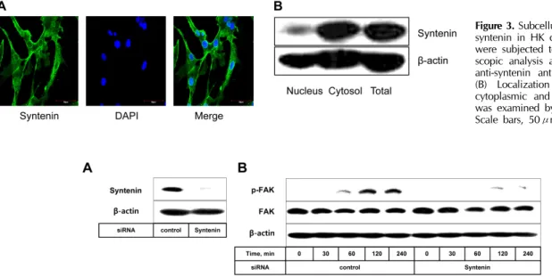
Figure 3. Subcellular localization of syntenin in HK cells. (A) HK cells were subjected to confocal micro- scopic analysis after staining with anti-syntenin antibody and DAPI.
(B) Localization of syntenin in cytoplasmic and nuclear fractions was examined by immunoblotting.
Scale bars, 50μm.
Figure 4. Syntenin knockdown impairs FAK activation in HK cells. (A) Syntenin molecules in HK cells were knocked down by transfection with syntenin siRNA as evaluated by immunoblotting analysis. (B) HK cells transfected with syntenin or control siRNA were allowed to attach to tissue-culture treated plastics. At the indicated time points, cells were harvested to measure the phosphorylation degrees of FAK proteins.
Representative of two reproducible data.
co-localized cells (Fig. 1A). The co-localization of syntenin with CD19 was more frequent than that with CD3 (Fig. 1B), implying that a significant fraction of B cells expresses syntenin. However, inasmuch as anti-CD19 antibody is also reactive with FDC (13) only small subsets of B cells appear to express syntenin. Most FDCs expressed syntenin as con- cluded by the intensive overlapping of CNA.42 and syntenin (Fig. 1C). Control sections that were stained with iso- type-matched antibodies did not exhibit noticeable binding to antibodies (data not shown). These in situ data prompted us to explore syntenin expression with isolated cells from the tonsil; B, T, and HK cells. HK cells were used due to the paucity of bona fide FDCs obtainable from a tonsil. The ex- pression of syntenin in T and B cells was confirmed by im- munoblotting and flow cytometry. Only a small fraction of T and B cells expressed syntenin (Fig. 2). However, syntenin was strongly expressed in HK cells, in line with confocal mi- croscopic findings in situ. At subcellular levels, most syntenin molecules were localized in the cell membrane, nuclear mem- brane, and cytoplasm (Fig. 3A). Syntenin was not expressed in the nuclei of HK cells except the nuclear membrane, and a significant overlapping of syntenin and DAPI was not observed. These data obtained by confocal microscopy were confirmed by immunoblotting. Syntenin molecules were largely restricted to the cytoplasmic fraction (Fig. 3B).
Syntenin promotes phosphorylation of focal adhes- ion kinase
Given that HK cells express syntenin strongly in the cell membrane (Fig. 3A), we investigated its role in adhesion of HK cells to tissue-culture treated plastics. This reaction mim- ics focal adhesion (FA) that is a close contact of adherent cells with extracellular matrices (ECM), a dynamic process requir- ing coordinated rearrangement of cytoskeletons. The siRNA technology was adopted to knock-down syntenin molecules in HK cells (Fig. 4A). HK cells adhered to plastics in a time-dependent fashion, which correlated with FAK phos- phorylation (Fig. 4B). The phosphorylation peaked 2 h post adhesion, and then remained at the similar level. In contrast, FAK phosphorylation kinetics were markedly impaired when syntenin was knocked down. The phosphorylation level ob- tained after 4 h of incubation was only comparable to that observed 1 h post-incubation in control HK cells. However, syntenin knockdown did not change cell morphology or de- lay plastic adhesion (data not shown), implying that plastic adhesion of HK cells is a complicated process involving many molecules in addition to syntenin. These results suggest that syntenin takes part in the FA of HK cells by promoting phos- phorylation of FAK molecules.
DISCUSSION
This study revealed that syntenin was expressed and played a significant role in normal cells as well as in some malignant cells. Although there was weak or no expression of syntenin in T and B cells, it displayed a strong expression in FDC.
The syntenin expression patterns in lymphocytes and FDC were consistent in those in situ and ex vivo. Syntenin protein levels in HK cells were comparable to those in the MDA- MB-231 cell line (14) (data not shown).
Syntenin expression in FDC and HK cells at high levels is an interesting finding. FDCs are found only in B cell follicles and believed to play a critical role in the course of B cell differentiation. However, their functional characterization has not been fully carried out, particularly at the molecular level.
Accumulating evidence indicates that FDCs are derived from bone marrow mesenchyme cells, unlike other immune cells which originate from hematopoietic stem cells (15). They pro- liferate poorly in situ and do not express the proliferation marker Ki-67 (16). Therefore, precursor cells such as fibro- blasts in the connective tissue may migrate into B cell follicles to differentiate into FDCs. In this migration process and after their arrival at follicles, syntenin may play an important role when FDCs come into contact with ECM and neighboring B/T cells. Syntenin expression levels correlate with the migratory capability of expressing cells (6).
Since FDC/HK cells are adherent cells that show FA to the ECM or interacting lymphocytes, we explored the potential role of syntenin in FA of HK cells by utilizing siRNA technology. FAK is an essential non-receptor tyrosine kinase and serves as the major modulator of FA. The molecular structure of FAK explains its dual activity as tyrosine kinase and scaffold protein. The C-terminal region enables FAK to interact with other proteins (17). It accumulates near the points of FA, phosphorylates paxillin, regulates microtubule stability, and eventually transduces extracellular signals (18).
FAK activation involves phosphorylation of Tyr397 with the mediation of integrin (17). Our results suggest that FAK phos- phorylation depends on the presence of syntenin. Although molecular mechanisms for the role of syntenin in FA and FAK activation in FDC is currently unknown, a potential mecha- nism can be speculated from recent findings of other investigators. Syntenin interacts with c-Src via PDZ domain, and then activated c-Src induces additional phosphorylation of FAK in melanoma and breast carcinoma cells (8,14).
Whether similar molecular events operate in FDCs or HK cells
is the subject of our future investigation.
In conclusion, our results suggest that syntenin is stongly expressed and take part in the activation of FAK in normal human FDCs. More efforts to explore the biological sig- nificance of strong syntenin expression in FDCs may uncover important functions of syntenin in normal physiology in addi- tion to cancer pathology.
ACKNOWLEDGEMENTS
This research was supported by the “Leaders of INdustry-uni- versity Cooperation” Project through the National Research Foundation of Korea (NRF) funded by the Ministry of Education, Science, and Technology. This study was also sup- ported by 2013 research grant from Kangwon National University (No. C1009781-01-01).
CONFLICTS OF INTEREST
The authors have no financial conflict of interest.
REFERENCES
1. Grootjans, J. J., P. Zimmermann, G. Reekmans, A. Smets, G.
Degeest, J. Dürr, and G. David. 1997. Syntenin, a PDZ pro- tein that binds syndecan cytoplasmic domains. Proc. Natl.
Acad. Sci. USA. 94: 13683-13688.
2. Beekman, J. M. and P. J. Coffer. 2008. The ins and outs of syntenin, a multifunctional intracellular adaptor protein. J.
Cell Sci. 121: 1349-1355.
3. Gibferrer, I., A. Ibanez, M. Farnos, M.-R. Sarrias, R. Funutria, S. Rosello, P. Zimmermann, G. David, J. Vives, C.
Serra-Pages, and F. Lozano. 2005. The lymphocyte receptor CD6 interacts with syntenin-1, a scaffolding protein contain- ing PDZ domains. J. Immunol. 175: 1406-1414.
4. Latysheva, N., G. Muratov, S. Rajesh, M. Padgett, N. A.
Hotchin, M. Overduin, and F. Berditchevski. 2006. Syntenin-1 is a new component of tetraspanin-enriched microdomains:
mechanisms and consequences of the interaction of synte- nin-1 with CD63. Mol. Cell. Biol. 26: 7707-7718.
5. Chen, F., Y. Du, Z. Zhang, G. Chen, M. Zhang, H.-B. Shu, Z. Zhai, and D. Chen. 2008. Syntenin negatively regulates TRAF6-mediated IL-1R/TLR4 signaling. Cell Signal. 20: 666- 674.
6. Koo, T. H., J.-J. Lee, E.-M. Kim, K.-W. Kim, H. D. Kim, and J.-H. Lee. 2002. Syntenin is overexpressed and promotes cell migration in metastatic human breast and gastric cancer cell lines. Oncogene 21: 4080-4088.
7. Gangemi, R., V. Mirisola, G. Barisione, M. Fabbi, A.
Brizzolara, F. Lanza, C. Mosci, S. Salvi, M. Gualco, M. Truini, G. Angelini, S. Boccardo, M. Cilli, I. Airoldi, P. Queirolo, M.
J. Jager, A. Daga, U. Pfeffer, and S. Ferrini. 2012. Mda-9/
Syntenin Is expressed in uveal melanoma and correlates with metastatic progression. PLoS One 7: e29989.
8. Boukerche, H., Z.-z. Su, C. Prevot, D. Sarkar, and P. B.
Fisher. 2008. mda-9/Syntenin promotes metastasis in human melanoma cells by activating c-Src. Proc. Natl. Acad. Sci.
USA. 105: 15914-15919.
9. Choe, J., H.-S. Kim, R. J. Armitage, and Y. S. Choi. 1997.
The functional role of BCR stimulation and IL-4 in the gen- eration of human memory B cells from germinal center B cells. J. Immunol. 159: 3757-3766.
10. Kim, H.-S., X. Zhang, and Y. S. Choi. 1994. Activation and proliferation of follicular dendritic cell-like cells by activated T lymphocytes. J. Immunol. 153: 2951-2961.
11. Kim, J., S. Lee, Y.-M. Kim, D.-I. Jeoung, and J. Choe. 2013.
Human follicular dendritic cells promote germinal center B cell survival by providing prostaglandins. Mol. Immunol. 55:
418-423.
12. Cho, W., S. H. Hong, and J. Choe. 2013. IL-4 and HDAC inhibitors suppress cyclooxygenase-2 expression in human follicular dendritic cells. Immune Netw. 13: 75-79.
13. Clark, E. A., K. H. Grabstein, and G. L. Shu. 1992. Cultured
human follicular dendritic cells: Growth characteristics and in- teractions with B lymphocytes. J. Immunol. 148: 3327-3335.
14. Hwangbo, C., J. Kim, J. J. Lee, and J.-H. Lee. 2010.
Activation of the integrin effector kinase focal adhesion kin- ase in cancer cells is regulated by crosstalk between protein kinase C and the PDZ adapter protein mda-9/syntenin.
Cancer Res. 70: 1645-1655.
15. Munoz-Fernandez, R., F. J. Blanco, C. Frecha, F. Martin, M.
Kimatrai, A. C. Abadia-Molina, J. M. Carcia-Pacheco, and E.
G. Olivares. 2006. Follicular dendritic cells are related to bone marrow stromal cell progenitors and to myofibroblasts.
J. Immunol. 177: 280-289.
16. Lindhout, E. and C. de Groot. 1995. Follicular dendritic cells and apoptosis: life and death in the germinal centre. Histo- chem. J. 27: 167-183.
17. Franchini, K. G. 2012. Focal adhesion kinase - The basis of local hypertrophic signaling domains. J. Mol. Cell. Cardiol.
52: 485-492.
18. Stehbens, S. and T. Wittmann. 2012. Targeting and transport:
How microtubules control focal adhesion dynamics. J. Cell Biol. 198: 481-489.