J. of Korean Shoulder and Elbow Society Volume 9, Number 2, December, 2006
※통신저자: 김 명 선*
광주광역시 동구 학동 8번지 전남대학교병원 정형외과학교실
Tel: 062) 227-1640, Fax: 062) 225-7794, E-Mail: rhamses@chol.com
쇄골 골절의 관혈적 정복 및 내 고정술 후 발생한 쇄골하 동맥의 혈전증 - 증례 보고 -
전남대학교 의과대학 정형외과학교실
문은선∙김명선*∙정광철∙임근영
—
— Abstract ——
The Subclavian Artery Thrombosis after Open Reduction and Internal Fixation of Clavicular Fracture
- A Case Report -
Eun-Sun Moon, M.D., Myung-Sun Kim, M.D.*, Kwang-Cheul Jeong, M.D., Keun-Young Lim, M.D.
Dept. of Orthopaedics, Chonnam National University Hospital, Gwangju, Korea
Reported in this article is a case report of a patients who developed limb threatening ischemia as a consequence of a subclavian artery thrombosis resulting from screws. The subclavian artery thrombosis after open reduction and internal fixation of clavicle fracture, when it occurs, should be treated promptly by plate removal and claviculectomy.
It can be prevented by placing screws in locations away from the underlying neurovascular structures.
Key Words: Subclavian artery, Clavicle fracture, Thrombosis
서 론
쇄골 골절은 비교적 흔히 발생하는 골절로 보존 적 방법으로도 성공적인 치료가 가능하며 대부분 의 경우 심각한 동반 손상이나 합병증을 수반하지 않는다. 하지만 쇄골 골절과 함께 발생하거나 지 연성 또는 의인성(iatrogenic)으로 발생하는 혈전
에 의한 원위부 동맥 폐색은 매우 심각한 합병증 으로 간과되기 쉽다. 저자들은 쇄골 골절의 관혈 적 정복 및 금속판을 이용한 내 고정술 후 발생한 혈전에 의한 원위부 동맥 폐색을 경험하여 이에 대한 증례를 보고하고자 한다.
증례 보고
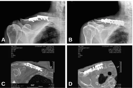
63세 남자 환자로 내원 약 3개월 전부터 발생 한 우측 상지의 동통 및 수지 말단부의 체온 저하 를 주소로 내원하였다. 과거력 상 6개월 전 우측 쇄골 간부 골절에 대해 타병원에서 관혈적 정복 및 금속판 내 고정술을 시행하였으며, 이후 골절 의 불유합 및 금속판의 파단(breakage)이 발생 하여 술 후 3개월째 금속판과 강선을 이용한 내 고정을 시행하였다(Fig. 1). 증상은 2차 수술 직 후부터 서서히 발생하였으며, 누워있을 때에는 호
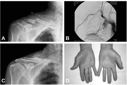
전되고 앉거나 엎드려 있을 경우 심해지는 양상을 보였다. 본원 내원 당시 요골 동맥이 촉지되지 않 았으며, 혈관 조영 검사 상 우측 쇄골하 동맥 (subclavian artery)이 척추 동맥(vertebral artery) 기시부의 원위 2 cm까지만 조영되었고 상완 동맥(brachial artery)은 측부 혈류(col- lateral circulation)를 통해 유지되고 있었다 (Fig. 2). 단순 방사선 사진 및 컴퓨터 단층 촬영 상 쇄골에 삽입된 모든 금속 나사못이 하방으로 돌출된 것을 확인할 수 있었다(Fig. 1).
경흉골 도달법(transsternal approach,
Fig. 1. At postoperative 3 months from initial operation, metal failure developed at the fracture site (A). Open anatomi- cal reduction and internal fixation with the reconstruction plate and tubular plate was performed at local hospi- tal (B). Note the all medial screws penetrated the inferior (C) and posterior (D) cortex of the clavicle.
A B
C D
Fig. 2. A 63-year male developed paresthesia, aching pain and skin color change at right hand (A). Runoff arteri- ogram demonstrated an occluded right subclavian artery, distal to vertebral artery origin site. Note the opaci- fied ipsilateral brachial artery by rich collateral network from remnant subclavian artery (B).
A B
median sternotomy)을 통하여 수술을 시행하 였다. 기존 골절은 유합되어 있었으며, 쇄골 후방 및 하방으로 금속 나사못이 약 0.5 cm 가량 돌출 되어, 직접적인 혈관 압박 소견을 관찰할 수 있었 다(Fig. 3). 기존 골절선을 기준으로 하였을 때 내측에 시술된 3개의 강선 주위로 심한 흉터조직 이 형성되어 있었으며, 이로 인하여 쇄골하 동맥 등 내부 구조물들이 외측으로 전위되어 있었다.
주관부(cubital fossa)에서 상완동맥을 찾아 풍 선 도자술을 시행하였으며, 상부로 약 30 cm 가 량부터 풍선 도자가 진입하지 못하였다. 쇄골하
동맥을 촉지하였으며, 협착이 일어난 부위를 확인 하였다. 수술 시야를 확보하기 위해 금속판 및 강 선을 제거하였으며, 협착 부위에서 쇄골의 절골술 을 시행하였다. 다량의 혈전을 제거하였으며, 이 후 상지의 혈액 순환이 이루어졌다. 하지만, 흉골 을 닫은 후 쇄골에 대해 정복을 시행하려 하였으 나, 정복 후 다시 혈액 순환이 악화되는 양상을 보여, 쇄골 정복을 하지 않은 채 수술을 마쳤다.
술 후 시행한 혈관 조영 검사 상 쇄골하 동맥의 혈전의 소실 및 상완 동맥으로의 정상 혈류가 유 지되고 있었으며 우측 상지 동통 및 수지 말단의
Fig. 3. Intraoperative photograph, obtained after claviculectomy. Note the scar (long arrow) caused by penetrated screw was seen at the subclavian artery with distal dilatation (A). Resected subclavian artery. Note the throm- botic debris in and around the perforation (B). *: carotid artery
A B
Fig. 4. The displacement at the claviculectomy site was seen in immediate postoperative radiograph (A). The postop- erative arteriogram demonstrated patency of the subclavian aryery and distal runoff (B). The postoperative 1 year radiograph show union (C) and recover the skin color (D).
A B
C D
체온 저하 상태는 호전되었다(Fig. 4). 최종 1년 추시 상 우측 쇄골의 부분적인 골 유합이 관찰되 었으며 수지 말단의 피부 색 등의 혈류 상태는 양 호하였다.
고 찰
쇄골 주위의 혈관들은 쇄골과 매우 가까이 위치 해 있으나 쇄골하근(subclavius muscle)과 심부 경부 근막(deep cervical fascia)에 의해 보호되 어 있으므로 상당한 전위를 보이는 골절에서도 혈 관 손상을 경험하는 경우가 거의 드물다. 또한 해 부학적으로 쇄골 주위의 신경, 혈관들은 쇄골의 내측 하방에 위치하며 골절 후 원위 골편은 상지 의 무게에 의해 전하방으로 전위되고, 근위 골편 은 승모근에 의해 후상방으로 전위되기 때문에 쇄 골하 신경 혈관의 골절편에 의한 직접적인 압박은 피할 수 있다. 따라서 쇄골 골절 시 쇄골 주위의 혈관 손상은 간과되기 쉬우며 Strum과 Cicero6) 는 첫 번째 늑골의 골절이 있을 때, 요골 동맥의 맥박이 촉지 되지 않을 때, 쇄골 상부로 혈종이 형성되어 있을 때, 흉부 단순 방사선 사진 상 종 격동이 넓을 때(wide mediastinum) 그리고 상 완 신경총 손상이 있을 때 쇄골 주위의 혈관 손상 을 의심해야 한다고 하였다.
쇄골 골절 후 발생한 혈관 손상 및 혈류의 차단 은 부정 유합이나 불유합으로 인한 지나친 가골의 형성, 쇄골하 동맥의 동맥류 형성, 골절 당시의 심한 전위 후 생기는 지연성 손상 및 수술 시 사 용된 K-강선이나 나사에 의한 의인성 손상 등에 의해 발생한다3,5). 이 중 쇄골 골절 및 불유합의 금속판 고정술 후 발생한 흉곽 출구 증후군(tho- racic outlet syndorme)은 Casselman1) 등에 의해 보고된 바 있다. 그들의 보고에 의하면 환자 는 수상 후 8년 후에 불유합에 대해 금속판 고정 술을 시행하였으며 술 후 해당 상지의 허혈 증세 를 보여 혈관 조영술 후 쇄골하 동맥의 부분 절제 및 8 mm Gore-Tex를 이용한 재건술(interpo- sition grafting)과 제 1늑골 절제술을 시행하였 다. Shackford4)은 금속판 고정술 후 나사못에 의한 가성 동맥류(pseudoaneurysm)에 대하여 쇄골 절제술(claviculectomy) 및 복재 정맥
(saphenous vein)을 이용한 재건술을 시행하여 좋은 결과를 보고하였고 쇄골의 내고정술 후 흉곽 출구 증후군이 발생하였을 경우 즉시 금속판 절제 술과 쇄골 절제술을 시행해야 한다고 기술하였다.
Jupiter와 Leffert2)는 쇄골의 간부의 경우 회전 력(torsional force)를 받으며 해면골(cancel- lous bone)이 적어 금속판의 압박력과 나사못의 움직임이 지속적으로 발생하여 나사못이 반대측의 피질 골을 침범한다고 주장하였다. 본 환자의 경 우 첫 번째 금속판 고정술 후에는 특별한 증세가 없다가 금속판의 파단이 발생하여 재수술을 시행 한 후 체위의 변화에 따른 해당 상지의 저체온과 색깔 변화, 동통 등의 증상이 나타났고 단순 방사 선 사진과 혈관 조영술 및 수술 소견 상 나사못에 의한 쇄골하 동맥 압박 소견을 관찰할 수 있었으 며 이로 인한 혈전의 형성과 혈류 차단으로 증상 이 발생하였을 것으로 생각된다. 또한 금속판 제 거술과 다량의 혈전 제거술 후 쇄골 절제술 없이 쇄골 절골술만으로도 상지의 혈류를 유지할 수 있 었으며 쇄골 절제술을 시행하더라도 상지의 정상 기능에 큰 장애는 없다는 보고도 있으나 정상에 가까운 쇄골의 길이를 유지함으로써 상지의 정상 적인 구조를 보존할 수 있을 것으로 생각한다.
따라서 쇄골 골절에 대하여 부득이하게 내 고정 술을 시행해야 할 경우 쇄골의 내측 하방으로 주 행하는 신경, 혈관을 고려하여 나사못의 방향 및 길이를 결정해야 하며 내 고정술 후 혈관 손상이 의심되었을 경우 적절한 검사 후 금속판 제거술과 함께 추가적인 재건술도 고려해야 할 것으로 사료 된다.
REFERENCES
01) Casselman F, Vanslembroek K and Ver- ougstraete L: An unusual case of thoracic outlet syndrome. J Trauma, 43:142-143, 1997.
02) Jupiter JB and Leffert RD: Non-union of the clavicle. J Bone Joint Surg, 69-A:753-760, 1987.
03) Olin T: Arterial complications in thoracic outlet compression syndrome. Acta Radiol, 56:97-112, 1961.
04) Shackford SR: Taming of the screw: A case report and literature review of limb-threatening
complications after plate osteosynthesis of a calvicular nonunion. J Trauma, 55:840-843, 2003.
05) Steinberg I: Subclavian vein thrombosis associ- ated with fractures of the clavicle. Report of two
cases. New England J Med, 264:686-688, 1961.
06) Sturm JT and Cicero JJ: The clinical diagnosis of ruptured subclavian artery following blunt thoracic trauma. Ann Emerg Med, 12:17-19, 1983.