J. of Korean Bone & Joint Tumor Soc.
Volume 13, Number 2, December, 2007
※통신저자: Han-Soo Kim, M.D.
Department of Orthopaedic Surgery, Seoul National University College of Medicine 28 Yongon-Dong, Chongro-Gu Seoul 110-744, KOREA
Tel: 02) 2072-2362, Fax: 02) 764-2718, E-mail: hankim@snu.ac.kr
Giant Cell Tumor involving the Ulnar Diaphysis
Department of Orthopaedic Surgery, Seoul National University College of Medicine
Ji Hyeung Kim, M.D., Ilkyu Han ,M.D., Hyun Guy Kang, M.D., Han-Soo Kim ,M.D.
Giant cell tumor of bone is relatively common neoplasm usually involving epi- physis of long bone. And rarely it involves the diaphysis or metaphysis without epiphyseal extension. We report on an 18-year-old girl with giant cell tumor of ulnar diaphysis. She was treated with wide excision and reconstuction with non- vascularized autogenous fibular graft. We harvested fibular fragment preserving fibular continuity to reduce donor site morbidity. Surgical outcome and func- tional result was excellent.
Key Words: Giant cell tumor, Diaphysis, Ulna, Wide excision
Giant cell tumor of bone is a benign but locally aggressive tumor that usually involves the end of long bone. Giant cell tumors of bone were first described by Jaffe et al6) in 1940. It is characterized by varying numbers of multinucleated giant cells dis- persed in a stroma of round, ovoid, or spin- dle-shaped mononuclear cells that fuse to form the giant cells of the lesions. Giant cell tumors account for about 5% of primary bone tumors and 21% of benign skeletal tumors1). The tumors occur more frequently in women than men, are most common in patients of 18~40 years. The typical location
is epiphyseal-metaphyseal regions of the long bones2), which is very characteristic.
Diaphyseal location of this tumor is very rare and just a few cases of diaphyseal giant cell tumor without epiphyseal involvement have been reported13,14). In larger series study, the incidence of nonepiphyseal loca- tion varied from 0.9~2.6%4,5). Giant cell tumor of the ulnar diaphysis is extremely rare. We present a case of giant cell tumor with a diaphyseal location of the ulna.
Case report
An 18-year-old, right-hand dominant girl
presented with a 1 month history of painful swelling of left forearm. The pain was locat- ed in the distal half of the left forearm. It initially was moderate and aggravated over several weeks. Her past medical history and review of systems were unremarkable.
Physical examination of the left forearm showed tenderness to palpation of distal one third area of ulna. There was mild soft tis- sue prominence about the distal ulna. There was no sign of infection such as lym- phadenopathy, erythema or local heat. Full range of motion was possible on left wrist and elbow. No neurovascular abnomal find- ing was observed. Calcium, phosphorus and PTH levels were within normal limits. And other laboratory study showed no abnormal findings.
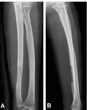
Plain radiographs and magnetic resonance imaging of left forearm were checked. The radiograph of the forearm showed an intramedullary osteolytic lesion of distal ulnar diaphysis (Fig. 1). But no sclerotic rimming or periosteal new bone formation Fig. 1. Preoperative antero-posterior and lateral radi-
ographs is shown.
A B
Fig. 2. Coronal T-1 weighted (A), T-2 weighted MRI scans are presented. Axial T-1 weighted scans before (C) and after (D) gadolinium enhancement is also presented.
A B
C
D
was observed. On magnetic resonance imag- ing, the lesion was 3.6×1.7×1.9 cm sized mass in the distal ulnar diaphysis showing of homogeneously intermediate signal inten- sity on T1-weighted image and heteroge- neously high signal intensity on T2-weighted image (Fig. 2). Cortical destruction, periosteal elevation and medullary canal involvement were observed in the distal ulnar diaphysis. The mass showed soft tissue involvement causing marked soft tissue mass in the flexor compartment of the forearm.
Ulnar neurovascular bundles were preserved.
After gadolium contrast media injection, the lesions were homogeneously well enhanced.
Bone scan showed increased uptake of radioisotope in distal one third area of left ulna. Chest CT revealed no abnormality in lung parenchyma, endobronchial lesion and mediastinum. Periosteal osteosarcoma, Ewing’s sarcoma, lymphoma, giant cell tumor and brown tumor were included for in the radiological differential diagnosis.
Incisional biopsy was done via volar approach retracting ulnar neurovasculature radially. Gross appearance of the submitted tissue was yellow-brown colored soft tissue mass. Permanent histologic section showed a
large population of giant cells (Fig. 3).
Necrosis was observed in the central portion of mass and 3 mitoses were present per 10 high power fields.
Because of the huge extraosseous soft tis- sue mass, wide excision was performed for definitive surgery rather than curettage.
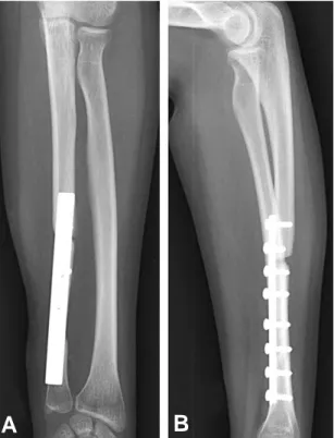
Including previous biopsy incision, en bloc excision was done. Distal ulnar osteotomy was done 4 cm proximal from the ulnar sty- loid process and at the 5 cm proximal from that level, proximal ulnar osteotomy was done. For reconstruction of the ulna, we used left fibular diaphysis as autograft. For reducing donor site morbidity, 5 cm length and half the width of fibula was harvested preserving continuity of fibula. Fibular autograft was fixed to the ulna by 8 hole plate and screws (Fig. 4). Postoperatively, compression dressing and long arm splint
Fig. 3. Histological section of the tumor showing typical stromal and giant cells is presented. (HE, ×200)
Fig. 4. Postoperative antero-posterior and lateral radi- ographs is shown.
A B
were applied. One week after surgery, long arm cast was applied. At 1.5 year follow- up, full range of motion was obtained in left wrist and elbow. And there were no evi- dence of local recurrence or distant metasta- sis. The donor site was completely remodeled.
Discussion
Giant cell tumors (GCT) commonly involve epiphysis of long bones. Tumor often extends to the articular subchondral bone or even abuts the cartilage. The most frequent locations are the distal femur, the proximal tibia, the distal radius, and the sacrum5,12). We presented nonepiphyseal giant cell tumor involving ulnar diaphysis. Of the 1229 GCT of bone from a collection of series by Campanacci1), Huvos5), and Unni12), GCT of distal ulna was 17 cases. Of the 14 cases of nonepiphyseal GCT reported by Fain3), 5 patients were younger than 15 years of age.
And seven of the 14 tumors were in patients with open growth plates. In his cases, the portion of younger patient in nonepiphyseal GCT was greater than in conventional GCT.
Only a limited number of cases of GCT involving the diaphysis of a long bone has been reported in the literature. Of the 1682 cases of GCT treated at Mayo clinic, only 2 cases were identified as being located in the diaphysis of a long bone3). Both cases were located in the tibia and were treated by curettage. One of the cases developed local recurrence 4 years after operation and was successfully treated with repeat curettage.
Visscher et al. reported a case of GCT involving the diaphysis of ulna in a 7 months old patient13). En bloc resection and reconstruction with fibular autograft was performed. A GCT occurring from cortical surface of tibial diaphysis was reported by
Wilkerson et al.14). The lesion presented as a soft tissue mass and was treated by margin- al excision.
Pain is the leading symptom in GCT and relates to the mechanical insufficiency resulting from bone destruction. Pathologic fracture is seen in about 12% of patients at the time of diagnosis2,4). A bump or a soft tissue mass can occasionally be seen and results from the cortical destruction and tumor progression outside bone. Our patient showed similar clinical findings. Painful swelling was aggravated and soft tissue mass was palpated at the distal one third area of left ulna.
Radiographically, GCT shows purely lytic and eccentric features within the bone. The tumor appearance is geographic, with ill- defined borders and often without any iden- tifiable sclerosis. Cortical and cancellous bones likewise appear destroyed. Bone con- tour can be expanded with faint and thin periosteal new bone formation. Tumor matrix is devoid of any ossification or calci- fication and of similar density to the sur- rounding soft tissues. Campanacci1) described 3 stages of GCT based on radiological appearance. Stage 1 is the least frequent and shows features of latent or slow-growing tumor. Stage 2 shows features of an active lesion with ill-defined borders and without sclerosis. Stage 3 shows extreme aggressive- ness, with a tumor of large volume that destroys bone and invades the surrounding soft tissues. Our case showed intramedullary osteolytic lesion with cortical destruction and periosteal elevation compatible with stage 2.
Histologically GCT shows increased cellu- larity, with numerous multinucleated giant cells uniformly dispersed among a large pop- ulation of mononuclear cells. There is little or no intercellular substrate other than a
few collagen fibers. Mitotic figures can be numerous but devoid of abnormalities. On histology, the differential diagnosis must include giant cell reparative granuloma and brown tumor of hyperparathyroidism.
Curettage has been the preferred treat- ment for most cases of GCT. But historically local recurrence rates were reported 25% to
50%4,10,12). Recent series of study with modern
imaging techniques and curettage through the use of power burr revealed improved recurrence rates(10~20%)9,12). And varient adjuvants such as phenol, liquid nitrogen, bone cement, hydrogen peroxide, zinc chlo- ride, and argon beam cauterization were introduced. Recurrence rate was after curet- tage was 18% and 3% after wide excision.
Due to diaphyseal location, wide excision preserving adjacent joint was possible in our case. So we selected en bloc excision to reduce recurrence rate without functional deficit. And we used fibular autograft for reconstruction and fixed by 8 hole plate and screw. Lackman7) reported fibular recon- struction for GCT of the distal radius and Mendicino8) performed en bloc resection and used autogenous middle fibular strut graft for the treatment of GCT of the first metatarsal bone.
In the presented case, the location of the tumor was very unusual for GCT but gross appearance and histologic findings were com- patible with GCT. For local control, en bloc excision was performed due to the soft tissue involvement and fibular hemicortical auto- graft was used. We report a case of giant cell tumor involving diaphysis of ulna.
REFERENCES
01) Campanacci M. Giant cell tumor. In: Gaggi A,
editor. Bone and soft-tissue tumors. Bologna, Italy, Springer-Verlag, 117-153, 1990.
02) Campanacci M, Baldini N, Boriani S, Sudanese A: Giant cell tumor of bone. J bone Joint Surg, 69A:106-114, 1987.
03) Fain JS, Unni KK, Beabout JW, Rock MG:
Nonepiphyseal giant cell tumor of the long bones.
Clinical, radiologic, and pathologic study. Cancer, 71(11):3514-3519, 1993.
04) Goldenberg RR, Campbell CJ, Bonfiglio M:
Giant-cell tumor of bone: An analysis of two hun- dred and eighteen cases. J Bone Joint Surg, 52A:
619-664, 1970.
05) Huvos AG: Bone tumors: diagnosis, treatment and prognosis. 2ndedition. Philadelphia: WB saunders Co, 1991.
06) Jaffe HL, Lichtenstein L, Portis RB: Giant cell tumor of bone: Its pathologic appearance, grading, supposed variants and treatment. Arch Pathol, 30:993-1031, 1940.
07) Lackman RD, McDonald DJ, Beckenbaugh RD, et al: Fibular reconstruction for giant cell tumor of the distal radius. Clin Orthop, 218:232-238, 1987.
08) Mendicino SS: Giant cell tumor of the first metatarsal bone en bloc resection with autogenous middle fibular strut graft. J Foot Ankle surg, 32(4):405-410, 1993 .
09) O’Donnel RJ, Springfield DS, Motwani HK, et al: Recurrence of giant cell tumors after curettage and packing with cement. J Bone Joint Surg Am, 76(2):1827-1833, 1994.
10) Trieb K, Bitzan P, Dominkus M, et al: Giant-cell tumors of long bone. J Bone Joint Surg Am, 82:1360-1361, 2000.
11) Turcotte RE, Davis AM, Wunder J, et al: Giant cell tumour of long bone: a Canadian Sarcoma Group Study. Clin Orthop Rel Res, 397:248-258, 2002.
12) Unni KK: Dahlin’s bone tumors: general aspect and data on 11087 cases. 5th edition. Philadelphia:
Lippincott-Raven, 1998.
13) Visscher DW, Alexander RW, Dempsey TR:
Heretical giant cell tumor in the diaphysis of the ulna in a 7 month old boy. Skeletal Radiol, 17(4):285-288, 1988.
14) Wilkerson JA, Cracchiolo A: Giant-cell tumor of the tibial diaphysis. J Bone Joint Surg[Am], 51:1205-1509, 1969.