Bile duct preserving pancreatic head resection (BDPPHR):
Can we conclusively define the extent of head resection in surgery for chronic pancreatitis?
Pagadala Naga Balaji Nitesh and Biju Pottakkat
Department of Surgical Gastroenterology, Jawaharlal Institute of Post Graduate Medical Education and Research (JIPMER), Pondicherry, India
The role of surgical management for chronic pancreatitis in providing pain relief and improving quality of life is significant. Surgical techniques involving pancreatic head resection scored more over the drainage procedures. Among the resection procedures, Frey’s procedure received widespread acceptance. However, the exact extent of pancreatic head resection to be performed and the limits of the resection are still debatable. The present report of bile duct pre- serving pancreatic head resection (BDPPHR) describes an innovative approach to the pancreatic head and conclusively defines the extent of head resection. The simplicity, feasibility and easy reproducibility of the BDPPHR is also reiterated.
(Ann Hepatobiliary Pancreat Surg 2020;24:309-313)
Key Words: Chronic pancreatitis; Surgery; Pancreatic head resection; Posterior pancreatic capsule
Received: January 19, 2020; Revised: April 10, 2020; Accepted: April 12, 2020 Corresponding author: Biju Pottakkat
Department of Surgical Gastroenterology, Jawaharlal Institute of Post Graduate Medical Education and Research (JIPMER), Pondicherry 605006, India
Tel: +91-7598566982, Fax: +91-8179705152, E-mail: bijupottakkat@gmail.com
Copyright Ⓒ 2020 by The Korean Association of Hepato-Biliary-Pancreatic Surgery
This is an Open Access article distributed under the terms of the Creative Commons Attribution Non-Commercial License (http://creativecommons.org/
licenses/by-nc/4.0) which permits unrestricted non-commercial use, distribution, and reproduction in any medium, provided the original work is properly cited.
Annals of Hepato-Biliary-Pancreatic Surgery ∙ pISSN: 2508-5778ㆍeISSN: 2508-5859
INTRODUCTION
The concept of early surgical management for chronic pancreatitis (CP) has significant impact on prevention of deterioration in the quality of life of patients. Resection procedures are associated with long-lasting pain relief and will prevent further complications associated with chronic pancreatitis, when compared to drainage procedures. In view of associated morbidity, extensive resection proce- dures like total pancreatectomy should be avoided in pa- tients with disease process localised to head of pancreas.
Currently, Frey’s procedure, Beger procedure and Whipple pancreaticoduodenectomy are the common resection pro- cedures performed in patients with CP. In spite of sig- nificantly increased acceptance of the Frey’s procedure, the debate continues regarding the exact extent of re- section of head of pancreas performed in this procedure.
Due to varied reasons, the original Frey’s procedure de- scribed in 1987 has been modified in the context of extent of head resection into modified Frey’s and minimum
Frey’s procedure.1-3 Amount of diseased pancreatic head tissue retained after these modified procedures is a matter of concern. To standardise the resection procedure for CP, we describe a technique of bile duct preserving pancreatic head resection (BDPPHR).
SURGICAL TECHNIQUE
The abdominal cavity is usually exposed through bi- lateral subcostal incision with the use of self-retaining retractor. A complete Kocher maneuver till the identi- fication of superior mesenteric vein (SMV) is performed with the intent of manual palpation of gland for assess- ment of consistency. This mobilisation of head of pan- creas also aids during the coring of head of pancreas to secure hemostasis with palpating hand beneath the head of pancreas. We recommend the plane of kocherisation to be slightly away from the duodenum in order to avoid any breach in the posterior pancreatic capsule. The anterior surface of entire pancreas is exposed by releasing the gas-
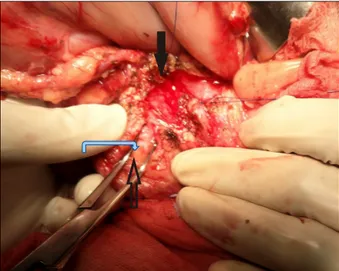
Fig. 1. Image showing technique of blunt dissection through anterior surface of the pancreatic parenchyma with the at- tempt to define posterior pancreatic capsule in head of the pancreas (marked with transparent arrow). Entire pancreatic duct in the body and tail region of pancreas was exposed (pointed with Black arrow). The Left hand of operating sur- geon behind the kocherised duodenum and head of pancreas is guiding the procedure. Exposed posterior pancreatic capsu- le is marked with blue arrow.
terior surface of the head and neck of the pancreas. No further dissection is warranted at the superior border of the pancreas. Meticulous dissection is performed along the anterior and right lateral surface of SMV to provide opti- mal exposure of uncinate process. It facilitates complete coring of uncinate process of pancreas with SMV in view and also prevents any inadvertent injury to SMV. This might require even ligation of the draining veins from un- cinate process to SMV. We never initiate pancreatic head resection before complete delineation of SMV as a pre- caution to avoid SMV injury. The inferior border of body and tail of the pancreas is mobilised.
Dilated pancreatic duct is often palpated as groove along the axis of the gland. In case of difficulty with duc- tal identification by palpation, we routinely perform intra- operative ultrasound for the identification of pancreatic duct. Exposure of pancreatic duct is ensured with 21- gauge needle with syringe placed in duct and incising the anterior pancreatic tissue over the needle using electroco- agulation. Ductal probe or fine right-angled clamp is used to probe the direction of the duct. Pancreatic duct is ex- posed till distally within 0.5 cm of the end of the tail of the pancreas. Proximally in the head of pancreas, pancre- atic tissue anterior to duct of Wirsung is incised till within 0.5 cm of the ampulla of Vater. The pancreatic duct pa- tency near the ampulla of Vater is assessed by passing 8 Fr infant feeding tube through the ampulla and palpating the tube within the duodenal lumen. All the ductal calculi, including branch duct calculi, identified during ductal ex- posure are removed thoroughly.
We recommend resection of head of the pancreas with posterior pancreatic capsule as the limit of resection. In our observation, posterior pancreatic capsule is thickened and is a tough structure in patients with chronic pan- creatitis due to inflammation. Attempts to delineate poste- rior pancreatic capsule is made by blunt dissection through the anterior surface pancreatic parenchyma as shown in Fig. 1 and also by graded progression of re- section of slices of pancreatic head using electrocoagu-
can be easily stripped off posterior pancreatic capsule from below upwards with the help of electrocoagulation. After excising each slice of pancreatic tissue, the remaining amount of pancreatic tissue needed to be removed is accu- rately assessed and proceeded. With progressively pro- ceeding the resection in a similar manner, complete pan- creatic head resection can be accomplished. In this techni- que, authors want to reiterate that resection proceeds in plane almost perpendicular to the surface of pancreas and the entire pancreatic parenchymal tissue including ducts of Wirsung, ducts of Santorini and its branch ducts in the head of pancreas, beyond its posterior wall till the posteri- or pancreatic capsule, are excised as shown in Fig. 2.
The right lateral extent of resection is limited by leav- ing a 0.5 cm rim of pancreatic parenchyma in order to preserve pancreaticoduodenal vascular arcade. Since the bile duct is in the right superolateral part of pancreatic head resection, we specifically focus in this technique to
Fig. 2. Line diagram showing perpendicular plane of trans- ection in BDPPHR as compared with Frey’s procedure.
Fig. 3. Image showing completed bile-duct preserving pancre- atic head resection (BDPPHR) technique. This image also highlights the possibility of leaving significant amount of pancreatic tissue by performing pancreatic head resection when posterior wall of pancreatic duct is considered as the limit of resection.
Fig. 4. Line diagram showing the view of pancreas from in- ferior aspect following BDPPHR with posterior pancreatic capsule as the posterior limit of pancreatic head resection.
preserve pancreatic parenchyma in the superolateral aspect of head resection to safeguard the intrapancreatic portion of bile duct from injury. The superior and inferior extent of resection is limited by sparing 5 mm of thickness of pancreatic parenchyma in order to facilitate further pan- creaticojejunostomy suturing. The medial extent i.e., left lateral extent of resection is highlighted by sparing a 5 mm rim of uncinate tissue along the medial wall of supe- rior mesenteric vein to facilitate for placement of sutures during pancreaticojejunostomy and also to avoid any in- advertent injury to SMV during head resection. Rest of the medial extent of resection is limited by pancreatic duct in the neck region of pancreas. Posterior pancreatic capsu- le itself will be the posterior layer of pancreatic partition as shown in Figs. 3, 4. In this technique of BDPPHR, complete resection of pancreatic parenchyma in the head region of pancreas including the main and minor pancre- atic ducts and uncinate ducts thoroughly with posterior pancreatic capsule as limit is performed.
The jejunum is divided at 25 cm distal to the duodeno- jejunal flexure and Roux limb is created. The Roux limb is passed through the mesocolic route to accommodate freely over the entire pancreas. A single layered pancrea- ticojejunostomy is usually performed using running 3-0 non-absorbable suture approximating jejunal mucosa to the capsule of the pancreas, instead of the mucosa of pan- creatic duct, to facilitate drainage of small pancreatic ducts. The continuity of gastrointestinal tract is restored by side–to–side jejunojejunostomy in isoperistaltic fashion at 40 cm distal to pancreaticojejunostomy. All the mesen- teric defects are closed with 3-0 non-absorbable sutures to prevent internal hernia formation. A 24 Fr drain is rou- tinely placed near the pancreaticojejunostomy site.
Abdomen is closed in layers with continuous 1-0 absorb- able sutures for inner layer and 1-0 non-absorbable sutures for outer layer.
DISCUSSION
Surgical management will be required in about 40-75%
of patients with chronic pancreatitis during the course of their disease.4 A systematic review by Yang et al.5 high- lighted that early surgical management for pain in chronic pancreatitis was associated with improved postoperative pain relief and reduced risk of pancreatic insufficiency.
Currently on-going ESCAPE trial by Dutch study group will provide more information on importance of early sur- gical management for chronic pancreatitis.6 Among the re- section procedures described for management of chronic
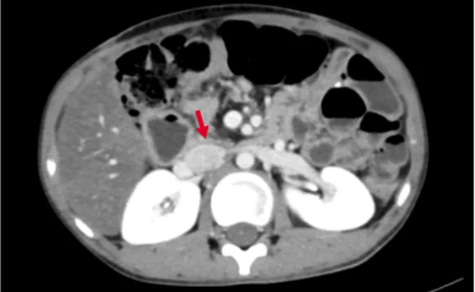
Fig. 5. CECT abdomen image of postoperative patient – six months following bile duct preserving pancreatic head re- section with posterior pancreatic capsule as the limit of re- section (pointed with red arrow).
cinate process and the inferior vena cava. He concep- tualised that this shell of pancreatic tissue will prevent egress of pancreatic secretions into retroperitoneum. The exact extent of pancreatic tissue to be retained was not defined. He also proposed to expose the anterior surface of the intrapancreatic common bile duct in order to relieve constriction by fibrosis, which may not be the required scenario always.1 Frey and Amikura2 reported the results of the proposed Frey’s procedure in 1994 after follow-up of 37 months. In this article, Frey and Amikura2 described that local resection of head of pancreas is achieved by ex- cising the head of the pancreas overlaying the ducts of Wirsung and Santorini, and duct to the uncinate, along with their tributary ducts. The extent of resection de- scribed by Frey and Amikura2 in 1994 study has been as- sumed by many as modification of Frey’s procedure by himself to minimise the risk of penetration of posterior pancreatic capsule. This varied description of the proce- dure has created some confusion to conclude the limit of posterior extent of pancreatic resection. Many further modifications of the proposed procedure of local resection of head of pancreas have been described in the literature.
Tan et al.7 conducted a retrospective review to compare the outcomes of original and modified Frey’s procedure.
He concluded that original Frey’s procedure, inspite of be- ing safe, achieved better pain relief and equal preservation of pancreatic function as compared to the modified Frey’s procedure. Sakata et al reported technique of modified
“minimum Frey’s procedure” involving limited coring of pancreatic head to include only the area anterior to the main pancreatic duct. His findings stated that “minimum Frey’s procedure” is sufficient for resolving intractable pain and improving nutritional status in most patients with chronic pancreatitis.3
Gloor et al.8 reported a modified technique of Beger and Frey procedure in which retropancreatic dissection and transection of the pancreas is avoided in contrast to Beger procedure. In contrast to Frey’s procedure, Gloor et al.8 advocated deliberate opening of common bile duct
leaving behind a thin bridge of pancreatic tissue, similar to original Frey article. The exact extent of remnant pan- creatic tissue after pancreatic head excision is definitely a matter of concern. Hence, the present report of BDPPHR is contemplated to accurately define the posterior limit of pancreatic head excision and also to alleviate concerns about the remnant pancreatic tissue in the head region.
In the present report, the posterior pancreatic capsule is considered as the posterior limit of pancreatic head resection. The authors wanted to reiterate the finding of thickened posterior pancreatic capsule observed in the pa- tients of chronic pancreatitis during surgery. In order to avoid any inadvertent injury to posterior pancreatic capsu- le during kocherisation of duodenum, the authors recom- mend the plane of dissection to be close enough to in- ferior vena cava rather than to the posterior pancreatic capsule. In BDPPHR technique, perpendicular plane of pancreatic head excision is advised instead of saucer- ization of pancreatic head as shown in Fig. 2. This ap- proach is facilitated by blunt dissection through the pan- creatic tissue till the posterior pancreatic capsule is reached. Once the posterior pancreatic capsule is identi- fied, slices of pancreatic tissue held with tissue holding forceps can be easily cleared from the pancreatic capsule
using even monopolar electrocoagulation applied at an an- gle parallel to posterior pancreatic capsule. This technique does not require utilisation of an ultrasonic aspirator and dissector as described by Andersen and Topazian9 for his technique of pancreatic head excavation. In the event of any breach in the posterior pancreatic capsule, re-approx- imation of the capsule with interrupted monofilament non-absorbable sutures is enough. In the author’s experi- ence, ten patients of chronic pancreatitis underwent the described technique of BDPPHR and none had any breach in the posterior pancreatic capsule during pancreatic head excision. None of the patients had developed pancreatic fistula during the postoperative period and in further fol- low up. During the follow up, none of the patients re- quired admission for pain related to chronic pancreatitis.
Among these 10 patients, three patients were known to have diabetes mellitus preoperatively. None of the patients developed new-onset diabetes in the post-operative follow up of one year. However, much longer follow up is re- quired to assess the accurate efficacy of the procedure, es- pecially regarding functional outcomes. Post-operative CECT image of patient following BDPPHR at six months follow up is shown in Fig. 5.
CONCLUSION
Therefore, BDPPHR is a safe, feasible and easily re- producible technique of pancreatic head excision. This technique can be accomplished with widely available mo- nopolar electrocoagulation itself. The authors wanted to recapitulate the practicability of BDPPHR technique with the posterior pancreatic capsule as the limit of resection.
ORCID
Pagadala Naga Balaji Nitesh:
https://orcid.org/0000-0002-9522-7149
Biju Pottakkat: https://orcid.org/0000-0002-8474-0270
AUTHOR CONTRIBUTIONS
Conceptualization: BP. Data curation: PNBN, BP.
Formal analysis: PNBN, BP. Methodology: PNBN, BP.
Project administration: BP. Visualization: PNBN, BP.
Writing - original draft: PNBN, BP. Writing - review &
amp; editing: PNBN, BP.
REFERENCES
1. Frey CF, Smith GJ. Description and rationale of a new operation for chronic pancreatitis. Pancreas 1987;2:701-707.
2. Frey CF, Amikura K. Local resection of the head of the pancreas combined with longitudinal pancreaticojejunostomy in the man- agement of patients with chronic pancreatitis. Ann Surg 1994;
220:492-504; discussion 504-507.
3. Sakata N, Egawa S, Motoi F, Goto M, Matsuno S, Katayose Y, et al. How much of the pancreatic head should we resect in Frey's procedure? Surg Today 2009;39:120-127.
4. Issa Y, van Santvoort HC, van Goor H, Cahen DL, Bruno MJ, Boermeester MA. Surgical and endoscopic treatment of pain in chronic pancreatitis: a multidisciplinary update. Dig Surg 2013;
30:35-50.
5. Yang CJ, Bliss LA, Schapira EF, Freedman SD, Ng SC, Windsor JA, et al. Systematic review of early surgery for chronic pan- creatitis: impact on pain, pancreatic function, and re-intervention.
J Gastrointest Surg 2014;18:1863-1869.
6. Ahmed Ali U, Issa Y, Bruno MJ, van Goor H, van Santvoort H, Busch OR, et al. Early surgery versus optimal current step-up practice for chronic pancreatitis (ESCAPE): design and rationale of a randomized trial. BMC Gastroenterol 2013;13:49.
7. Tan CL, Zhang H, Yang M, Li SJ, Liu XB, Li KZ. Role of original and modified Frey's procedures in chronic pancreatitis.
World J Gastroenterol 2016;22:10415-10423.
8. Gloor B, Friess H, Uhl W, Büchler MW. A modified technique of the Beger and Frey procedure in patients with chronic pancreatitis. Dig Surg 2001;18:21-25.
9. Andersen DK, Topazian MD. Pancreatic head excavation: a var- iation on the theme of duodenum-preserving pancreatic head resection. Arch Surg 2004;139:375-379.