Received October 17, 2016, Revised January 12, 2017, Accepted for publication January 16, 2017
Corresponding author: Bong Seok Shin, Department of Dermatology, Chosun University Hospital, 365 Pilmun-daero, Dong-gu, Gwangju 61453, Korea. Tel: 82-62-220-3130, Fax: 82-62-222-3215, E-mail: derm75@
chosun.ac.kr
This is an Open Access article distributed under the terms of the Creative Commons Attribution Non-Commercial License (http://creativecommons.
org/licenses/by-nc/4.0) which permits unrestricted non-commercial use, distribution, and reproduction in any medium, provided the original work is properly cited.
Copyright © The Korean Dermatological Association and The Korean Society for Investigative Dermatology
Ann Dermatol Vol. 29, No. 5, 2017 https://doi.org/10.5021/ad.2017.29.5.578
ORIGINAL ARTICLE
A Study of the Changes of T Helper 17 Cells and Regulatory T Cells in Herpes Zoster
Min Sung Kim, Dong Jin Kim, Chan Ho Na, Bong Seok Shin
Department of Dermatology, Chosun University School of Medicine, Gwangju, Korea
Background: Immunosuppression and age-related defi- ciencies in cell-mediated immunity are important factors for the reactivation of latent varicella-zoster virus (VZV).
CD4+CD25+Foxp3+ regulatory T (Treg) cells and T helper 17 (Th17) cells are closely associated with various viral infections. Objective: We analyzed Treg cells and Th17 cells in patients with herpes zoster and investigated their relation- ship with the reactivation of latent VZV. Methods: Treg and Th17 cells in peripheral blood and the ratio of Th17 to Treg cells were examined in patients with herpes zoster and healthy controls. Changes between pre-treatment and post-treat- ment estimates of Treg and Th17 cells and clinical parame- ters in patients with herpes zoster were also analyzed.
Results: The proportion of circulating Th17 cells and the Th17/Treg cell ratio were significantly higher in patients with herpes zoster than controls (p=0.012, 0.013), but there was no significant difference in the proportion of Treg cells be- tween groups. There was no significant difference in the pro- portions of Treg and Th17 cells and the Th17/Treg cell ratio before and after treatment and between the non-postherpetic neuralgia and postherpetic neuralgia groups. Changes in Treg and Th17 cells and the Th17/Treg cell ratio were not sig- nificantly correlated with changes in the visual analog scale.
Body surface area was significantly correlated with Treg
cells, Th17 cells, and the Th17/Treg cell ratio (p=0.022, 0.002, 0.004). Conclusion: An imbalance between Th17 and Treg cells is associated with the reactivation of VZV, which may contribute to pathogenesis of herpes zoster. (Ann Dermatol 29(5) 578∼585, 2017)
-Keywords-
Herpes zoster, Regulatory T cells, Th17 cells, Varicella-zoster virus
INTRODUCTION
Varicella results from primary infection with varicella-zos- ter virus (VZV), and herpes zoster is caused by the re- activation of latent VZV in the sensory ganglia1. The mechanisms underlying the reactivation of latent VZV are unclear. However, reactivation is related to immunode- ficiency, irradiation of the spinal column, and local trau- ma, and the most important factor for reactivation is de- creased VZV-specific cell-mediated immunity with ad- vancing age1. A recent study confirmed that VZV-specific T cells in the peripheral blood decrease with advancing age, whereas regulatory T (Treg) cells increase2. T cell-me- diated immunity may play an important role in the re- activation of VZV via changes in CD4+ T cells and cyto- toxic CD8+ T cells after the development of herpes zos- ter3,4.
Tissue injury in common viral infections can be directly caused by viral replication or immunopathology; it also occurs as a result of an increased inflammatory response associated with viral replication or the inhibition of the in- flammatory response to the virus5. Treg cells in viral in- fections can control anti-viral inflammation and prevent immunopathology, but they inhibit antiviral immunity, fa- cilitate viral replication, and cause persistent viral in-
fections5. T-helper 17 (Th17) cells play an important role in the host defense against some viral infections, but they can cause detrimental immunopathological responses5. A number of studies have examined the roles of Treg cells and Th17 cells in chronic viral diseases, such as chronic hepatitis B and C virus infections, and have shown that the relationship between Treg cells and Th17 cells, rather than each of the T cell subsets alone, may play an im- portant role in disease progression and virus persistance6,7. Although recent studies have examined changes in Treg cells in the peripheral blood and postherpetic neuralgia (PHN) in patients with herpes zoster, no study has eval- uated the correlation between Treg cells and Th17 cells.
In the present study, Treg cells, Th17 cells, and the Th17/Treg cell ratio were examined in the peripheral blood from patients with herpes zoster and normal con- trols using flow cytometry to investigate their correlations with the pathogenesis of herpes zoster.
MATERIALS AND METHODS
Subjects
The subjects consisted of 25 patients with herpes zoster, including 13 inpatients who visited the Dermatology De- partment outpatient care clinic at this hospital and were diagnosed with herpes zoster from September 2015 to March 2016 and 21 healthy controls recruited during the same period. The present study was approved by the Institutional Review Board of Chosun University Hospital, Gwangju, Korea (IRB no. CHOSUN 2015-07-013-001).
All subjects agreed to participate in the study. Patients were excluded if they were taking any antiviral drug pre- scribed at another hospital prior to their visit to this hospi- tal, if they took any anti-inflammatory analgesic drug for the purpose of pain control prior to treatment, and if they were lost to follow-up. Controls with similar age and gen- der distributions were recruited among general individuals who did not have herpes zoster, received no vaccination against herpes zoster within one year, and had no past his- tory or family history of autoimmune diseases, as identi- fied in an interview.
Methods
Blood was collected once from all subjects before treat- ment, and blood was collected one additional time from the hospitalized patients after treatment for 5 days or be- fore discharge from the hospital to measure changes in Treg and Th17 cells before and after treatment. The hospi- talized patients received treatment with acyclovir at a dose of 5 mg/kg intravenously three times daily for 5 days with analgesics and didn’t take any systemic agents in-
cluding systemic glucocorticosteroid that may affect im- mune cells. The affected body surface area (BSA) was measured using clinical photographs obtained at admission.
Pain scores before and after treatment were assessed on a scale from 0 (no pain) to 10 points (the most severe pain) in patients with herpes zoster using the visual analog scale (VAS). PHN was defined as a pain index of greater than 30% of the initial pain even after 4 weeks of skin rash8, and the development of PHN was examined after 4 weeks of hospital treatment. The proportions of Treg cells and Th17 cells and the Th17/Treg ratio were compared in pa- tients with herpes zoster before and after treatment to evaluate the effects of treatment. The relationships be- tween the proportion of each of T cell subset in the pe- ripheral blood and herpes zoster-related clinical parame- ters, such as disease duration, pain scores, presence or ab- sence of PHN, and affected area of skin (BSA), were evaluated.
Cell isolation
Venous blood (10 ml per subject) was collected from pa- tients with herpes zoster and normal controls in hep- arin-treated tubes. Ficoll-PaqueTM PLUS (10 ml) (GE Heal- thcare Bio-Sciences AB, Uppsala, Sweden) was added to a 50-ml tube and the blood was then carefully added to the top of Ficoll-PaqueTM PLUS, followed by centrifugation for 40 min (400×g, 20oC). After centrifugation, 3 ml of the peripheral blood mononuclear cell (PBMC) fraction was pipetted using a micropipette, and was transferred to a 15-ml tube, mixed thoroughly with phosphate-buffered saline, and washed twice (200×g, 10 minutes, 20oC) to prepare PBMCs.
Flow cytometric analysis
Th17 cells were defined as helper T cells that produce in- terleukin (IL)-17. The IL-17 Secretion Assay-Detection Kit (Miltenyi Biotec, Bergisch Gladbach, Germany) was used to determine the number of cells that produce IL-17. Two samples of 5×10⁶ PBMCs cultured in 500 μl of RPMI 1640 with 5% human serum were prepared according to the manufacturer’s protocol, and each sample was added to a 48-well plate. T cells were stimulated with CytoStimⓇ (Miltenyi Biotec) (10 μl) in one 48-well plate, and were cultured in a 5% CO2 incubator at 37oC for 4 hours.
CytoStimⓇ induces the activation of T cells. Activated CD4+ T cells may secrete effector cytokines within several hours or begin to express activation markers on cell surfaces. After cultivation, each sample was stained with monoclonal antibodies against CD4-fluorescein iso- thiocyanate (FITC) and IL-17-allophycocyanin (APC). The sample without CytoStimⓇ was treated as the negative con-
Table 1. Baseline characteristics of the herpes zoster patients and control
Characteristic Herpes zoster (n=25)
Control
(n=21) p-value
Age (yr) 0.416
Mean±standard deviation
53.40±13.74 50.38±10.63
Range (min∼max) 30∼76 34∼72
Sex (n, %) 0.669
Male 7 (28.0) 8 (38.1)
Female 18 (72.0) 13 (61.9)
trol, and the proportions of CD4+IL-17+ T cells relative to CD4+ cells were then converted to percentages using the Navios Flow Cytometer (Beckman Coulter, Krefeld, Germany) to quantify T cell subsets.
Treg cells were defined as CD4+CD25+Foxp3+ T cells. In order to determine Treg cells, Treg cell surfaces were stained for cell surface antigens of PBMCs using CD4-FITC and CD25-APC (Miltenyi Biotec) monoclonal antibodies.
Then, cells were subjected to fixation and permeabiliza- tion processes. Subsequently, intracellular staining was performed with Anti-Foxp3-PE (phycoerythrin; Miltenyi Biotec). Stained cells were analyzed by three-color fluo- rescence activated cell sorting (FACS) using a flow cy- tometer and Kaluza Analysis Software (Beckman Coulter, Brea, CA, USA), and the proportions of CD4+CD25+Foxp3+ T cells relative to total CD4+ cells were converted to per- centages to obtain the number of Treg cells.
The ratios of Th17 cells to Treg cells were calculated to in- vestigate the relationship between Th17 and Treg cells in the development and treatment of herpes zoster.
Statistical analyses
Mann-Whitney U test (Wilcoxon rank sum test), Wilcoxon signed rank test, Student’s t-tests and χ2–test were used to evaluate differences in Th17 cells, Treg cells, and Th17/Treg cell ratios between herpes zoster and control. Their rela- tionships with various clinical parameters in patients with herpes zoster before and after treatment and controls were analyzed using Pearson’s correlation tests. All statistical analyses were performed using IBM SPSS Statistics ver.
22.0 (IBM Co., Armonk, NY, USA). Graphs were plotted using GraphPad Prism 7.02 software. A p-value of less than 0.05 was statistically significant.
RESULTS
Age and gender distributions
The patients with herpes zoster ranged from 30 years old to 76 years old (mean age, 53.40±13.74 years). Individuals in the control group ranged from 34 years old to 72 years old (mean age, 50.38±10.63 years), and there was no sig- nificant difference between the two groups (p=0.416, Table 1). In terms of gender distribution, the subjects with herpes zoster included 7 males (28.0%) and 18 females (72.0%), and the control group included 8 males (38.1%) and 13 females (61.9%); there was no statistical difference between groups with respect to gender (p=0.669, Table 1).
Comparison of Treg and Th17 cell counts in the peripheral blood before treatment between patients with herpes zoster and the control group
The patients with herpes zoster visited this hospital 3.44±1.55 days after the occurrence of skin lesions and underwent a blood test. In a comparison of the numbers of Treg cells and Th17 cells in the peripheral blood before treatment between the patients with herpes zoster and the control group, there were fewer Treg cells in the patients with herpes zoster (1.26±0.88%) that in the control group (1.79±1.27%), but this difference was not statistically sig- nificant (p=0.193, Fig. 1A). In contrast, the number of Th17 cells was higher in the group of patients with herpes zos- ter (0.63±0.36%) than in the control group (0.38±
0.19%), and this difference was statistically significant (p=0.012, Fig. 1B). In order to investigate the imbalance between the two T cell subsets, the ratios of Th17 cells to Treg cells were calculated. These Th17/Treg cell ratios were statistically significantly higher in the group of pa- tients with herpes zoster (0.92±1.07) than in the control group (0.38±0.36) (p=0.013, Fig. 1C).
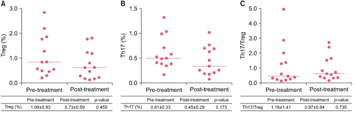
Comparison of Treg and Th17 cells before and after treatment in patients with herpes zoster
Mean time duration from pre-treatment to post-treatment sample of blood was about 5.2 days.
In a quantitative comparison of Treg cells and TH17 cells in the peripheral blood from 15 hospitalized patients with herpes zoster between before and after treatment, there were fewer Treg cells after treatment (0.73±0.59%) than before treatment (1.09±0.83%), but this difference was not statistically significant (p=0.455, Fig. 2A). There were fewer Th17 cells after treatment (0.45±0.29%) than be- fore treatment (0.61±0.33%), but a statistically significant difference was not observed (p=0.173, Fig. 2B). To inves- tigate the imbalance between Treg cells and Th17 cells, the ratios of Th17 cells to Treg cells were calculated and there
Fig. 1. Percentages of circulating regulatory T (Treg) cells (A), T helper 17 (Th17) cells (B) and the ratio of Th17 to Treg cells (C) in patients with herpes zoster and healthy controls. Data were expressed as the means±standard deviations in the table. The inset lines represented Median. Statistically significant differences were determined by Mann-Whitney U test. *p<0.05.
Fig. 2. Percentages of circulating regulatory T (Treg) cells (A), T helper 17 (Th17) cells (B) and the ratio of Th17 to Treg cells (C) in patients with herpes zoster between pre-treatment and post-treatment. Data were expressed as the means±standard deviations in the table. The inset lines represented Median. Statistically significant differences were determined by Wilcoxon signed rank test.
was no statistically significant difference between this ratio before treatment (1.16±1.41) and after treatment (0.97±0.84) (p=0.735, Fig. 2C).
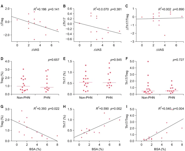
Correlations between clinical parameters and the numbers of Treg and Th17 cells in patients with herpes zoster
The mean VAS before treatment in the hospitalized pa- tients with herpes zoster was 5.8, and the mean VAS after treatment was 2.53, indicating that pain score improved after treatment. There were no significant correlations be- tween the change of the pain score and those of Treg cells, Th17 cells, or Th17/Treg ratio before and after treat- ment (Fig. 3A∼C). Additionally, no significant difference was observed between Treg cells, Th17 cells, or the Th17/Treg ratio before treatment with respect to the pres- ence or absence of PHN (Fig. 3D∼F). However, there was a statistically significant negative correlation between
BSA and Treg cells in the patients and a statistically sig- nificant positive correlation between BSA and Th17 cells and, the Th17/Treg ratio (Table 2, Fig. 3G∼I).
DISCUSSION
Herpes zoster caused by the reactivation of VZV is charac- terized by grouped vesicles accompanied by severe pain, and PHN, i.e., pain persisting for several months or years after the rash has resolved, can develop as a conse- quence9,10. The cellular mechanisms of the reactivation of the latent virus and the precise causes of PHN are un- clear3. However, in view of the fact that treatments or dis- eases that affect the functions of T cells increase the risk of herpes zoster and HIV-infected patients with a progressive decrease in CD4+ cells are at high risk for herpes zoster, cell-mediated T cells are thought to play an important role in the development of herpes zoster3,9.
Fig. 3. The correlations between changes (Δ) of regulatory T (Treg) (A), T helper 17 (Th17) (B), ratio of Th17 to Treg (C) in peripheral blood and changes (Δ) of visual analog scale (VAS) of herpes zoster patient. Percentages of circulating Treg cells (D), Th17 cells (E) and the ratio of Th17 to Treg cells (F) in patients with non-postherpetic neuralgia (PHN) group and PHN group. The correlations between body surface area (BSA) and Treg (G), Th17 (H) and the ratio of Th17 to Treg (I). (A∼C) Linear regression analysis was used to fit lines on graphs. Statistically significant differences were determined by Pearson’s correlation analysis. (D∼F) The inset lines represented Median. Statistically significant differences were determined by Mann-Whitney U test. (G∼I) The inset lines are linear regression lines. Statistically significant differences were determined by Pearson’s correlation analysis.
Table 2. Pearson correlations between BSA, Treg cells, Th17 cells, the ratio of Th17 to Treg cells
BSA Th17 cells Treg cells Th17/Treg BSA
Th17 cells 0.768**
Treg cells −0.627* −0.388
Th17/Treg 0.735** 0.795** −0.621*
BSA: body surface area, Treg: regulatory T, Th17: T helper 17.
*Correlation is significant, p<0.05 (two-tailed), **Correlation is significant, p<0.01 (two-tailed).
Naïve CD4+ T cells can differentiate into Th1, Th2, Treg, and Th17 cells under certain conditions and produce a va- riety of cytokines that mediate immune responses11. Trans- forming growth factor (TGF)-β induces the differentiation of naïve CD4+ T cells into Treg cells to produce TGF-β and IL-10, maintain immunological tolerance, and control inflammatory responses. TGF-β and IL-6 induce the dif- ferentiation of naïve CD4+ T cells into Th17 to produce IL-17, -21, and -23 and promote inflammatory responses11. Treg differentiation and Th17 differentiation are related processes; the two T cell subsets play an important role in maintaining immunological homeostasis and there may exist an important plasticity of the two T cell subsets7.
A recent study has reported that during viral infection, CD4+CD25+ Treg cells inhibit the activation and func- tions of effector T cells and, in turn, control antiviral re- sponses3. Tissue damage in viral infection can be caused directly by viral propagation in the tissue, and this is regu- lated by the antiviral immune response, which acts to sup- press viral replication5.
Although Treg cells can be beneficial in viral infections by acting to suppress tissue damage caused by virus-specific T cells, Treg cells can suppress host immunity, which is instrumental in eradicating viruses, leading to persistent viral infection and disease progression12,13. CD4+ cells and CD8+ cytotoxic cells play an important role in con- trolling VZV replication in the ganglia during the acute phase of herpes zoster4. CD8+ cytotoxic T cells play a key role in the control of intracellular viral replication by elim- inating infected cells. When cytotoxic T cell responses are impaired or the virus alters cytotoxic responses, a chronic viral infection can result. Treg cells are also involved in the maintenance of a persistent viral infection5,12. In a study on herpes zoster-related antigens, Treg cells increased in the peripheral blood and skin tissue with increasing age; when the skin was sensitized to the VZV antigen, CD4+Foxp3+ T cells increased and there was an inverse correlation between CD4+Foxp3+ T cells and VZV skin reactions. Thus, CD4+Foxp3+ T cells were thought to sup- press antigen-specific responses14.
Th17 cells that produce IL-17 are associated with in- flammatory tissue damage, lead to a variety of auto- immune diseases, and play a key role in host defense against bacterial infections or fungal infections15. Th17 cells in viral infections generally cause immunopathology, with detrimental effects on the host15. IL-17 secreted by Th17 cells suppresses the production of IL-2 and interfer- on-γ and, in turn, inhibits the differentiation of Th1 cells, which have cytotoxic and antiviral functions, thus causing persistent viral infections5,15. If Th17 cells suppress Th1 immune responses, viral replication cannot be controlled or it can be detrimental to the host by mediating im- munopathology12. Although Th17 cells induce immuno- pathological responses in some infections, they are essen- tial and protective in the host response to intracellular bacteria or viruses5. Deficiencies in Th17 cells caused by viral infections, such as HIV infection, can exacerbate dis- eases caused by extracellular bacteria or viruses, and lead to increased opportunistic infections5. In 2016, Zajkowska et al.16 first reported that Th17-related cytokines (IL-17, 21, 23) levels were significantly higher in herpes zoster than controls. However, they stated that further studies are needed to investigate that. This result indirectly suggests that Th17 cells may be high in herpes zoster.
An imbalance between Treg cells and Th17 cells is asso- ciated with the progression or prognosis of autoimmune diseases, such as psoriasis and atopic dermatitis, or in- flammatory diseases. Recently, studies have examined the imbalance between the two T cell subsets in chronic viral diseases, such as hepatitis B virus (HBV) and hepatitis C virus (HCV) infection6,7,11. According to a study by Xue-Song et al.6, Th17 cells are involved in acute and chronic HBV infection, and an imbalance in the Treg/Th17 ratio was observed for chronic hepatitis B and acute-on-ch- ronic HBV-related liver failure patients, which was linked to disease progression and continuous HBV infection. In addition, in a study of chronic HCV-infected patients by Hao et al.7, Treg cell proportions were significantly ele- vated in HCV-infected patients, but there was no sig- nificant difference in the proportion of Th17 cells between infected and uninfected individuals. Furthermore, when HCV replication was inhibited, there was a reduction in Treg cells, leading to a significant decrease in the Treg/Th17 ratio. An imbalance in the ratio of Treg cells to Th17 cells might play an important role in persistent HCV infection.
Herpes zoster may have acute and chronic responses, be- cause of acute viral infection and chronic exposure of VZV antigen during periodic episodes of subclinical reactivation.
In a recent study of Treg cells in patients with herpes zos- ter by Xing et al.3, the number of CD4+CD25+Foxp3+ T cells in the peripheral blood of patients with herpes zoster was significantly higher than that of normal controls and tended to increase as pain increased in severity. Furthermore, the proportions of CD4+CD25+Foxp3+ T cells were sig- nificantly higher in the group with PHN than the group without PHN. These findings suggest that T cell immunity was impaired in patients with herpes zoster, and increased activation of Treg cells might suppress the antiviral im- mune response of CD4+ T cells. Accordingly, they as- serted that Treg cells may play an important role in the pathogenesis of herpes zoster and progression toward PHN. But, the results of our study revealed that Treg cell quantities were decreased compared to those in the con- trol, although it was not statistically significant. Our find- ings conflict with the results of Xing et al.3 and suggest that antiviral immune response increased in response to a reduction in Treg cells, leading to the decrease of VZV af- ter outbreak of the disease, furthermore inflammatory re- sponses increased via an increase in Th17 cells. We think there may be a difference between authors in herpes zoster. Perhaps, this may be discrepancy of blood sam- pling time in accordance with disease activity. For exam- ple, blood sample taken before outbreak of herpes zoster showed higher Treg cells count than herpes zoster neg-
ative control17. In addition, these observations may be ex- plained by the plasticity of the two T cell subsets, in which Treg cells are converted to Th17 cells by inflammatory cy- tokines owing to increased inflammatory responses, al- though this is controversial recently18,19. When analyzing the imbalance between the two T cell subsets or each T cell subset and the development of herpes zoster, the ratio of Th17 cells to Treg cells was significantly elevated in pa- tients with herpes zoster compared with normal controls.
The results of the present study confirm that the balance between Treg cells and Th17 cells is critical for the devel- opment of herpes zoster, similar to observations that an imbalance between Treg cells and Th17 cells, rather than each T cell subset alone, in chronic viral diseases, such as chronic HBV and HCV infections, affects progression and treatment6,7. The analysis of herpes zoster-affected BSA, Treg cells, Th17 cells, and Th17/Treg ratios showed that significant positive correlations between BSA and Th17 cells and the Th17/Treg ratio and a significant negative correlation between BSA and Treg cells. Accordingly, the affected area may reflect cellular changes in the peripheral blood. So, we thought that Th17 cells may be associated with herpes zoster per se but also with the extent of lesions.
This study had several limitations. The number of partic- ipants was relatively small, and the onset time of herpes zoster differed among patients at the time of blood collec- tion; this may explain differences in cellular changes. Treg cells generally tend to increase with age. Although the mean age of controls was higher than that of patients with herpes zoster, age-related changes were not considered in this study. In addition, when the proportions of Treg cells and Th17 cells in the peripheral blood were compared be- fore and after treatment, blood was collected during a short period of time, i.e., 1 week, to observe changes in each cell type, based on the fact that that viral replication is generally regulated within 1∼2 weeks9.
In the present study, the imbalance between Treg and Th17 cells and the development of herpes zoster was ex- amined to determine the effects of Treg and Th17 cells on the reactivation of VZV and to examine the imbalance in Treg and TH17 cells and the development of herpes zoster.
In conclusion, the number of Th17 cells and the ratio of Th17 to Treg cells were significantly higher in patients with herpes zoster than in healthy controls. And, there was a significant correlation between the proportion and ratio of Treg and Th17 cells and the affected BSA.
Therefore, Th17 was involved in herpes zoster and an im- balance between Treg cells and Th17 cells was observed in patients with herpes zoster. But, additional studies are
needed to examine the mechanism by which Treg and Th17 cells influence the development of herpes zoster.
ACKNOWLEDGMENT
The present study was supported by grants from the Clinical Medicine Research Institute at Chosun University Hospital, 2015.
CONFLICTS OF INTEREST
The authors have nothing to disclose.
REFERENCES
1. Schmader KE, Oxman MN. Varicella and herpes zoster. In:
Goldsmith LA, Katz SI, Gilchrest BA, Paller AS, Leffell DJ, Wolff K, editors. Fitzpatrick's dermatology in general medicine. 8th ed. New York: McGraw-Hill, 2012:2386.
2. Vukmanovic-Stejic M, Sandhu D, Seidel JA, Patel N, Sobande TO, Agius E, et al. The characterization of varicella zoster virus-specific T cells in skin and blood during aging. J Invest Dermatol 2015;135:1752-1762.
3. Xing Q, Hu D, Shi F, Chen F. Role of regulatory T cells in patients with acute herpes zoster and relationship to postherpetic neuralgia. Arch Dermatol Res 2013;305:715- 722.
4. Steain M, Sutherland JP, Rodriguez M, Cunningham AL, Slobedman B, Abendroth A. Analysis of T cell responses during active varicella-zoster virus reactivation in human ganglia. J Virol 2014;88:2704-2716.
5. Martinez NE, Sato F, Kawai E, Omura S, Chervenak RP, Tsunoda I. Regulatory T cells and Th17 cells in viral infections: implications for multiple sclerosis and myocarditis.
Future Virol 2012;7:593-608.
6. Xue-Song L, Cheng-Zhong L, Ying Z, Mo-Bin W. Changes of Treg and Th17 cells balance in the development of acute and chronic hepatitis B virus infection. BMC Gastroenterol 2012;12:43.
7. Hao C, Zhou Y, He Y, Fan C, Sun L, Wei X, et al. Im- balance of regulatory T cells and T helper type 17 cells in patients with chronic hepatitis C. Immunology 2014;143:
531-538.
8. Falaki F, Nejat AH, Dalirsani Z. The effect of low-level laser therapy on trigeminal neuralgia: a review of literature. J Dent Res Dent Clin Dent Prospects 2014;8:1-5.
9. Arvin A, Abendroth A. VZV: immunobiology and host response. In: Arvin A, Campadelli-Fiume G, Mocarski E, Moore PS, Roizman B, Whitley R, et al, editors. Human herpesviruses: biology, therapy, and immunoprophylaxis.
Cambridge: Cambridge University Press, 2007:700-712.
10. Roh NK, Park YM, Kang H, Choi GS, Kim BJ, Lee YW, et al.
Awareness, knowledge, and vaccine acceptability of herpes zoster in Korea: a multicenter survey of 607 patients. Ann Dermatol 2015;27:531-538.
11. Chen Y, Fang J, Chen X, Pan C, Liu X, Liu J. Effects of the
Treg/Th17 cell balance and their associated cytokines in patients with hepatitis B infection. Exp Ther Med 2015;
9:573-578.
12. Li S, Gowans EJ, Chougnet C, Plebanski M, Dittmer U.
Natural regulatory T cells and persistent viral infection. J Virol 2008;82:21-30.
13. Rushbrook SM, Ward SM, Unitt E, Vowler SL, Lucas M, Klenerman P, et al. Regulatory T cells suppress in vitro proliferation of virus-specific CD8+ T cells during persistent hepatitis C virus infection. J Virol 2005;79:
7852-7859.
14. Vukmanovic-Stejic M, Sandhu D, Sobande TO, Agius E, Lacy KE, Riddell N, et al. Varicella zoster-specific CD4+Foxp3+ T cells accumulate after cutaneous antigen challenge in humans. J Immunol 2013;190:977-986.
15. Hou W, Kang HS, Kim BS. Th17 cells enhance viral
persistence and inhibit T cell cytotoxicity in a model of chronic virus infection. J Exp Med 2009;206:313-328.
16. Zajkowska A, Garkowski A, Świerzbińska R, Kułakowska A, Król ME, Ptaszyńska-Sarosiek I, et al. Evaluation of chosen cytokine levels among patients with herpes zoster as ability to provide immune response. PLoS One 2016;11:e0150301.
17. Weinberg A, Huang S, Song LY, Fenton T, Williams P, Patterson J, et al. Immune correlates of herpes zoster in HIV-infected children and youth. J Virol 2012;86:2878-2881.
18. Xu L, Kitani A, Fuss I, Strober W. Cutting edge: regulatory T cells induce CD4+CD25-Foxp3- T cells or are self-induced to become Th17 cells in the absence of exogenous TGF- beta. J Immunol 2007;178:6725-6729.
19. DuPage M, Bluestone JA. Harnessing the plasticity of CD4(+) T cells to treat immune-mediated disease. Nat Rev Immunol 2016;16:149-163.